In modern society, migration flows are extremely high. The existence of multiple accessible modes of transportation has led to the fact that infectious diseases from certain territories migrate throughout the globe. Cases of imported tropical diseases are increasing. Currently, any trip or travel abroad can result in an encounter with an infection. One of these diseases is Q fever.
- 2 Forms of the disease: acute, subacute, chronic
- 3 The causative agent of the disease
- 4 How the infection is transmitted to humans
4.1 Some aspects of infectious diseases - video
- 6.1 Microbiology comes to the rescue
6.1.1 Immunological methods for diagnosing Q fever - photo gallery
- 9.1 Vaccination is a method of specific prevention
Pathogenesis[ | ]
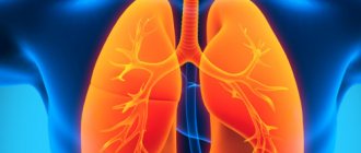
A low dose (1 to 10 bacteria) can cause infection. The incubation period is approximately two to three weeks (range: one to six weeks). After infected particles enter the host’s body, the microorganism multiplies in the phagolysosomes of macrophages and monocytes, which allows them to avoid phagocytosis. If the bacterium enters by inhalation, it is transported in the body mainly by pulmonary macrophages to the liver, spleen and bone marrow. Systemic invasion of bacteria into the host body leads to the appearance of symptoms and various clinical manifestations, which depend on the infecting dose and the response of the host body. In immunocompetent patients, the inflammatory response may be caused by immune mechanisms that manifest as non-necrotizing granulomas in the liver or bone marrow, known as donut granulomas.
In a small proportion of patients, primary infection results in persistent focal infection. Such changes depend on both host and bacterial factors. For example, in a small number of patients, macrophages are unable to kill the microorganism due to increased secretion of interleukin (IL)-10, which is produced by infected monocytes. Patients with persistent focal infection have high levels of interleukin-10. Patients at risk of developing persistent focal infection include pregnant women and patients with pre-existing valvulopathy or vasculopathy, as well as immunocompromised patients due to HIV infection or chemotherapy for cancer.
Although the life cycle of the pathogen remains unclear, there are two forms of the microorganism (small and large) that can be easily distinguished using electron microscopy. The small form of the microorganism ("pseudospore") is resistant to heat, drying and multiple disinfectants, which allows the pathogen to remain viable for a long period of time. For example, bacteria can survive at low temperatures in stored meat for one month and in skim milk at room temperature for 40 months.
C. burnetii
has two antigenic states. Bacteria isolated from patients or laboratory animals are in antigen phase I, and the organism is considered virulent. The bacterium, isolated from subcultures of cells or eggs with embryos, has an antigenic shift and is in antigen phase II, and is an avirulent form. Bacteria that are in antigen phase I and II contain plasmids, but their role in the pathogenesis of the disease is not well understood. To confirm the diagnosis of the disease, antibodies to phase I and II antigens of the microorganism are determined. Only 1-2% of patients die from acute infection. Those who fully recover may have lifelong immunity against reinfection (immunity is non-sterile; no cases of reinfection have been reported). However, up to 65% of people with untreated endocarditis may die from the disease[6].
Diagnostics
Although Q fever is rarely diagnosed, it should always be considered when evaluating a child with fever of unknown origin, atypical pneumonia, or endocarditis and negative culture results if the child lives in a rural area and is exposed to livestock, cats, or animal products.
The easiest way to diagnose Q fever is to compare the antibody titer in sera taken during the acute period of the disease and during the recovery period. A 4-fold increase in titer is diagnostically significant.
C. burnetii grows in cell cultures, signs of growth can sometimes be determined after 48 hours, however, culture for C. burnetii, as well as antibiotic sensitivity testing, due to the high danger, are carried out only in specialized laboratories.
Differential diagnosis . The range of diseases for differential diagnosis of Q fever depends on the clinical picture. If the respiratory system is affected, it is necessary to exclude mycoplasma pneumonia, Legionnaires' disease, psittacosis, and infection caused by the Epstein-Barr virus. For granulomatous hepatitis, the differential diagnosis is made with mycobacterial infections, salmonellosis, visceral leishmaniasis, toxoplasmosis, lymphogranulomatosis, ehrlichiosis, brucellosis and autoimmune diseases, including sarcoidosis. Endocarditis with negative culture results may be caused by Brucella, Bartonella, or have a nonbacterial etiology.
Clinical picture[ | ]
Incubation period:
from 3 to 32 days, more often 12-19 days.
In most cases, the disease begins acutely. Complaints are varied:
headache, pain in the lower back, muscles, joints, feeling of weakness, dry cough, sweating, loss of appetite, sleep disturbance.
Upon examination, facial hyperemia, injection of scleral vessels, and pharynx hyperemia are revealed. Most patients develop hepatolienal syndrome early. Temperature - 39-40°, the temperature curve is varied - constant, remitting, wavy, irregular. Duration of fever:
usually within 2 weeks, but relapses and prolonged low-grade fever are possible. In some patients, pneumonia and tracheobronchitis are detected (due to airborne dust infection). The blood picture is not very characteristic; Leuko- and neutropenia, relative lymphocytosis, and a moderate increase in ESR are more often observed. There are acute (up to 2-3 weeks), subacute (up to 1 month) and chronic (up to 1 year) forms, as well as an erased form, which is diagnosed only in lesions during laboratory examination. Complications are rare.
Treatment[ | ]
21-day course of oral antibiotic therapy[ | ]
- Acute infection is usually mild, tends to be self-limiting, and usually resolves spontaneously within two weeks.
- Treatment is not recommended for patients with acute infection without valvulopathy who are asymptomatic. However, if symptoms are present, oral antibiotic therapy should be given as it may shorten the duration of illness and reduce the risk of hospitalization.
- The recommended first-line therapy is doxycycline (see Tetracyclines). If the patient cannot tolerate doxycycline, other antibiotics (eg, fluoroquinolones or trimethoprim/sulfamethoxazole) may be used.
- The course of treatment ranges from 14 to 21 days.
Plus - Maintenance therapy[ | ]
- Patients with acute infection should remain in bed and drink plenty of fluids.
- If you have a cough, you can use antitussives
- Paracetamol or non-steroidal anti-inflammatory drugs (NSAIDs) are not recommended for the treatment of fever and discomfort as they may impair liver function and thus worsen the disease[6].
Prevention[ | ]
Prevention of Q fever:
consists in carrying out a complex of sanitary-veterinary and sanitary-preventive measures. They are aimed at preventing the introduction of infection into livestock farms and include inspection and examination of new animals entering the farm, isolation and treatment of sick animals, disinfection of their feces and amniotic fluid, as well as premises (stalls, feeding troughs, etc.) 3-5% creolin solution or 10-20% bleach solution. When working with sick animals (care, treatment), personal preventive measures should be observed - use special clothing (rubber boots, gloves, aprons, gauze respirators and disinfect them after work). Milk from disadvantaged farms must be sterilized; preparing kefir, cottage cheese, butter, etc. from unboiled milk is unacceptable. In the epidemiological focus of Q fever, current and final disinfection is carried out, and according to epidemic indications, people are vaccinated. Hygienic education of the population regarding the prevention of Q fever is important.
Prevention of Q fever
Have questions or something is unclear?
Ask the article editor - . Once Q fever is detected in livestock or pets, workers must be warned about the risk of infection.
Milk from herds containing diseased animals should be pasteurized at a temperature that ensures the destruction of C. burnetii. These microorganisms persist in the external environment for a long time, but die when exposed to a 1% Lysol solution, 1% formaldehyde solution and 5% hydrogen peroxide solution. There is no need to isolate patients, since transmission of Q fever from person to person is extremely rare (the exception is contact with infected tissues of the placenta). There is a vaccine against Q fever; Immunization, for example, of slaughterhouse workers provides protection against this infection for at least 5 years. The vaccine is highly reactogenic and has not been tested in children, so it should only be administered to children if there is an exceptionally high risk of infection. The article was prepared and edited by: surgeon I.B. Pigovich.
Video:
Healthy:
Related articles:
- Acute rheumatic fever Acute rheumatic fever is most common among children from 5 to 15 years old, in adults…
- Transient fever of newborns Sometimes newborns on the 2-3rd day of life experience an increase in temperature to 38-39 ° C without obvious ...
- Trench fever The causative agent of trench fever was originally named Rickettsia quintana, then it was assigned to the family Rochalimaea, and subsequently ...
- Marseilles fever The infection caused by R. conorii has received different names depending on the place of origin: Mediterranean spotted fever,…
- Dengue fever Dengue fever, a benign form of the disease, is caused by several arboviruses and manifests itself as a two-wave fever,…
- Yellow fever Yellow fever is an acute infection that, in its most severe forms, is accompanied by fever, jaundice, proteinuria...
Use as a bacteriological weapon[ | ]
The causative agent of Q fever
used in the USSR in World War II as a bacteriological weapon. The development was carried out at the Research Institute of Epidemiology and Hygiene (currently the Research Institute of Microbiology of the Ministry of Defense of the Russian Federation) in Kirov[7]. According to the assumption of a certain lieutenant colonel mentioned in the book by Kanatzhan Alibekov, the outbreak of Q fever in the ranks of German troops in the Crimea was caused by the use of the corresponding rickettsia [8]. Before this incident, Q fever was not known on the territory of the Soviet Union[7].
Treatment of coxiellosis
Treatment of the disease is carried out in a hospital setting under the guidance of an infectious disease specialist. The main measure is the prescription of antibacterial drugs. Can be used:
- tetracycline antibiotics: Tetracycline, Doxycycline, Levomycetin;
- fluoroquinolone antibiotics: Ofloxacin, Ciprofloxacin;
- macrolide antibiotics: Clarithromycin, Azithromycin, Erythromycin, Roxithromycin.
The drug Azithromycin is an effective treatment for Q fever
Notes[ | ]
- Disease ontology database (English) - 2020.
- E.P.Shuvalov, E.S.
Belozerov. Infectious diseases. — 2020. - E. P. Shuvalova.
Infectious diseases. - Medicine, 2005. - ISBN 522-504-00-63. - R.F. Sosov and others.
Epizootology. - M.: Kolos, 1969. - 400 p. - Flu that is not flu (Russian). Retrieved December 15, 2009.
- ↑ 1 2
Coxiella burnetii infections: symptoms, diagnosis, prevention and treatment
(unspecified)
. - ↑ 12
Soviet biological weapons: history, ecology, politics, L. A. Fedorov, Moscow 2005 - Alibek, Ken.
Biohazard : the chilling true story of the largest covert biological weapons program in the world, told from the inside by the man who ran it. — 1st ed. - New York: Random House, 1999. - P. 36. - xi, 319 pages, 8 unnumbered pages of plates p. — ISBN 9780375502316.