Tropical diseases at the present stage of development of civilization have ceased to be the lot of exclusively local residents of equatorial regions. Business travel and international tourism contribute to the emergence of imported cases and further spread of infection. Similar diseases include Ebola hemorrhagic fever.
- 2 Causes and development factors
- 3 Signs and symptoms of Ebola
3.1 Basic clinical aspects of Ebola fever - video
- 5.1 Drugs for the symptomatic treatment of Ebola
General information
Ebola fever is an endemic disease (spread over a limited area) that is characteristic of the countries of North-West Africa. An earlier name was Ebola hemorrhagic fever. Refers to acute, especially dangerous infectious diseases, which is accompanied by the development of a systemic inflammatory reaction and subsequent development of DIC syndrome and severe multiple organ failure (Wikipedia).
The distribution area of the virus is the countries of Central/West Africa (Nigeria, Liberia, Sudan, Zaire, Gabon, Ethiopia, Central African Republic, Senegal, Cameroon), but it may shrink or expand in different years. Below is the distribution area of the virus as of February 2020.
The first cases of the disease were recorded back in 1976 in Sudan and Congo in villages located in the Ebola River basin, where the disease got its name. Since 1976, 6 large (regional) and about 20 local epidemic outbreaks have been registered, the largest of which was the 2014 outbreak, during which a total of 23,200 people fell ill (in Sierra Leone, Guinea, Congo, Liberia, Senegal, Nigeria), and the number of fever victims reached 8,290 people. At the same time, the mortality rate in different countries varied from 55 to 90%.
The current epicenters of the outbreak are Katwa and Butembo health zones, which account for the majority (71%) of reported cases, but isolated cases continue to occur simultaneously in different countries with wide geographical dispersion. Thus, as of February 5, 2020, a total of 789 confirmed cases of Ebola disease were registered, of which 488 cases resulted in the death of patients (the overall case fatality rate is 62%). According to WHO (2019), the risk of regional and national spread of the outbreak is very high, but the risk of international spread is low, due to active population movement for personal/economic reasons between affected areas and other parts of the country, as well as neighboring countries. The Ebola virus has not been registered in Russia, with the exception of 2 cases of laboratory infection in 1996 and 2004. In addition, in Russia there are no carriers of the Ebola virus and there are no endemic foci, which makes it impossible for an outbreak of the disease to occur in the country except in cases of importation of the disease.
The main threat to the spread of Ebola disease to other parts of the world comes from passengers infected with the virus traveling from the region's main hub in Nigeria to various international routes (Figure below).
Below are photos of people infected with the Ebola virus.
According to space sensing data, all outbreaks of Ebola disease are associated with landscape zones of gallery tropical forest (along river banks in Sudan) or continuous tropical forest (in Congo and Gabon). Outbreaks occur primarily in spring and summer.
Since the incidence of Ebola in different years can vary widely: from the absence of cases or sporadic incidence to epidemic outbreaks, many during the period of absence of cases of the disease are trying to understand where Ebola disease went? Unfortunately, Ebola disease does not go away, nor does it appear out of nowhere. It, like all natural focal diseases, exists in nature, and its causative agent is a member of the ecosystem. And the incidence appears when a person invades the territory of a natural outbreak and comes into contact with a carrier of the virus.
Story
Infection occurred not only in densely populated cities in hot countries with insufficient hygienic living conditions, but also in northern regions, and even in laboratories.
Ebola virus
The Ebola virus was recorded at the end of the twentieth century in Zaire, Sudan, and Gabon. Three strains were identified that were different from each other (Lassa, Marburg and Ebola). Even earlier, closer to the middle of the century, the disease was noted in Nigeria, Senegal, and Ethiopia.
A few facts from the history of the twenty-first century:
- 2003 Congo, 128 people died. And outbreaks are currently occurring, the last one was recorded in the twelfth year. Fourteen people died then.
- West Africa. Over the winter, eight thousand registered people died, and approximately another two thousand unregistered. In the fifteenth year, it was announced that there was no more virus, but still more than three hundred residents died in the early summer of that year.
- America, England, Canada and some other countries in the northern hemisphere also have infected people. Infected tourists who arrived could infect their loved ones. Therefore, in the fourteenth year, WHO staff recognized the disease as a worldwide threat. At the same time, the first deaths were recorded.
In Russia, there were also cases of infection in laboratory conditions:
- At the end of the twentieth century, while studying a virus at the center of a research institute of the Russian Federation, a laboratory assistant died from exposure to the microorganism through a skin puncture. She was injecting rabbits and accidentally hit her finger with the needle.
- In two thousand and four, a senior laboratory assistant at a scientific research center died from a skin cut while working with guinea pigs.
Pathogenesis
Entry points for infection can be areas of damaged skin and mucous membranes. At the same time, there are no local changes at the site of virus introduction. The virus infects several types of cells (epithelial cells, macrophages, monocytes, dendritic cells, fibroblasts, endothelial cells, hepatocytes, adrenal cortex cells). The virus, after entering the body, migrates first to the regional lymph nodes, and then to the spleen, liver, and adrenal glands, which leads to the development of hepatocellular necrosis and disruption of the processes of synthesis/regulation of blood coagulation factors with the development of coagulopathy.
Necrosis of adrenal tissue leads to disruption of the synthesis of steroid hormones and hypotension . The Ebola virus suppresses the interferon production system, and the activation of immunocompetent cells does not contribute to the complete elimination of the virus. The leading role in tissue damage is played by direct cytopathic damage to cells by the virus with an increase in the production of cytokines against the background of an insufficient humoral immune response caused by the immunosuppressive effect of soluble Gp-antigens of the virus. It is Gp antigens that provide suppression of neutrophils and inhibit the immune response.
An uncontrolled and excessive inflammatory reaction acquires a pronounced systemic character (the so-called “cytokine storm”), which ultimately leads to massive damage to the endothelium of blood vessels, the development of disseminated intravascular coagulation syndrome and severe multiple organ failure. In cases of an active immune response, Ebola disease ends in recovery.
Pathogenetic mechanism
Once in the body, the Ebola virus concentrates in the lymph nodes and spleen. The acute febrile stage coincides with the spread of the microorganism through the lymphatic tissues to different parts of the body. Damage to cells and tissues occurs under the influence of the virus itself and the resulting immune cells that destroy healthy ones. The vessels begin to let blood through the walls, fluid accumulation around them is noted, and a disseminated blood clotting disorder occurs. This symptom is the main one.
When opening the patient's body, frequent necrotic changes in organs and tissues, as well as hemorrhages, are noted. Immunity is reduced and does not form antiviral cells. If a person has lived long enough with the infection inside, then the immune system works, but still, it is already very late.
Causes
Etiology
Ebola virus is an RNA genomic virus belonging to the genus Filovirus of the Filoviridae family. The Ebola virus exists in 5 subtypes: Bundibugyo (BDBV); Reston (RESTV); Zaire (EBOV); Thai Forest (TAFV) and Sudan (SUDV). All except Reston (RESTV) are highly pathogenic and capable of causing Ebola disease in humans. RESTV viruses can infect humans, but do not lead to the development of clinical manifestations of the disease, that is, they cause an asymptomatic course of the disease with the formation of specific antibodies .
Epidemiology
The main natural reservoir of the Ebola virus is presumably the fruit bat (hammerhead, epaulette, collared fruit bat), which lives only in the subtropical/tropical zones of the Eastern Hemisphere, in whose blood RNA of the Ebola virus and antibodies to it were found. This is confirmed by the coincidence of the places of epidemic manifestations of the disease and the habitats of fruit bats. It is highly likely that the Ebola virus can infect other mammals (rodents, primates, antelopes, pigs), and dogs with the development of viremia . Ebola infection is a natural focal infection, and its pathogen can circulate for a long time in natural biocenoses through a continuous epizootic process among animals that are donors and recipients.
The Ebola virus can be transmitted from infected animals to people through direct contact with secretions, blood and other fluids. The virus can also spread through contact from person to person when biological fluids contaminated with the Ebola virus (saliva, blood, semen, urine) enter damaged skin/mucous membranes and through indirect contact with all environments contaminated with costume fluids. Ebola patients/survivors are dangerous to others as long as their fluids, including breast milk and semen, contain the pathogen (up to 7 weeks). That is, after the virus penetrates the human population, Ebola disease behaves, in essence, like anthroponosis .
The main risk group in African republics are relatives and friends caring for the patient, as well as people who have direct contact with him (hospital staff/patients and participants in funeral ceremonies) if security is not strictly observed. Specific local funeral rites with the participation of shamans are important in the spread of infection, during which people present at the funeral are in direct contact with the body of the deceased (joint washing of the body by all relatives, crowded funerals with hugging and kissing the deceased, wearing the clothes of the deceased by relatives, etc.) , since for 50 days the deceased may pose a danger (at the peak of viremia, the corpse releases the virus with all biological fluids). The spread of the Ebola virus through water, air and food has not been reliably proven.
Mostly adults are affected. Susceptibility to the virus is high, the contagiousness index is 95%. The risk of intrafamilial infection varies between 5-17%, and nosocomial infection - more than 50%. Almost 7% of the population in regions endemic for Ebola disease have antibodies to the virus, which indirectly indicates the possibility of a mild/subclinical course of infection. People suffering from Ebola in cases of recovery acquire a relatively stable viremia , that is, cases of recurrent disease are extremely rare.
Epidemiologists distinguish 3 types of epidemic outbreaks of Ebola fever - forest, rural and urban (table below):
- The forest type is found mainly in forest villages located in the subequatorial forests of Central Africa. Fruit bats living in forests often infect monkeys, and contaminated fruits that fall to the ground can infect terrestrial animals of the adjacent ecological complex (ungulates), which suffer predominantly in a clinically pronounced form. Such animals with reduced mobility are often the prey of local hunters, who first of all become infected themselves and bring the virus to forest villages. Given the lack of any medical care due to the isolation of such settlements, uncontrolled epidemic outbreaks of Ebola fever occur with a high mortality rate (70-90%).
- Village type . It occurs in cases when plantations of agricultural crops and fruits come close to forests, and fruit bats “raid” them, contaminating the fruits and becoming a direct target of hunting. As a result, epidemic outbreaks occur in villages adjacent to the vicinity of plantations.
- Urban type of epidemic. Occurs in large populated areas, especially in cities with high population density. The source of the virus here is directly sick people and it is transmitted to healthy people through household/contact through various biological fluids (urine, feces, blood, vomit, sweat, semen, saliva, tears).
A characteristic feature of the Ebola epidemic is its protracted nature, which is facilitated by a high rate of population growth, which was accompanied by an acceleration of anthropogenic impact on the habitat (deforestation brought virus carriers closer to populated areas), porous borders, extreme poverty and illiteracy, forcing people to frequently move in search of food/work, the impossibility of carrying out a full range of sanitary and anti-epidemic measures, as well as low availability/lack of medical care. All this often leads to a loss of control over the epidemic process at the regional level in West African countries.
The duration of the incubation period is 3-9 days, varying from 2 to 21 days. It is also important to take into account that a significant proportion of cases of Ebola disease in endemic regions are asymptomatic in people (according to various sources, up to 35% of those examined). Despite the fact that these individuals do not pose a danger to others, they may have protective immunity , which is of great epidemiological significance.
Who is the source of infection for this infection?
It has not yet been established which animals are the natural reservoir and source of infection. Under natural conditions, the release of the virus has been recorded in various animals; the main role is assumed to be in monkeys and various rodents. However, at the same time, these animals are not considered as the main natural reservoir, since Ebola disease in them is severe, with high mortality, and carriage is rare. A person with a fever becomes extremely dangerous to others within three weeks after the first symptoms of the disease appear. The virus is detected in various biological fluids: blood, mucus, urine, semen, etc., which causes a high probability of infection upon contact. During the incubation period, virus shedding does not occur and the patient is not infectious. The severity of the disease and its outcome are directly dependent on the strain; the highest mortality rate (90%) is characterized by Ebola from Zaire.
Symptoms
Symptoms of the Ebola virus are determined by the stage of development of the disease. The initial period is characterized by the sudden appearance of body temperature and its rapid increase to 39–40°C, against the background of which intense headache , often in the frontal/occipital regions, pain in muscles and joints. Characterized by soreness and severe dryness in the throat, dry cough. On days 2–3, severe abdominal pain, frequent vomiting, and melena (loose stools with blood) appear. Cracks and symptoms of ulcerative pharyngitis with the formation of yellowish filmy deposits on the pharyngeal mucosa.
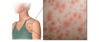
The height of the period - there is a deterioration in the general condition of patients, and the symptoms of general intoxication increase. The dry, painful cough intensifies, as well as abdominal pain. Profuse diarrhea with bloody stool appears In most patients, on days 5–7 of the disease, a maculopapular rash appears first on the face, then on the chest, extensor surface of the limbs and on the lower half of the body, accompanied by peeling of the skin on the palms/soles, which persists until 10–14 days of illness. During the same period of the disease, and sometimes earlier, hemorrhagic syndrome , appearing in the form of hematuria , nasal, uterine and gastrointestinal bleeding, and hemorrhages in the skin.
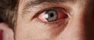
The appearance of patients is extremely characteristic - sunken eyes, conjunctival hyperemia , sluggish/immobile face. As the disease progresses, dehydration and severe weight loss develop. Some patients develop acute pancreatitis . Shock develops and progresses quickly, which leads to the death of patients from massive blood loss and circulatory failure.
At a later stage, damage to the central nervous system is often observed, manifested by delirium , drowsiness , or coma .
With a favorable course of Ebla disease, from the 10th to 12th day the patient’s body temperature begins to decrease, other clinical manifestations of the disease decrease, and a long (protracted) period of convalescence begins, which lasts several weeks. During this period, many patients may experience decreased hearing, loss of vision, behavioral disturbances, and the development of psychosis .
In terms of clinical symptoms, it is important to consider that in most patients, early/persistent symptoms include high fever , severe headache , vomiting , bloody diarrhea , and/or abdominal pain. Hemorrhagic syndrome and bleeding occur in only a third of patients.
Much less often in patients, on the contrary, the dominant symptoms are sore throat, conjunctivitis , exanthema , hiccups, bloody vomiting/diarrhea. Mortality is caused by sharply increasing massive blood loss. These differences are likely due to different virulence of the virus.
Many patients experience secondary complications of bacterial flora. As a rule, on the 12th day of illness, such patients develop bacterial sepsis with the subsequent development of infectious-septic shock and an increase in multiple organ failure.
The main signs of an unfavorable outcome of Ebola disease are dizziness / confusion , abdominal pain , profuse diarrhea . The addition of severe watery diarrhea quickly leads to dehydration of the body with the development of metabolic disorders, which sharply worsens the patient’s condition and the prognosis of the disease.
Lassa hemorrhagic fever
- an acute zoonotic natural focal viral disease, characterized by the development of hemorrhagic syndrome, ulcerative necrotizing pharyngitis, pneumonia, myocarditis, kidney damage and a high mortality rate.
The causative agent is Lassa virus of the Arenavirus genus of the Arenaviridae family; belong to the LChM/Lassa complex of Old World arenaviruses. It is antigenically related to other arenaviruses (the causative agents of lymphocytic choriomeningitis and HF in South America). The virus has a spherical capsid, with a particle diameter of 50–300 nm, covered with a lipid shell, including glycoproteins (G1 and G2).
The nucleocapsid consists of a protein (N) and RNA, two fragments of which (L and S) encode the synthesis of virion components in the infected cell; There are no hemagglutinins.
Epidemiology
The source and reservoir of the pathogen is the Mastomys natalensis rat, which lives in most African countries near human habitation. The virus was also isolated from other African rodents (M. erythroleucus, M. huberti). Animals release the virus into the environment through excreta and saliva.
Mechanisms of pathogen transmission: aerosol, fecal-oral, contact. Routes of transmission: airborne, food, water, contact.
Transmission factors: food, water, and objects contaminated with rodent urine. Infection of people in natural foci can occur by inhalation of an aerosol containing rodent excreta; drinking water from infected sources; insufficiently heat-treated meat from infected animals.
A sick person poses a great danger to others. The main transmission factor is blood, but the virus is also contained in the patient’s excreta.
Infection occurs through airborne droplets, contact and sexual contact. The virus can be shed by patients for up to a month or more.
Infection occurs through microtraumas when the patient’s blood or secretions come into contact with the skin. Cases of illnesses among medical personnel have been recorded when using instruments contaminated with the pathogen, performing surgical operations and dissecting corpses.
Susceptibility is high.
Post-infectious immunity is intense and long-lasting; repeated cases of the disease have not been described.
Pathogenesis
The entry gate for the pathogen is the mucous membranes of the respiratory and digestive organs, damaged skin. At the site of virus introduction, after its primary replication in the lymphoid elements, viremia develops with hematogenous dissemination of the pathogen, damage to many organs and systems. The virus has a tropism for various human organ systems and causes necrotic changes in the cells of the liver, myocardium, kidneys, and endothelium of small vessels, which determines the course of the disease. In severe cases, due to the cytopathic effect of the virus and cellular immune reactions, damage to endothelial cells in combination with impaired platelet function leads to increased “fragility” and permeability of the vascular wall. Profound disorders of hemostasis occur with the development of disseminated intravascular syndrome.
coagulation and consumption coagulopathy.
Clinical picture
The incubation period lasts 3–20 days, most often 7–14 days.
There is no generally accepted classification. There are: mild, moderate and severe course of the disease.
Main symptoms and dynamics of their development
The onset of the disease is subacute or gradual. General malaise, moderate muscle pain and headaches, low fever, and conjunctivitis are detected. During this period, the majority of patients (80%) experience a characteristic lesion of the pharynx in the form of ulcerative necrotizing pharyngitis, as well as enlargement of the cervical lymph nodes. By the end of the first week of the disease, body temperature reaches 39–40 °C; symptoms of intoxication increase; nausea, vomiting, chest and abdominal pain occur; diarrhea develops, leading to dehydration. From the second week, a maculopapular rash may appear; identify hemorrhagic manifestations (subcutaneous hemorrhages, nasal, pulmonary, uterine and other bleeding). Bradycardia and arterial hypotension occur; Possible hearing loss, seizures and focal neurological clinical manifestations. If the course of the disease is unfavorable, swelling of the face and neck occurs, free fluid is detected in the pleural and abdominal cavities, and hemorrhagic syndrome increases. In severe cases, death occurs on the 7th–14th day. In surviving patients, body temperature decreases lytically after 2–4 weeks. Recovery is slow. General weakness persists for several weeks, in some cases hair loss occurs and deafness develops; relapses of the disease are possible.
Clinical diagnosis
Early clinical diagnosis of Lassa fever is difficult due to the lack of specific symptoms of the disease. Of the clinical manifestations, the greatest diagnostic significance is: subacute onset; a combination of fever, ulcerative pharyngitis, hemorrhagic syndrome and renal failure.
Epidemiological data (stay in an epidemic focus) in conjunction with the results of virological and serological studies are of great importance.
Specific and nonspecific laboratory diagnostics
The absolute diagnostic sign of the disease is the isolation of the virus from the blood, throat wash, saliva, urine and exudates (pleural, pericardial, peritoneal) of the patient; and also from the dead - from samples of internal organs. Effective diagnostic methods: ELISA and RNIF. The diagnosis is confirmed serologically (with an increase in antibody titers to the Lassa virus by 4 times or more). The setting of the complement fixation reaction is retrospective.
Treatment
Drug treatment
Antiviral treatment is carried out by intravenous administration of ribavirin for 10 days (the initial dose of the drug is 2 g, then 1 g is administered every 6 hours for 4 days and 0.5 g every 8 hours for the next 6 days). In the early stages of the disease, convalescent plasma is used in a number of endemic regions.
Pathogenetic treatment is aimed at combating shock, hemorrhagic syndrome, cardiac and DN, as well as detoxification measures and infusion rehydration with saline solutions. Antibiotics are used for bacterial complications.__
Tests and diagnostics
The diagnosis of Ebola virus disease is made based on the results of laboratory diagnostics, including:
- PCR with reverse transcription.
- Biological method (virus isolation in cell cultures).
- ELISA (detection of Ebola virus antigens/detection of specific IgM).
- Biochemical blood tests.
- If necessary, FGDS, ultrasound of the abdominal organs, ECG, radiography.
- Differential diagnosis is carried out with shigellosis , malaria , relapsing/ typhoid fever , leptospirosis , rickettsiosis , cholera , plague , typhus , meningitis and other hemorrhagic fevers of viral etiology.
What is a virus?
Ebola disease is one of the most dangerous to human life in our time. The powerful virus, which infects humans, monkeys and a number of small mammals, is almost immune to treatment. Its destructive effect is transmitted to all parts of the body, from internal organs to the skin and human brain. The fatal process begins when the blood becomes more viscous and clots begin to form, which clog capillaries throughout the body. The result of all this is cell death and organ decomposition. Cracks form on the skin, from which blood oozes, the insides decompose, paralysis occurs, and in the worst case, death.
Diet
Dietary nutrition is prescribed that corresponds to Table No. 4 according to Pevzner, which provides a diet containing the physiological norm of protein with a limitation of fats and carbohydrates. All products that enhance the processes of rotting/fermentation in the intestines and the secretion of the digestive organs are excluded: liquid, semi-liquid and mashed dishes, steamed/boiled in water are indicated. The power mode is fractional. During the recovery period, a diet with a high content of animal protein is indicated.
Prevention
Until recently, there were no means of specific prevention of Ebola disease (vaccine). However, in 2020, the first vaccine, Ervebo (Merck), was approved in the United States and the European Union to prevent Ebola. It is a live genetically modified vaccine derived from the Ebola virus. Tested and evaluated in Africa on approximately 15 thousand people and found to be an effective and safe means of preventing Ebola disease. It is administered once as an injection. As evidenced by the latest news about the Ebola virus, its use has significantly reduced the incidence in the Congo. Common side effects include redness, pain and swelling at the injection site, fever , headache , fatigue , and muscle/joint pain.
Also, according to the statement of Scientists of the Russian Federation in 2020 at the Federal Research Institute of Epidemiology and Microbiology named after. N. F. Gamaleya, based on his own production, created and produced the Ebola vaccine “Gam-Evac” and “Gam-Evac Combi” (Fig. below), which is approved for clinical use.
However, there are no statistical data on its testing (efficacy and safety) in the specialized literature.
Nonspecific prevention includes:
- Isolation of patients in special boxes or glass-metal isolation cabins with autonomous life support.
- Identification and hospitalization of all persons who have contact with the patient.
- Strict monitoring and control of the surveillance area for up to two incubation periods from the moment of discharge of the last patient/last death.
- Strict adherence to precautions, including personal hygiene, use of various personal protective equipment when in contact with infected material, safe injections, disinfection of corpses and safe burial of the deceased.
- In order to reduce the risk of infection as a result of close (closer than 1 m)/direct contact with patients, with secretions from the body, medical personnel are required to work in special personal protective equipment (protective suit with mask, gloves, goggles).
- Thorough sterilization of medical instruments and equipment.
- Elimination of contact with wild/domestic animals in endemic areas and restriction of their movement. Thorough heat treatment of livestock products.
How does the Ebola virus spread among people?
Considering the isolation of the virus from almost all biological fluids of the body, there are many methods of infection: contact (through the skin), airborne (with splashes of saliva), parenteral (through blood), sexual (with infected semen), fecal-oral, household sharing common household items and sharing meals. Infection most often occurs as a result of direct contact with a sick person and infected material, the most dangerous being blood. The disease is highly contagious, Ebola epidemics occur easily, and the greatest risk is seen among medical personnel caring for patients, as well as those involved in eliminating natural outbreaks of the disease among monkeys. Surviving patients remain infectious for three months after the illness, as the virus continues to be excreted in urine, semen and tear fluid during this time. The natural susceptibility of this infection among humans is very high. Immunity after an illness develops lasting; a person becomes ill again very rarely (no more than 5% of cases).
Consequences and complications
Frequent bleeding with the development of hemorrhagic syndrome, acute cardiovascular and adrenal insufficiency, and cerebral edema . Less commonly, acute liver failure . During pregnancy - spontaneous abortion. The addition of a bacterial infection with the development of bacterial sepsis and infectious-septic shock , multiple organ failure .
Marburg hemorrhagic fever
Marburg hemorrhagic fever is an acute zoonotic highly lethal viral disease manifested by intoxication and severe symptoms of universal capillary toxicosis.
Etiology
The causative agent is Marburgvirus of the Marburgvirus genus of the Filoviridae family.
Epidemiology
The reservoir of the Marburg virus has not been reliably established at this time.
The source of the pathogen is monkeys, in particular African monkeys Cercopithecus aethiops. Mechanisms of pathogen transmission: aerosol, contact, artificial. Routes of transmission: airborne droplets, contact, injection. The virus is contained in the blood, nasopharyngeal mucus, urine and semen (up to 3 months). Infection of humans occurs through direct contact with the blood and organs of monkeys, also through damaged skin (through injections, cuts), and when the virus enters the conjunctiva. A sick person is contagious to others.
Human susceptibility to Marburg virus is high. Post-infectious immunity is long-lasting. There is no information about recurrent diseases. The entrance gate is damaged skin, mucous membranes of the oral cavity and eyes. Primary replication of the virus occurs in cells of the monocyte-macrophage lineage. Then viremia develops, accompanied by suppression of the functions of the immune system and generalized microcirculation disorders, which leads to the occurrence of disseminated intravascular coagulation syndrome and multiple organ lesions. Foci of necrosis and hemorrhage are found in the lungs, myocardium, kidneys, liver, spleen, adrenal glands and other organs.
Clinical picture
Incubation period 3–16 days.
Main symptoms and dynamics of their development
The onset of the disease is acute: high fever for 2 weeks, severe intoxication, headache, myalgia, pain in the lumbosacral region.
On examination, conjunctivitis, enanthema, vesicular-erosive changes in the oral mucosa, and bradycardia are revealed. The muscle tone is increased, their palpation is painful. From the 3rd–4th day of the disease, vomiting and watery diarrhea occur, leading to rapid dehydration of the body.
On the 5th–6th day, a maculopapular rash may appear, followed by peeling of the skin. From 6–7 days, hemorrhagic manifestations are detected in the form of skin hemorrhages, nasal, gastrointestinal and other bleeding, as well as signs of hepatitis, myocarditis, and kidney damage. Damage to the central nervous system is characterized by adynamia, lethargy and meningism. At the end of the first week, signs of ITS and dehydration are revealed. The deterioration of the patients' condition occurs on the 8–10th day and on the 15–17th day of the disease (sometimes ending in death).
During the period of convalescence, which lasts 3–4 weeks, prolonged diarrhea, severe asthenia, mental disorders and baldness may occur.
Specific laboratory diagnostics are carried out using the same virological and serological methods (virus culture isolation, PCR, RNIF, ELISA, RN, RSK, etc.) as for Ebola fever. In the deceased, the virus is detected by electron microscopy or using RNIF. All studies are carried out in a laboratory with the maximum level of protection.
Drug treatment
Etiotropic treatment
Not developed.
Pathogenetic treatment
Has basic meaning. Aimed at combating dehydration, ITS, hemorrhagic syndrome. There is evidence of the effectiveness of convalescent serum, plasmapheresis and large doses of interferon.
List of sources
- Shchelkanov M.Yu., Magassuba NF, Boiro MY, Maleev V.V. Causes of Ebola fever in West Africa. Attending doctor. 2014; 11:30-6.
- On the epidemiological situation regarding the incidence of Ebola hemorrhagic fever / Letter of the Russian Federation dated August 7, 2014 N 32-024/424.
- Borisevich I.V., Markin V.A., Firsova I.V., Evseev A.A., Khamitov R.A., Maksimov V.A. Epidemiology, prevention, clinical picture and treatment of hemorrhagic fevers (Marburg, Ebola, Lassa and Bolivian). Question virusol. 2006; 5:8–16.
- Petrov A.A., Lebedev V.N., Plekhanova, L.F. Stovba T.M., Borisevich G.V., Sidorova O.N., Chukhralya O.V. Current state of development of means of emergency prevention and treatment of disease caused by the Ebola virus//Central Research Institute" of the Ministry of Defense. Russian Federation, Sergiev Posad.
Treatment
At the moment, there is no etiotropic treatment for Ebola fever . Patients are prescribed symptomatic supportive therapy, which can only delay the onset of death. At the same time, several countries have announced the creation and testing of a vaccine against the Ebola virus.
The most extensive developments in the field of creating a vaccine against Ebola are being carried out by the American scientific institute Mapp Biochemical. The antiviral cocktail called MB-003 includes three different artificially created monoclonal antibodies.
Each is a protein that blocks the Ebola virus at three locations on its surface. Pharmacologists tested the drug MB-003 on laboratory macaques and mice.
Virologists from Liberia and Russia are also working on creating a vaccine. A number of vaccines have been created that have already shown a positive effect on patients . However, almost every person who becomes infected with this deadly virus dies. An effective vaccine is expected to emerge.
Prevention
Patients with Lassa, Marburg and Ebola hemorrhagic fevers are subject to immediate hospitalization in ward wards, subject to the strict regime recommended in cases of particularly dangerous infections such as plague and smallpox.
Those who have recovered are discharged no earlier than the 21st day from the onset of the disease if their condition is normalized and virological tests are 3 times negative. All patient household items must be strictly individual and labeled. They are stored and disinfected in a box.
Disposable instruments are used for treatment. After use, reusable instruments are autoclaved, disposable instruments are burned.
During the period of current disinfection, a 2% solution of phenol, iodoform (450 g per 1 ml of active iodine with the addition of 0.2% sodium nitrate) is used. Discharges from patients are also treated accordingly. Maintenance personnel must work in type 1 anti-plague suit.
Special plastic boxes have been developed in which, using an exhaust system equipped with a decontamination unit, air flow is ensured in one direction - inside the box.
Such boxes are equipped with a conventional system to ensure complete safety of personnel during medical procedures. Particular care should be taken when examining blood and other biological materials from patients with hemorrhagic fevers and persons suspected of these diseases.
People who were in direct contact with the patient (or a person suspected of developing the disease) are isolated in a box and observed for 21 days.
In all cases of suspected infection with the Ebola virus, specific immunoglobulin is administered from the serum of hyperimmunized horses. The onset of immunoglobulin action from the moment of administration is 7-10 days.


