What is exostosis
Exostosis is a benign superficial bone or cartilage tumor that resembles a thorn. The pathology occurs before adolescence against the background of trauma, with prolonged healing of fractures, which were accompanied by damage to the periosteum.
Single exostoses are more often detected on the following bones:
- lower femur;
- upper tibia;
- brachial.
Multiple growths are located bilaterally on the metaphyses of long bones, on the ribs and clavicles.
Trauma stimulates the growth of exostosis (improper healing of a fracture in the area of hemorrhage if the periosteum is damaged).
The size of the growth ranges from 1-2 to 10 or more centimeters.
Exostosis grows slowly throughout skeletal growth and rarely manifests itself. With multiple lesions, various deformations occur due to impaired bone formation:
- varus position of the knees;
- curvature of the tibia;
- fracture of the femoral head;
- curvature of the forearm from the elbow.
The rapid growth of a benign tumor should suggest malignant transformation.
The term “exostosis” means a bone pathology, the occurrence of which is caused by other diseases . Multiple cartilaginous exostosis is considered an independent type.
Exostoses account for 10-15% of all bone tumors and 36% of benign neoplasms. They arise from unusual cartilaginous epiphyseal tissue growing in the zone of endochondral ossification. They are distinguished by their stem-like structure.
What does it look like
The occurrence of exostoses is typical for people 10-30 years old. Tumors appear with equal frequency in men and women.
There are single and multiple growths. They resemble thorns, mushrooms, cauliflower. Another etiology is growth plate dysplasia.
The disease is often hereditary. The photo shows that the growth may resemble a small bump or be located in the thickness of soft tissue.
The formation manifests itself as hard, immobile, near the ends of long bones.
The symptoms of the lesion are associated with irritation of the overlying tissues, which can become filled with inflammatory exudate. Externally, the area will resemble an inflamed bursa.
The fluid inside the capsule is sometimes mistaken for a cyst.
Attention! Tumors in the knee area are more likely to become malignant. The risk of sarcoma is 1% for single exostoses and 10% for multiple exostoses.
Prevention
Prevention, as such, comes down to identifying exostoses at the earliest stages. Regular medical examinations help achieve these goals. Given the risk of skeletal deformation, early diagnosis is especially important for children and adolescents. An examination is also necessary after injuries to the musculoskeletal system, because even a minor bruise or fracture can trigger the occurrence of pathology. And as mentioned above, it is highly advisable to regularly monitor calcium levels in the body, because people with high calcium levels are at risk.
By and large, regardless of the etiology, exostosis does not belong to the group of dangerous diseases. Transformation of a tumor into a malignant one occurs extremely rarely. This neoplasm does not pose a serious threat to human life and health. In children, there are often cases of healing spontaneously, without medical intervention.
Tsygankova Yana Aleksandrovna, medical observer, therapist of the highest qualification category
27, total, today
( 204 votes, average: 4.71 out of 5)
Bursitis - symptoms, causes of development and treatment methods
Dislocation of the leg in the ankle area: symptoms and treatment
Related Posts
Causes and consequences of bone exostosis
The origin of exostosis has a different nature. Tumors can develop against the background of regeneration after trauma, chronic inflammation of the periosteum, mucous and fibrous membranes. The causes and consequences of osteochondral growths are under study.
Any violation of bone integrity can provoke growth:
- chronic joint pathologies;
- surgical interventions;
- birth defects and bone deformities.
Exostoses develop as a side effect of benign tumors and chondromatosis.
The etiology of the development of solitary osteochondromas is currently unknown. Multiple osteochondromatosis has a genetic basis and is inherited in 70% of cases. The disease is associated with pathologies of genes with EXT 1 and EXT 2.
Possible complications of exostoses include:
- rupture of the muscle tendons above the tumor;
- bursitis;
- pathological fracture (most occur in the knee - distal femur and proximal tibia);
- osteomyelitis;
- muscle infarctions due to acute disruption of blood supply.
There is a loss of sensitivity and a change in skin color.
Diagnostics
Exostosis is most often diagnosed by visual examination of the patient, and the disease is confirmed after X-ray examination.
A mandatory laboratory test is a general blood test.
To confirm the exact picture, the following may be additionally prescribed:
- ultrasonography;
- Magnetic resonance imaging.
If a malignant process or infection is suspected, a tissue biopsy and biochemical blood test are prescribed. Osteochondral exostosis is a benign tumor, but under certain circumstances, especially in the area of the spine and auricle, it can become malignant.
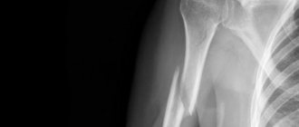
Exostosis of the knee joint
Main symptoms of exostosis
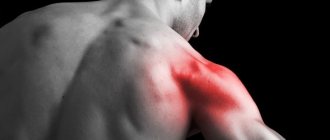
Osteochondromas are most often found as a painless lump near a joint - the knee or shoulder. Other symptoms are related to the location of the tumor:
- If osteochondromas are located under a tendon, this can cause a locking effect on the joint when the muscle contracts.
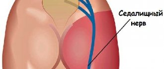
- When located near nerves and blood vessels, numbness, tingling may be observed, and blood flow and color of the limb are periodically disturbed due to secondary compression of the nerve and blood vessels.
The clinical manifestations of exostoses differ significantly. Some of them are asymptomatic, others cause pain and limit the mobility of the limbs. Sometimes the growths turn into tumors and undergo malignancy.
The main signs of exostosis:
- painless growth near the joint;
- limited range of motion;
- loss of pulse in the arteries of the lower limb.
When exostosis appears in the vertebrae, neurological symptoms of compression of the nerve roots appear, depending on the level of the lesion.
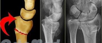
Diagnostic manifestations of exostoses are varied:
- X-ray shows flat, mushroom-like or stalk-like lesions;
- pedunculated osteochondral growths are oriented in the proximal direction (up).
The image clearly shows tumors in the metaphyses of the bones. They look like growths of tissue with a motley pattern. The disease manifests itself as calcification, and areas of calcification are detected on the cartilaginous plates. Calcifications increase with age.
Microscopic examination shows that the growths are of the nature of cartilaginous tissue, but less structurally organized. Elements of ossification (ossification) and calcification of cartilage are observed.
The cells are distinguished by small round or elongated nuclei. If the growth reaches a size of 1 cm in adolescence, then a tumor larger than 3 cm indicates chondrosarcoma.
Causes of the disease
Can appear:
- during the recovery process after injuries;
- due to bruise or blow;
- with inflammatory processes in the mucous membranes;
- if there are chronic inflammatory processes in the bones;
- due to aseptic necrosis;
- in case of disruption of the functionality of the endocrine system;
- when ligaments are torn at the place of their attachment;
- as a complication of non-malignant formations;
- after some operations.
Treatment of exostosis
Treatment for osteochondroma is usually not indicated, since in most cases the condition is asymptomatic.
Pain medication is used only when pain is present. However, surveillance and regular monitoring are very important.
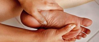
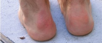
Exostosis of the calcaneus
Called a heel spur or retrocalcaneal exostosis, it refers to the formation of a bone spur on the heel bone. At the site of calcification is the Achilles tendon, which passes into the gastrocnemius muscle.
Calf tightness is secondary to weakness of the hip flexors - biceps muscle. Calcaneal exostosis develops in people with impaired posture and correct walking biomechanics:
- wearing high heels causes spasm of the Achilles tendon;
- weakness of the gluteal muscles provokes overstrain of the calf muscles;
- lack of emphasis on the heel and pushing off with the toe leads to tension in the plantar fascia;
- weakness of the peroneal and tibialis anterior muscles, rectus femoris muscle - causes overload of the calf muscles.
The listed factors together influence the places of muscle attachment, where deposits appear - growths.
To treat exostosis of the calcaneus, in addition to insoles and steroid injections, it is necessary to restore the biomechanics of the step.
Try rolling your sole with a tennis ball, strengthen your gluteal muscles, and restore the muscles in the front of your body: rectus abdominis and rectus thighs.
Osteochondral exostoses
Cartilaginous exotosis or chrondodystrophy is a disease associated with excessive cell proliferation. Growths appear at the metaphyses of long bones.
The pathology is more often found in men and is hereditary. Women more often act as carriers of the gene, but do not have signs of the disease.
Symptoms of osteochondral exostosis are associated with defects in bone growth and the location of tumors:
- decreased skeletal height (dwarfism or short limbs);
- bone deformities;
- short stature;
- early osteoarthritis;
- impaired joint mobility;
- compression of peripheral nerves.
Exostosis disease is confirmed by carriage of the EXT1 and EXT2 genes in 70-90% of cases. The analysis is carried out only after the patient reaches the age of 4 years, when symptoms and abnormalities become obvious.
For painful lesions, surgical excision of tissue is performed to slow the development of additional growths and abnormalities.
Most often, the cartilage growth and perichondrium, the connective tissue around the cartilage, are removed. Large deformations are eliminated by cutting off the exostosis.
Attention! The disease can lead to skeletal abnormalities, joint damage, and limb immobility. Patients are constantly at risk of relapse.
Exostosis of the knee
Exostosis of the knee joint develops in the distal part of the femur - the growth plate. When detected at an early age, before 10 years, it is usually located near the diaphysis (thickening of the tubular bone), and in adolescence it moves to the middle.
In childhood, growths are detected by chance. Sometimes you can feel them. Due to the flexibility of the muscles and the small growth of exostosis, there is no effect on the function of the joint.
Surgeons recommend active tactics - removing tumors at an early stage. At a later age, the formation can disrupt the function of the rectus femoris muscle and injure its tendon if it is located on the anterior surface of the femur.
When located on the posterior surface, the risk of pinching blood vessels and nerves increases. Flexion of the knee joint is limited.
Exostoses of the tibia in the knee area are more often formed after injury or fracture or insufficiency of the perichondral ring - due to impaired blood supply.
The growth of exostoses is explained by the theory of growth zones until the child’s skeletal maturity.
Exostosis of the femur
Growths on the femur are most often localized in the lower growth plate and are called exostotic dysplasia.
Most often they are identified by secondary symptoms. Growths on the femur can look different:
- wide base and pointed end;
- narrow base and spherical cartilaginous end;
- rounded bone growth with a cartilaginous cap.
Femoral growths account for almost 50% of all exostoses. In rare cases, they will lead to hip deformity - usually such complications are associated with multiple tumors and genetic predisposition.
Reference. Most often, single exostoses are associated with bruises, sprains, and microtraumas in childhood, which no one considers significant.
Any marginal femoral exostoses seen on x-ray in adults with joint pain are caused by muscle weakness and instability. They do not belong to the typical growth of cartilage tissue at a young age.
Features of treatment in children
Treatment approaches depend on symptoms and various factors, such as age, size of the growth, extent of the disease, and overall health.
For exostoses that do not affect the function of the joint, only regular monitoring is carried out.
If symptoms are present, use:
- anesthesia;
- surgical intervention.
Complications that may arise during surgery become the reason for refusing the operation.
If osteochondroma appears in close proximity to a bone growth area, then surgery will affect future bone growth. The choice of treatment method depends on the competence of the doctor.
Single osteochondral exostosis in children usually does not require surgery and can be monitored with x-rays.
Surgery is suggested if the patient has reached skeletal maturity and the osteochondroma has completed growth during observation.
Other prerequisites for intervention are:
- pain during physical activity;
- pressure on a nerve or vessel;
- large cartilaginous cap;
- bone maturity.
Multiple osteochondral exostoses rarely need to be removed unless they cause symptoms. In case of bone deformities, tumor resection is accompanied by reconstructive techniques.
Physiotherapy is necessary in the postoperative period. It is impossible to influence exostoses using massage and other methods.
The goal of treatment is to restore functional mobility. Therefore, it is important to restore proper biomechanics, muscle tone and strength.
Symptoms
The onset of the disease does not manifest itself in any way and is asymptomatic, and signs begin to appear when the tumor rapidly increases in size.
General symptoms include:
- headache;
- dizziness;
- trauma to the tumor, which can lead to painful wounds;
- impaired joint mobility;
- inflammatory processes;
- nerves and tendons may be compressed;
- numbness of certain parts of the body;
- impaired blood circulation;
- edema.
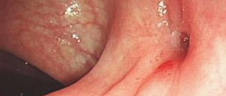
Exostoses of the external auditory canal contribute to hearing loss, with tinnitus appearing and the patient begins to hear his voice more clearly. In this case, headaches and inflammatory processes very often occur, which can cause frequent otitis media.
The most severe manifestations of a tumor in the spine: they cause severe pinching of the nerve, including loss of consciousness, and there is also a high risk of damage to the spinal cord.
Heel exostosis, just like a tumor on the gum, is very traumatic, as it is constantly exposed to friction, as a result of which wounds and inflammations with suppuration can appear.