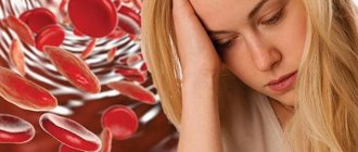
Reasons for ordering an examination
There are a number of indications for prescribing the procedure. Diagnostics is carried out for the purpose of:
- studies of frequent acute respiratory infections, acute respiratory viral infections;
- identifying pathological disorders of the respiratory system with a prolonged cough, respiratory failure, sputum production, chest pain;
- identifying the causes of deviations in the gas exchange process;
- analysis of the relationship between pulmonary diseases and external respiration function, the effectiveness of therapeutic measures in their treatment;
- prevention and early detection of abnormalities in people with an increased risk of developing pathologies: smokers and people whose work activity is associated with harmful substances;
- monitoring the course of bronchopulmonary diseases: pneumonia;
- flu;
- bronchitis;
- asthma;
- chronic obstructive pulmonary disease;
- pulmonary tuberculosis, etc.;
For persons over 40 years of age, who have smoked for 10 years or more, and who have a chronic cough or shortness of breath, examination is mandatory.
Preventive medical measures are recommended for workers associated with the regular use of harmful chemicals.
Indications for use
This procedure is painless and is recommended to be performed strictly for medical reasons. Additionally, to clarify the clinical picture, the doctor may prescribe electrocardiography, echocardiography and x-ray of the lungs. Such an integrated approach to the problem is appropriate for diseases of the respiratory system of various origins. The main indications for spirography are presented below:
- pressing pain in the chest area;
- long smoking history;
- persistent cough for 3–4 weeks;
- suspicion of obstructive bronchitis;
- impaired airway patency;
- bronchospasms of unknown etiology;
- more frequent attacks of bronchial asthma;
- hereditary predisposition to bronchopulmonary diseases;
- taking an incomplete breath, shortness of breath;
- work in hazardous production.
Spirography is performed if the following diseases of the body are suspected:
- oncology;
- COPD;
- pneumonia;
- pneumonia.
https://www.youtube.com/watch?v=upload
For chronic diseases of the cardiovascular system, not all patients are allowed to undergo spirography. Medical restrictions include the following pathological processes and diseases of the whole body:
- exacerbation of a chronic disease;
- arterial hypertension;
- angina pectoris;
- hypertensive crisis;
- myocardial infarction;
- pulmonary failure;
- disorders of the circulatory system;
- toxicosis during pregnancy.
The essence of spirography is to determine changes in lung volume during normal and increased breathing, as well as other indicators of their functioning. It is a mandatory method of examination for various bronchopulmonary pathologies, for example, when detecting symptoms of bronchial asthma. Also, with the help of a spirometric examination, the effectiveness of the treatment used is established, especially for asthma; medical examinations are carried out for athletes, workers in hazardous industries, smokers with many years of experience, and people with a hereditary predisposition to allergies or diseases of the respiratory system.
In addition, spirography is prescribed if the following symptoms are present:
- prolonged cough that does not stop for 1 month or more;
- frequent respiratory diseases;
- lung diseases detected during other examinations;
- compressive pain behind the sternum;
- sensations of incomplete breathing, shortness of breath;
- regularly exacerbating bronchitis;
- violations of gas exchange processes;
- chronic obstructive pulmonary disease in the early stages;
- bronchial asthma (to determine the effectiveness of treatment).
- acute allergic reactions.
Despite the safety and non-invasiveness of the procedure, it cannot be performed in the following cases:
- serious condition of the patient;
- the presence of toxicosis during pregnancy;
- angina pectoris, heart attack;
- disorders of the circulatory system;
- persistent increase in blood pressure, hypertensive crisis;
- serious pulmonary insufficiency.
That is, spirometry is the direct examination process, and spirography is the same procedure, but displaying the results obtained on a special diagram - a spirogram.
What is spirometry, is it really necessary?
Pulmonary function testing should be performed when:
- symptoms of respiratory diseases;
- identifying sources of abnormal gas exchange;
- assessing the risk of the therapy used for the patient;
- determining physical condition;
- determining the level of bronchial obstruction, especially in COPD (chronic obstructive pulmonary disease).
The results will indicate the correct choice of tactics for treating pathologies of external respiration functions. Spirometry performed in the initial stages increases the patient's chances of recovery. This method will be useful for assessing the health of athletes and smokers.
Spirometry for bronchial asthma detects signs of the disease, and for patients with asthma it monitors the effectiveness of treatment. Timely diagnosis of COPD will allow treatment to begin and death to be avoided. To correctly assess pathologies, in addition to clinical tests, the doctor must examine the patient and listen to his complaints.
The difference between spirometry and spirography
The difference between the two concepts lies in word formation according to the rules of Greek terminology. Medical names designated in Latin and Greek are translated according to the parts that form them.
The term “Spirometry” consists of two particles: “spiro-” and “-metry”. The first is translated as breathing, to breathe, and the second is dimension. The term “Spirography” has the same first part, and the second part “-graphy” means to write down.
For reference. It follows that spirometry refers to the process of measuring the function of external respiration. Spirography is an image or recording of the results obtained on paper. As a rule, the terms are considered synonymous, since during the research the result is always recorded on paper.
ESSENCE OF THE STUDY
What spirometry is becomes clear from the name of the procedure: spiro meter translates as “breathing measurement.” During the examination, the doctor determines the speed and volume of breathing using a spirometer.
To better understand the essence of the method, you need to turn to the anatomy of the respiratory system. Its 3 main elements are:
- The respiratory tract allows air to pass through.
- Lung tissue is responsible for gas exchange.
- The chest works like a pump.
If the functions of any department are disrupted, it upsets the functioning of the lungs. Spirometry evaluates breathing parameters, which makes it possible to identify respiratory diseases, learn about the severity of pathologies and the effectiveness of therapy.
In addition to the name “spirography”, “spirometry” is also used. This means the same study. These designations differ only in that doctors understand spirography as a method of examining the respiratory organs, and spirography as a graphical recording of measurements made by a spirograph.
Spirometry results
Spirometry results vary from person to person. Average results depend on a number of factors, including age, height, gender and race.
Obstructive airway disease
is when narrowing of the airways affects a person's ability to breathe quickly, but they are still able to inhale a normal amount of air. This is common in people with asthma and COPD.
In COPD, air inhalation is reduced because the lungs cannot expand fully, such as in pulmonary fibrosis.
Preparing for spirometry
Lung spirometry is performed in the morning on an empty stomach; a low-fat breakfast is allowed 2 hours before the procedure. To ensure the reliability of the test, you should adhere to the basic rules:
- quit smoking in a few hours;
- replace your morning coffee with a healthier drink, such as juice;
- in some cases, the attending physician can stop the patient from taking medications in a few hours;
- choose loose clothing that will make you as comfortable as possible.
20 minutes before the procedure, the patient will be asked to rest and restore respiratory functions while at rest. The doctor must find out whether the subject has diseases that may affect the study of pulmonary function (pneumothorax or myocardial infarction in the first two weeks of development). People after eye surgery or with hemoptysis should perform this test carefully, following the basic recommendations of a specialist.
How is spirometry performed?
The history of the technique begins in Ancient Rome: the Greek physician Galen studied the volumes of inhaled and exhaled air using simple bubbles. Today, a modern method of studying external respiration functions is popular.
Computer spirometry is a sterile procedure that begins with attaching a disposable mouthpiece to the device. After the patient has sat down, he is asked to press his mouth tightly against the disposable mouthpiece and follow the doctor’s recommendations: exhale as much as possible with or without effort, alternating with a calm exhalation. If you exhale at maximum speed for 15 seconds, then you should talk about lung pathologies. All indicators are recorded and performed 3 times. Then the most successful results are selected, with the help of which the pulmonologist makes a diagnosis or adjusts the therapy of an existing disease. The spirometer deciphers the data and automatically calculates respiratory function indicators.
Interpretation of results
Upon completion of data collection, they are analyzed in order to draw up a diagnostic conclusion. In the process of interpreting all the obtained indicators, an electronic spirogram is generated by a computer program. The following values are interpreted.
The number of respiratory movements in one minute is recorded. Its normal values do not exceed 16–17 times.
The volume of air filling the lungs with one breath is determined. Its normal values have a fairly wide range. The limits for healthy men can fluctuate between 300–1200 ml, and for healthy females values within the range of 250–800 ml are typical.
Shows the volume of air absorbed by the lungs in 1 minute. It also has a relatively large interval and ranges between 4–10 liters.
This value is studied when determining the maximum volume of air exhaled by the subject during a quiet exhalation after the deepest inhalation.
The maximum volume of air exhaled by the patient during the deepest (forced) exhalation after the same inhalation is diagnosed. Normally, for individuals this figure ranges from 2.5 to 7.5 liters.
The maximum volume of air exhaled by a person in 1 second during a deep exhalation after an extremely deep inhalation is determined. Its value is significantly influenced by the gender and age of the subject.
Tiffno Index (IT)
Its value is the ratio FEV1/FVC, and is expressed as a percentage.
This value is obtained by multiplying the average amplitude of extreme respiratory excursions and their frequency in 1 minute.
This value looks like MVL/VC, and is indicated as a percentage.
Contraindications to spirometry
Spirometry has no strict contraindications. Mild dizziness, which may occur, passes quickly and does not pose a health hazard. Forced or strong deep inspiration causes a short-term increase in intracranial and intra-abdominal pressure.
The procedure should be carried out with caution or abandoned for the following indications:
- recent operations on the abdominal organs or ophthalmic surgical procedures (less than 2 months ago);
- myocardial infarction or stroke (depending on the patient’s condition, but not earlier than 3 months after);
- previous respiratory tract infections (at least 2 weeks after their supervision);
- a history of pneumothorax;
- arterial or aortic aneurysm;
- severe attacks of bronchial asthma;
- presence of pulmonary hemorrhages;
- epilepsy;
- hypertensive crisis and other pathologies associated with blood pressure disorders;
- increased blood clotting;
- mental disorders;
- pregnancy;
- Age restrictions: up to 5 and after 75 years.
Even in the absence of obvious contraindications, consultation with a specialist is necessary before the study.
Method of spirometry
Portable spirometer
Before the advent of digital technology, mechanical spirometers, most often water-based, were widespread. In them, the exhaled air entered a cylinder placed in a vessel with water. When exhaling, the cylinder moved upward, and a recording device connected to it left a graph of volume versus time on the moving paper. Examination with such a device was labor-intensive and required manual calculation of parameters.
Currently, digital instruments are used, which consist of an air flow sensor and an electronic device that converts the sensor readings into digital form and performs the necessary calculations. There are many computer spirometers available, in which all calculations and information analysis are performed by a personal computer.
Spirometry classification
The way the procedure is performed determines its type. Spirometric tests are performed during the following maneuvers:
- normal calm breathing;
- exhale with effort (forced);
- with maximum ventilation;
- with physical activity (before and after it) - dynamic spirometry;
- using special substances - functional and provocative spirometry: with bronchodilators that dilate the bronchi. The method helps to identify hidden bronchospasms, correctly diagnose the disease, determines the reversibility of disorders and the effectiveness of therapeutic techniques;
- with methacholine, which helps to definitively diagnose asthma, identify a predisposition to bronchospasm and hyperresponsiveness.
Modern spirometers make it possible to determine the level of diffusion capacity of the lungs - the gas exchange of oxygen and carbon dioxide between the respiratory organs and the blood.
Additional examination - bronchospirometry. Allows you to separately record indicators in different lobes of the lungs.
FEATURES OF SPIROMETRY IN CHILDREN
Spirometric testing is carried out in children from 5 years of age. It is not prescribed at a younger age, since the rules for performing the procedure require taking a maximum breath. Otherwise, spirometry interpretation will be inaccurate.
At the adult level, a child can be examined from the age of 9 years. Before this, you need to try to create a positive atmosphere - with toys, affectionate attitude.
It is better for young patients to undergo spirometry in children's centers, and conventional laboratories do not adapt to their characteristics. Before the procedure, the child should be told in simple language how to inhale and exhale. For intense forced exhalation, images are sometimes used - for example, showing a candle on the screen, asking it to be blown out. The doctor should ensure that the baby's lips are pressed tightly against the mouthpiece. The protocol then indicates the number of successful cycles. Spirometry results are adjusted for age.
RESEARCH RESULTS
Spirometry indicators are the main source of information for diagnosing pulmonary diseases. The norms are average values calculated based on the results of a survey of healthy people. They vary based on gender, age, height, weight and lifestyle.
Spirometry standards are given in the table:
| Parameter | Description | Average rate |
| vital capacity | Vital capacity of the lungs, the main static indicator. All the air from exhalation is at maximum exhalation after the same inhalation. | There is no norm for vital capacity; other parameters are calculated on its basis. |
| FVC | Forced vital capacity, the main dynamic indicator. The volume of air entering the lungs during intense exhalation. This is necessary to clarify the patency of the bronchi: as their lumen decreases, FVC also decreases. | 70-80% VEL. |
| BH | Respiratory rate, number of inhalations and exhalations at rest. | 10-20/min. |
| BEFORE | Tidal volume (from inhalation and exhalation in 1 cycle). | 0.3-0.8 l (15-20% vital capacity). |
| MAUD | Minute volume of respiration, that is, passed through the lungs in 1 minute. | 4-10 l/min. |
| District Department of Internal Affairs | The inspiratory reserve volume, that is, the maximum inhaled during a normal inhalation. | 1.2-1.5 l (50% vital capacity). |
| ROvyd | Expiratory reserve volume. | 1-1.5 l (30% vital capacity). |
| FEV1 | Forced expiratory volume in 1 second. | > 70% FVC. |
| JEL | Proper vital capacity for a healthy person, based on physical parameters. Men: 0.052 * height (cm) – 0.028 * age – 3.2 Women: 0.049 * height – 0.019 * age – 3.76 | 3-5 l. |
| OOL | Residual volume of the lungs, that is, remaining after exhalation. | 1-1.5 l or 20-30% VOL. |
| OEL | The total capacity of the lungs, or how much air can be held in the lungs after inhalation. It is calculated as follows: VOL + VEL. | 5-7 l. |
| Tiffno index | FEV1 (ml) / Vital capacity (ml) * 100%. | > 70-75 %. |
Ventilation failure can be obstructive or restrictive. The first develops due to a decrease in the lumen of the bronchi with an increase in resistance to air flow. The second occurs due to a decrease in the ability of lung tissue to stretch.
When deciphering the results, the following parameters indicate the obstructive type:
- ELC is normal or higher;
- Tiffno index is underestimated;
- EOL increased.
- FEV1 decreased.
With restrictive insufficiency, TLC decreases.
Parameters used in spirometry
Peak flow metry norm indicators
- BH. This index shows the frequency of breathing movements performed in 60 seconds. The normal value varies around 16-18 units;
- DO, tidal volume. This is the air mass that enters the lung tissue in one breath. The norm is from 500 to 800 ml;
- MAUD. Breath volume per minute. This indicator indicates how much air passes through the lungs at rest in 60 seconds. The reflection of this parameter also shows gas exchange processes in the lung tissue. The MOD depends on the psychoneurological state of the patient at the time of the study, the level of fitness of the lungs, and metabolic processes. Based on this, the assessment of this indicator reflects the condition of the lung tissue only as an auxiliary research method;
- indicator of average volumetric velocity, SOS. Represents the speed at which a forced exhalation is made in the middle of the movement. This parameter reflects the condition of small airways. It provides more information, unlike FEV1, and makes it possible to identify earlier manifestations of obstructive pathology.
The vital capacity of the lung tissue (VC) is used to determine the vital capacity of the lungs. This is the air volume that enters the organ during the maximum inhalation after peak exhalation. During quiet breathing, a small part of the lung tissue is used.
When physical activity occurs after a quiet inhalation, the person performs breathing movements using the reserve air volume. Usually it is 1500 ml. After which, exhaling the usual amount of air, the patient exhales another 1500 ml. It turns out that when using reserve breathing, it becomes the deepest.
Vital vital capacity is calculated from the sum of the inspiratory reserve volume to the expiratory reserve volume
The normal value is 3500 ml. This parameter is most valuable for breathing control. It varies depending on the patient’s gender, age, weight, and height. Based on this, when measuring vital capacity, the doctor will need more accurate data from the patient. The average should be about 80% of the norm.
A decrease indicates pulmonary diseases and insufficient motor function of the lungs. A slight decrease develops as a result of bronchial obstruction. After maximum exhalation, the lung tissue contains a residual amount of air. The volume can vary from 800 to 1700 ml. These numbers, together with the vital capacity indicator, provide information about the total amount of air in the lungs.
The forced vital capacity of the lung tissue (FVC) is a parameter that determines the amount of accelerated vital capacity of the lung tissue. This is the amount of air that is exhaled when a person makes significant effort after taking a deep breath. The difference between the previous parameter is that the exhalation is performed most quickly.
FVC shows the state of tracheal patency. As you exhale, the pressure in the chest decreases, while the resistance to the air flow of the bronchi increases. Based on this, it is possible, by straining the respiratory muscles at maximum speed, to exhale not the entire volume, but only some of it. At this time, the residual part of the vital capacity is slowly exhaled with strong tension of the muscles involved in breathing.
If there is a violation of bronchial obstruction, then the bronchi begin to resist the air flow at the beginning of accelerated exhalation. Moreover, the resistance increases towards the end of its completion. Based on this, a small part of the air is forced out by a person. A standard exhalation of the entire volume of the lungs occurs in 2 seconds. when making a forced movement. In this case, FVC varies from 90 to 92% of the VC result.
For spirometry, it is also important to know what the forced expiratory volume per second (FEV1) is. This is the amount of air that is exhaled in 1 second. products of accelerated exhalation. Normal indicators include the border between 70 and 85% of the vital capacity parameter. If there is severe obstruction, the limit is reduced to 20%. A reduced parameter indicates a violation of bronchial patency.
The Tiffneau index (IT) provides an assessment of the type of obstruction. This study is conducted with bronchodilators. Increasing IT indicates the reason for the decreased OF1, which lies in bronchospasm. A negative test indicates the presence of other causes of obstruction. If there is a decrease in the FEV1 parameter, provided that vital capacity is normal, then the cause of the obstructive pathology lies in the weakened respiratory muscle of the patient. In persons suffering from bronchial asthma, this parameter is reduced to 25%.
The Tiffno index is calculated using the following formula
If there is a decrease in the FEV1 parameter simultaneously with vital capacity, then we are talking about pulmonary obstruction. This situation requires additional measurement of the residual volume of lung tissue. This indicator is taken during body plethysmography. For your information, the norm of the Tiffno index cannot accurately predict the absence of pathology. This indication should be simultaneously assessed with the patient’s symptoms.
During forced exhalation, the peak air flow velocity, POS, is recorded. This parameter shows what volumetric velocity the muscle flow has, the bronchial value. Normal values range from 25 to 75% depending on the patient’s condition.
Important! The results should be deciphered exclusively by a doctor. He will correlate the data with the patient's clinical picture.
CONTRAINDICATIONS
During the procedure, weakness and dizziness sometimes appear, which quickly disappear. An increase in pressure is also possible due to the load on the chest, since inhalation is made with effort.
Due to the possible deterioration of the patient's condition during spirometry, it is not prescribed in the following cases:
- operations on the eyes, sternum, and abdomen undergone during the last two months;
- pulmonary hemorrhage;
- metabolic disorders;
- heart attack or stroke that occurred less than a month ago;
- pneumothorax;
- uncontrolled hypertension;
- mental disorders;
- age less than 5 and more than 75 years.
The study is sometimes prescribed even if there are contraindications, but then doctors must be ready to provide emergency assistance to the patient.
IS IT POSSIBLE TO FICTION THE SpirOMETER?
To work in hazardous conditions, you must undergo a medical examination, including spirometry. The ability to continue working depends on whether the indicators are normal. In such cases, some try to deceive the device and the doctor, but this is not easy to do. During the procedure, the patient exhales 3 times, and if the specialist’s instructions are followed, this reduces the risk of errors to a minimum.
Inaccuracies in spirography occur when incorrect information about age, height and weight is provided in an attempt to obtain normal values, and also when the procedure is violated if the person breathes with insufficient intensity or takes a shallow breath.
Spirometry is a safe and informative method for diagnosing pathologies of the lungs and bronchi. During the examination, breathing parameters are measured, which allows you to identify the disease or find out the effectiveness of medications. By providing reliable information about weight, height, age and following the procedure, the results are accurate and the risk of errors is minimal.
SOURCE: https://proskopiyu.ru/metody/spirometriya.html https://tvojajbolit.ru/issledovaniya/spirometriya-spirografiya-chto-eto-takoe-pokazaniya-i-protivopokazaniya-rasshifrovka-rezultatov-fvd-funktsii-vneshnego -dyihaniya/ https://astmabronhit.ru/spirometriya-normalnie-pakazateli-tablitsa.html