The first symptoms of vulvar cancer
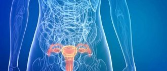
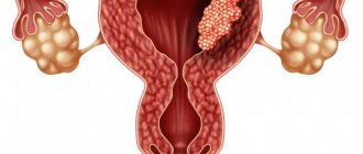
Vulvar cancer is an oncological disease on the genitals: outside the labia or clitoris, vestibule of the vagina or posterior commissure, urethra - in soft, skin or mucous tissues. These may be lumpy growths, a flat ulcer with ridge-like edges, or nodes in the vulva.
It is easy to diagnose the disease visually and manually, since all genital organs are accessible for examination. However, vulvar oncology has not yet been fully studied, so vulvar and vaginal cancer in women is one of the most difficult to treat neoplasms.
The occurrence of the disease is influenced by the characteristics of the blood supply to its organs, innervation and lymphatic drainage. The topographic proximity of adjacent organs and the high physiological and psychosexual vital significance of the female external genitalia are also important. The disease is rare, occurring in 2-5% of women with cancer. But at the same time, cancer of the labia and other components is induced by hormonal changes, which occurs during the postmenopausal and menopausal periods in 50% of older women 60-80 years old.
Symptoms and signs of pathology
In the early stages of vulvar cancer, patients may have no obvious complaints at all. Sometimes leukoplakia, along with kraurosis and lichen sclerosus, is a harbinger of the development of such a pathology. Often the first stage of the disease is asymptomatic.
The initial symptom of vulvar cancer is a feeling of itching with discomfort or burning in the genital tract. Painful sensations usually appear later. Externally, the first changes can be difficult to detect without examination, but sometimes you can see that a lump has appeared in the groin area in the form of an enlarged lymph node that does not hurt.
The primary visible manifestations are spots on the mucous membrane, which are not difficult to detect: it is enough to simply carefully examine the vulva. Unfortunately, this is done too late. Such spots have a whitish-grayish tint. Similar symptoms are observed against the background of leukoplakia.
Characteristic signs of vulvar cancer with exophytic growth are the appearance of inflammation, ulceration, and, in addition, purulent, bloody discharge, which indicate that the disintegration of the cancerous tumor has begun. Endophytic germinations are characterized by swelling along with compactions and infiltration. Later manifestations include weakness, fatigue and low temperature. At the terminal stage, symptoms progress rapidly, usually resulting in severe exhaustion. You may also experience rapid weight loss and a weak pulse with shortness of breath at the slightest exertion. The result most often is death.
You can see the symptoms of vulvar cancer in the photos presented in the article.
Precancerous diseases of vulvar cancer
The remaining statistical 50% of cancer cases occur in young women due to the herpes virus and papilloma. Kraurosis of the vulva affects the development of oncological tumors - these are precancerous diseases of the external genital organs. They give rise to dystrophic, atrophic and sclerotic changes in their skin, and subsequently – atrophy of the vulva. Leukoplakia often develops along with cancer.
According to the WHO classification (2003), precancerous diseases with changes in flat epithelial cells include:
Vulvar dysplasia (and carcinoma in situ):
- light (VIN*);
- moderate (VIN2);
- severe (VIN3) and carcinoma in situ.
Note: VIN refers to vulgar intraepithelial neoplasia of the vulva.
Causes of vulvar carcinoma
The exact causes of vulvar carcinoma have not been established, but experts can name a number of risk factors that significantly increase the likelihood of the disease occurring:
- infection with human papillomavirus;
- hormonal imbalance in the body;
- the presence of such background diseases as vulvar kraurosis, leukoplakia;
- decreased immune defense of the body;
- vulvar dysplasia;
- smoking;
- promiscuity.
↑ Top | Applying for treatment ↓
Classification of the disease
Squamous cell carcinoma of the vulva occurs in 70-90% of patients, since the epithelium contains squamous cells. It is divided into smaller and larger groups. A smaller group includes tumors that are induced by HPV. They are called aaloid-free and verrucous. The second large group includes an oncological process from a flat cell with an unknown etiology.
Cancer affects the vulva and its organs in different ways. The labia majora - 52%, minor - 7.1%, the clitoris is affected in 12-20%, the posterior commissure - in 6.4%, the periurethral zone - in 1.7%, the Bartholin gland - in 0. 2%.
Anatomy of the vulva
ICD code is C51, anatomical areas are designated as follows:
- labia majora – C51.0 and labia minora – C51.1;
- clitoris – C51.2;
- cancer of other localization – C51.8;
- unspecified areas of the vulva with oncological process – C51.9.
In accordance with the histological classification (WHO, 2003), they are also divided into: squamous cell keratinizing carcinoma of the vulva, non-keratinizing and basaloid, verrucous or verrucous and papillary, condylomatous and other forms.
Glandular malignant neoplasms include:
- extramamillary or Paget's cancer of the vulva;
- Bartholin gland: squamous cell, adenoid cystic, glandular squamous or transitional cell, adenocarcinoma;
- adenocarcinoma in situ;
- mammary-like anogenital glands (adenocarcinoma);
- squamous (or flat component);
- adenoid cystic;
- transitional cell;
- small cell;
- basal cell;
- from ectopic tissue cells of the mammary gland;
- from sweat gland cells.
Vulvar tumors in women are often located in soft tissue. These include according to WHO 2003:
- rhabdomyosarcoma;
- aggressive course of angiomyxoma;
- leiomyosarcoma;
- dermatofibrosarcoma protuberans;
- oncofibrous histiocytoma;
- epithelioid sarcoma;
- onkoshvanny;
- malignant hemangioendothelioma;
- Kaposi's sarcoma;
- hemangiopericytoma;
- liposarcoma;
- alveolar soft tissue sarcoma.
The new WHO histological classification (Lyon, 2003) of soft tissue tumors includes sarcomas such as:
- botryoid;
- leiomyosarcoma;
- epithelioid;
- alveolar soft tissue;
- liposarcoma;
- Dermatofibrosarcoma protuberans.
Other malignant processes include tumors in the form of:
- melanoma;
- hemoblastosis;
- lumps in the yolk sac, formations from Merkel cells;
- metastatic secondary fistulas.
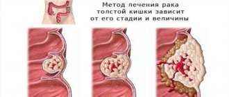
Stages of the malignant process
Clinical stages of vulvar cancer without surgical intervention data determine the degree of malignancy of the primary process.
- Stage 1 is a tumor that does not extend beyond the vulva, located on the skin tissue of the perineum: behind the vagina and up to the anus. Cone diameter - ≤2 cm;
- Stage 2 is determined by a lump with the same location, its diameter is >2 cm;
- Stage 3 indicates a node of any size. The spread occurs through the following channels: urethra and anus, covers the lower 2/3 of the vagina, metastasizes to the lymph nodes;
- Stage 4 is characterized by neoplasms of different diameters that spread through the urethra, vagina, bladder, rectum, and adhere to the pelvic bone tissue. There is metastasis to distant organs.
Data from surgical interventions (histological confirmation) are used for primary cancer in the FIGO and TNM classification (2009).
Classified as vulvar cancer (C 51)
Decoding the data of the primary tumor T:
- TX – cannot be assessed;
- T0 – cannot be determined;
- Tis – determine pre-invasive cancer (cancer in situ), grade III intraepithelial neoplasia (VINIII);
- T1 – the lump does not extend beyond the vulva and/or perineum and vulva;
- T1a – lump ≤2 cm in diameter, there is stromal invasion up to 1 mm1;
- T1b – lump larger than 2 cm or has stromal invasion > 1 mm1;
- T2 – the formation has a different diameter and is distributed to the perineal organs: vagina, urethra and anus;
- T32 – has a different size and covers: the urethra and 2/3 of the vagina, the walls of the bladder, the rectum, and is fixed to the bone tissue of the pubis.
Notes:
- 1 - deep invasion is determined by the distance from the epithelial-stromal junction of the dermal outgrowth on the surface to the deepest invasive point;
- 2 - FIGO does not use the T3 indicator, but only T4.
Interpretation of indicators of regional lymph nodes N - on the thigh and groin:
- NX – RLU – no assessment is possible;
- metastases:
- N0 – cannot be determined in the RLU;
- N1a – found in 1–2 lymph nodes (<5 mm);
- N1b – there are ≥ 5 mm in a single LN;
- N2a – in many lymph nodes <5 mm;
- N2b – in many lymph nodes > 5 mm;
- N2c – identified in the lymph nodes, there is capsule germination;
- N3 – there is a metastasis fixed or with ulcerations in the RLU.
Indicators of distant metastases M:
- M0 – no metastases in distant lymph nodes;
- M1 – metastases in distant lymph nodes and pelvis were identified.
Interpretation of indicators of the degree of histological differentiation G. The degree was established:
- GХ – cannot be installed;
- G1 – high;
- G2 – average;
- G3 – low or undifferentiated cancer;
- G4 – poorly differentiated vulvar cancer with a predominance of flat cells and keratinization. It is rare and has a poor prognosis.
In squamous cell carcinoma of the vulva, metastases are found in regional lymph nodes: in the groin (on both sides), on the thigh. Less commonly, in the pelvic area from a clitoral tumor. Hematogenous dissemination of cancer almost never occurs. Lymphogenous pathways of metastasis of vulvar cancer are characteristic. Due to the close connection of lymph vessels, bilateral and cross metastases occur.
Read here: Bladder cancer in men
Metastases from formations in the endometrium, urethra and bladder more often penetrate into the vulva and develop secondary cancer. Occasionally, tumors of the vagina, ovaries, mammary glands, kidneys, bronchi, cutaneous melanoma, lymphoma and choriocarcinoma metastasize to the vulva.
Regional lymph nodes are affected when the size of the nodes is:
- T1 – with a frequency of 8.9%;
- T2 – with a frequency of 25.3-35%;
- T3 – with a frequency of 31.1 – 55%.
Classification
According to the histological classification, the following types of this disease are distinguished:
- squamous cell carcinoma of the vulva;
- malignant melanoma of the vulva;
- primary adenocarcinoma.
Based on the type of growth of a malignant disease, the following types of pathology are distinguished:
- knotted;
- exophytic;
- ulcerative;
- hydropic.
Based on the degree of development, there are four main stages of development of this cancer. Surgery and concomitant therapy are performed only in the first, second and partially third stages. As for vulvar cancer at the fourth stage of pathology development, then, in this case, we are talking only about maintenance therapy, since surgery is impossible.
Causes
The causes of vulvar cancer in women most often include precancerous diseases, the presence of HPV DNA, and the presence of genital warts due to papillomavirus infection. It can cause papillomas or epithelial dysplasia in places such as the vulva, vestibule, anus, cervix, tongue, lips, palms and soles, and penis in men.
Attention! Women are at risk of developing cancer if they have HIV or VIN (vulgar intraepithelial neoplasia). The risk of the disease also includes frequent changes of sex partners, alcohol and smoking.
Main reasons
The etiology and main causes of this pathology have not yet been fully studied. True, fifty percent of patients have papillomatosis on their bodies. Its causative agent is the papilloma virus. It can be transmitted through contact or household contact. There are strains that cause the formation of warts and condylomas, which have a high degree of oncogenicity. Often this pathology begins with the appearance of papillomatous growth. Penetrating the epithelium, the virus changes the structure of human DNA, which can cause dysplasia with accelerated division. The relevance of this problem lies in the fact that if vulvar cancer was detected in a young woman, then with an eighty percent probability the papilloma virus is also detected. Today, more than ninety percent of the entire population of the Earth is infected with it.
Vulvar cancer can often be inherited. Hereditary predisposition is noted in thirty percent of cases. This is the so-called intraepithelial type neoplasia. The gene that provokes it has not yet been identified. Psychosomatics explains that malignant tumors are formed as a result of mental experiences, which are often unconscious. They can also arise due to lack of attention or love. Thus, according to some teachings, hatred with resentment, malice and envy towards other people can cause oncological processes.
Symptoms and manifestations of vulvar tumors
Early general signs of vulvar cancer are similar to those of vulgar intraepithelial neoplasia (VIN). As the problem area progresses, an inflammatory process appears:
- the mucous membrane becomes irritated and itchy, becoming rough;
- a red or white warty bump or abrasion appears;
- a long-term, non-healing painful ulcer forms;
- pain in the vulva becomes long-lasting and recurrent;
- urination causes pain in the urethra;
- discharge of blood with pus appears;
- the inguinal lymph nodes are enlarged.
Symptoms and manifestations of rare vulvar cancer
Melanoma is suspected if the following symptoms are present:
- rapid growth and increase in nevus density;
- pigment changes;
- ulcerations with slight bleeding and crusting;
- redness around the growth and new nodes with cracks;
- increasing the volume of lymph nodes.
Vulvar melanoma metastasizes to distant organs: bones, adrenal glands, liver, lungs and brain. Melanoma cells reach the papillary dermis (1 mm) through the basement membrane, and metastases occur in 5% of cases. If the tumor grows into the papillary layer with an invasion of 1.1-2 mm and spreads through the subcutaneous fatty tissue, then survival rates drop, and relapses occur in 78% of cases.
Paget's disease. The anogenital area is endowed with apocrine glands, so Paget's disease occurs in 2% of women during menopause. The tumor has an invasive component and often develops as a result of the screening of cancer cells from a primary tumor located nearby into the mucosa.
Symptoms:
- red, weeping eczematous lesion in the form of dermatosis;
- hyperkeratosis in the form of small lesions on the labia majora, skin of the abdomen and thighs;
- the presence of multicentric foci in the skin.
At the same time, Paget's disease can occur in the mammary glands and vulva. If pagetoid changes in the epidermis are found in the perianal zone, then cancer may develop in the rectum.
Sarcomas of the vulva
Botryoid sarcoma or embryonal rhabdomyosarcoma can be detected in girls under 10 years of age, and at any age - leiomyosarcoma in smooth muscles. Clinically, the symptoms of vulvar cancer in women are unpredictable. Subcutaneous painful nodules of high differentiation with the presence of necrosis are more often found. There is infiltration into surrounding tissues with a mitotic index > 10 mitoses in 10 fields of view. Metastases spread to the retroperitoneal space and peritoneal lymph nodes.
Symptoms of epithelioid sarcoma or malignant rhabdoid cancer are similar to soft tissue sarcoma of the extremities and large Bartholin gland cyst of the vaginal vestibule. Symptoms appear:
- discomfort during sexual intercourse, movement and wearing tight-fitting underwear, skirts and trousers;
- complications with the appearance of painful purulent abscesses;
- high temperature, intoxication: nausea, weakness, vomiting;
- a sharp deterioration in health.
When the tumor size is up to 10 cm, sharp painful pulsation and distension in the perineal area are possible. If the tumor opens on its own, then pus flows out and the sexual flora becomes infected. This can be seen in the signs of vulvitis, vaginitis and pathological vaginal discharge. Epithelioid sarcoma repeatedly recurs and metastasizes.
Dermatofibrosarcoma protuberans occurs at different ages; its symptoms are similar to low-grade cutaneous sarcoma. Fibrous cancer is most often located on the labia majora. It is located in the capsule, there is no invasion of the mucous membrane. After treatment it often recurs. Metastases spread to the lungs.
Metastatic tumors in the perineum occur in 8% of women. The cells are delivered by lymph from the primary cancer site: the cervix or endometrium of the uterus, ovary, breast, kidney, stomach or tongue. As a continuation of the tumor from the bladder, urethra or vagina, vulvar cancer is formed, the symptoms of which sharply reduce the quality of life of patients.
They suffer from the following clinical symptoms:
- inflammation and bleeding, especially after sexual intercourse;
- pain radiating to the lumbar region, sacrum, perineum;
- swelling of the extremities after metastasis to the lymph nodes, germination into neighboring organs;
- hyperthermia, general weakness and fatigue;
- disorders of the functional functioning of the gastrointestinal tract and urination.
Types of cancer
The following types of vulvar cancer are distinguished, which are based on its histological structure, and in addition, the direction of growth and other features. Thus, the cytological picture allows us to distinguish the following types of this disease:
- Squamous form.
- Basal cell variety.
- Presence of melanoma.
- Development of Paget's disease.
- The appearance of adenocarcinoma.
- The appearance of Bartholin gland carcinoma.
Squamous cell carcinoma accounts for about eighty percent of all cases today, and other types are much less common. According to the criterion of the direction of germination of the pathology, exophytic and endophytic forms are distinguished. The international classification shows the prevalence of this process, and it takes into account:
- Size of the primary tumor.
- The degree of damage to the lymph nodes.
- Presence of metastases.
Diagnosis of the disease
Diagnosis of cancer of the female genital organs is carried out:
- gynecological examination;
- palpating the RLU;
- vulvoscopy, vaginoscopy and colposcopy;
- cytological examination;
- incisional wedge biopsy to the lowest point of tumor formation and part of the tissue with healthy cells;
- histological examination of tumor biopsy;
- Ultrasound of lymph nodes in the groin, hips and ilium;
- Ultrasound of the peritoneal and pelvic organs;
- chest x-ray examination;
- ECG.
A total biopsy is used due to the rapid generalization of the tumor. Anamnesis and impression smears from the surface of the tumor are assessed. Vulvar cancer is confirmed by diagnosis using immunohistochemical studies (proteins: actin, desmin and myoglobin are determined) and electron microscopy to identify microfilaments.
Important! To correctly determine the prognosis and prescribe treatment, the expression of proteins p53, Ki-67, S-100, RE, RP and other tumor markers is studied at the molecular biological level.
In accordance with the indications, the following is carried out:
- fine-needle puncture biopsy of the lymph nodes of the groin and thigh and monitor the procedure with ultrasound (if metastasis to the lymph nodes is suspected);
- cystoscopy when the oncological process moves into the wall of the bladder (according to ultrasound indications);
- biopsy of the rectal mucosa and/or bladder;
- rectomanoscopy (if there are complaints and spread of cancer in the vulva);
- coagulogram before surgery;
- PET – positron emission tomography in late stages;
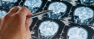
- CT, MRI to clarify the spread of the oncological process.
In the laboratory, urine (general analysis) and blood are examined:
- to determine the group and Rh factor;
- to determine the HBS-Ag antigen;
- for syphilis, determination of antibodies to hepatitis C virus, HIV antibodies and for HPV DNA testing;
- to determine general indicators;
- for total protein, urea, bilirubin, glucose by biological methods.
Additionally, the function of external respiration, kidneys with radioisotopes, excretory urography (according to indications), and radioisotope lymphography are examined.
Possible complications
Treatment of this pathology, which includes radiation with chemotherapy, can cause various side effects along with a deterioration in the quality of life of patients. For example, due to the removal of the genital organs, swelling of the legs may occur with inflammation and separation of postoperative sutures, as well as with the accumulation of fluid in the area of the affected area.
Negative surgical consequences can also occur when nerves are damaged. Patients may experience numbness with tingling in the damaged area of the skin. In addition, the suture may not heal well. As part of prevention, it is recommended to lubricate areas of the skin with a balm called “Vinilin”. Sea buckthorn oil is also suitable. Chemotherapy and radiation therapy for vulvar cancer can cause various complications, which include the following:
- Decreased immune strength of the body.
- Development of anemia.
- Significant hair loss.
- The appearance of disorders in the intestines.
- Observation of narrowing of the vagina.
- The appearance of a burning sensation during urination.
- The appearance of general weakness, irritability, apathy and anxiety.
Throughout the postoperative period, the patient needs special attention to herself, and in addition, care from the clinical staff and, of course, loved ones.
In the later stages of vulvar cancer (photos can be easily found), sick people may experience severe physical discomfort. In this regard, the question of how to relieve pain turns out to be the most pressing. At a certain point, the use of non-narcotic analgesics no longer brings relief. Patients are individually prescribed potent drugs, for example, Buprenorphine or Morphine, strictly according to a doctor’s prescription.
Painkillers can be somewhat addictive with some side effects such as brain fog, dizziness, nausea, low blood pressure, dry mouth and extreme thirst.
Not everyone knows what vulvar cancer looks like.
Vulvar cancer treatment
Treatment of vulvar cancer is carried out using the following methods:
- surgical;
- combined (surgical and radiation);
- complex and individual, combining radiation, medicinal and surgical techniques at the 4th stage and in case of recurrence;
- irradiation as an independent method or simultaneously with chemotherapy in case of absolute contraindications to surgical treatment.
Surgery on the vulva for cancer is performed by radical vulvectomy and femoroinguinal lymph node dissection. If the tumor is small, lymph node dissection is performed through separate incisions. If the tumor has spread over a large area, to hide the defects, tissue is removed from the vulva, pubis, perineum, thigh and groin on both sides using a one-piece plastic surgery method.
Read here: What is plasmacytoma: features of an oncological tumor
Treatment methods by stage
Squamous cell carcinoma of unknown etiology is most often described.
- Stage 0 (TisN0M0):
- the node is widely locally excised, departing from the edge 05-1.0 cm, and the operation is combined with laser treatment;
- skin vulvectomy is performed with tissue flap transplantation (or without);
- perform application chemotherapy with 5% Fluorouracil ointment.
- Stage IA (T1aN0M0):
- for microinvasive cancer, a tumor with d ≤ 2 cm and stromal invasion ≤ 1 mm is widely excised. If there is no severe diffuse dystrophy, inguinofemoral lymphadenectomy is not performed. The incision line is drawn at a distance of 1-2 cm from the edge of the formation circumference;
- carry out an urgent histological examination to determine the final extent of the operation;
- in case of multifocal lesions or the development of an oncological process in the vulva, cutaneous vulvectomy is performed simultaneously with dystrophic changes. Inguinal lymphadenectomy is not performed;
- Radiation exposure as an independent method is used if there are contraindications to surgery.
- Stage IB (T1bN0M0), carried out:
- radical hemi-vulvectomy and biopsy of sentinel lymph nodes are carried out when the diameter of the oncological node is <4 cm, inguinofemoral lymphadenectomy for metastases, the incision line from the edge of the node is 1-2 cm around the entire circumference;
- biopsy of sentinel lymph nodes on the affected side (lateral lesion) at a distance from the midline ≥ 1 cm, on one side inguinofemoral lymphadenectomy for metastases;
- bilateral biopsy of sentinel lymph nodes to determine the lesion: central (up to 1 cm from the midline), clitoris or posterior commissure; in case of metastases - bilateral inguinofemoral lymphadenectomy;
- radical vulvectomy and bilateral inguinofemoral lymphadenectomy with isolated removal of lymph nodes in the groin;
- radiation therapy for vulvar cancer: irradiation of inguinal lymph nodes (at N0) in women with medical contraindications or refusal of surgery;
- radiation methods using a radical program in case of a widespread tumor process or the impossibility of performing a radical vulvectomy.
- Stage II (T2N0M0)
They perform combined operations, combining chemistry and surgery for oncological processes in the lower part of the urethra and/or vagina, or anal ring.
The following methods are used:
- preoperative chemotherapy or chemoradiotherapy. At the same time, the operability of the node is increased and its size is reduced;
- radical local excision or vulvectomy and simultaneously bilateral inguinofemoral lymphadenectomy with a clinical incision line along the circumference up to 1-2 cm from the edge of the tumor masses;
- combined surgical intervention and simultaneously distal ureterectomy and/or distal vaginectomy for a tumor in the lower third of the vagina and/or urethra;
- treatment is prescribed as in the late stage of cancer, if the tumor has spread to the anal area;
- for the vulva area - postoperative irradiation and chemotherapy in the presence of: tumor > 4 cm, incision line < 8 mm, lymphovascular spread, tumor stromal invasion > 5 mm;
- for the area of regional and external iliac lymph nodes - postoperative chemotherapy and radiation if there is:
- macroscopic metastases in the RLU;
- more than 2 microscopic metastases in the RLU;
- radical radiation therapy if it is impossible to perform surgery due to too large areas of the oncological process or concomitant diseases.
- Stage III (T1,T2;N1a,N1b;M0), (T1, T2;N2a, N2b;M0)
Choose the following methods:
- neoadjuvant chemotherapy or radiation and excision of the remainder of the tumor masses;
- in case of oncological process in the lower part of the vagina and/or urethra - a combined operation: radical vulvectomy, inguinofemoral lymphadenectomy and simultaneously distal ureterectomy and/or distal vaginectomy, then postoperative chemotherapy;
- treatment as for the last stage of cancer, if the tumor has spread to the anal area.
- IVA stage (T1,T2;N3;M0), (T3NanyM0)
Choose treatment:
- radial;
- complex: superradical surgery + chemotherapy with Cisplatin at a dose of 40 mg/m² once every 7 days, using a neoadjuvant and/or adjuvant regimen;
- neoadjuvant chemotherapy and subsequent surgery or radiation.
- IVB stage (T any, N any, M1), (distant metastases)
Choose:
- chemoradiation methods according to a program compiled individually;
- individual palliative chemotherapy.
Relapses of the oncological process
Vulvar cancer can recur in the postoperative period, so treatment is carried out:
- surgical intervention in the presence of local relapses ± irradiation;
- radical vulvectomy and evisceration of organs in the pelvic area;
- chemoradiotherapy with or without surgery;
- if the iliac lymph nodes are affected and it is possible to operate them, radical lymphadenectomy and subsequent irradiation;
- palliative chemotherapy.
Important! Patients need to be observed and advised after treatment:
- 2 years – after every 3 months;
- 5 years – every six months after diagnosis;
- after 5 years - every year.
Radiation therapy
After surgery, the patient is irradiated for 21-28 days. External irradiation is performed on the primary tumor and areas of regional metastases (groin-thigh).
Carry out: conventional irradiation of primary tumor formation: ROD* 2-3 Gy, SOD** - 36-40 Gy (CT 2-2, 5D) and areas of regional metastases: ROD 2-3 Gy, SOD - 40 Gy (CT, 2- 2.5D) or conformal irradiation on the primary tumor: ROD 3 Gy, SOD 40 Gy (CT or MRI, 3D, electron accelerator with an MLC - multileaf collimator) and areas of regional metastases: ROD 2-3 Gy, SOD 40 Gy (CT , 2-2.5D, electron accelerator with the presence of MLC).
Therapy is carried out according to indications:
- intracavitary radiation using HDR brachytherapy devices (192Ir,60Co): SOD 28-30 Gy (3D with planning HR-CTV, IR-CTV);
- radical combined radiation;
- remote beam:
- conventional for the primary cancer node ROD 2-3 Gy, SOD 50-60 Gy (CT, 2-2.5D) and for areas of regional metastases - ROD 2-3 Gy, SOD 60 Gy;
- conformal irradiation to the primary oncological epicenter: ROD 2-3 Gy, SOD 50-60 Gy (CT or MRI, 3D, electron accelerator with the presence of MLC) and areas of regional metastases: ROD 2-3 Gy, SOD 60 Gy (CT or MRI, 3D, electron accelerator with MLC);
- intracavitary radiation, and use brachytherapy devices HDR (192Ir, 60Co): SOD 28-30 Gy (3D with planning HR-CTV, IR-CTV).
ROD is a single focal dose; **SOD is the total focal dose;
Chemotherapy
Chemistry is carried out according to one scheme:
- from 6 courses or until the oncological process begins to progress: neoadjuvant, 1st and subsequent lines;
- of 2-3 courses, using the neoadjuvant regimen.
For a minimal amount of chemotherapy, the following is administered:
- 50 mg/m² Cisplatin once, repeat 6 times every 21 days;
- 50 mg/m² Cisplatin on the first day + 500 mg/m² for 1-3 days. Repeat 6 times with an interval of 21 days.
Treatment is carried out with the optimal volume:
- Paclitaxel - 175 mg/m² + Cisplatin - 75 mg/m² on the first day, interval - 21 days;
- Paclitaxel -175 mg/m2 + Carboplatin AUC 5-6 on the first day, interval – 21 days;
- Cisplatin -50 mg/m2 on the first day + Gemcitabine 1000 mg/m2 on days 1.8, interval – 21 days;
- Xeloda-2500 mg/m2/day for days 1-14, interval – 21 days.
Systemic chemotherapy according to other regimens includes the following combinations of drugs:
- 500 mg/m² 5-fluorouracil – on day 1;
- 1.4 mg/m² Vincristine intravenously – on the 1st day;
- 15 mg of Bleomycin inside a vein or muscle – 5 days or 10 mg/m² inside a muscle – 2 times a week, for a total of 2-3 weeks. The interval between courses is 21 days.
Treatment of advanced or recurrent vulvar cancer is carried out according to the following polychemotherapy regimens:
- Cisplatin – 70-90 mg/m² – intravenous infusion (rate up to 1 mg/min) with pre- and post-hydration – on the 1st day. Vinorelbine – 25 mg/m² orally (injected over 6-10 minutes) – on the 1st and 8th days;
- Cisplatin – 75 mg/m² – intravenous infusion (rate up to 1 mg/min with pre- and post-hydration) – on the 1st day. Fluorouracil – 4 g/m², intravenous continuous infusion for 96 hours.
- Mitomycin C – 10 mg/m² – intravenously (injected over 20-30 minutes) – on the 1st day. Fluorouracil – 1 g/m², intravenous infusion for 24 hours. Begin 30 minutes after IV Mitomycin C on days 1, 2 and 3.
Read here: What causes skin cancer?
Consequences of complex cancer treatment
After a vulvectomy: partial, local or complete, the lymph nodes may become enlarged.
When they are removed, lymphadenoma occurs:
- legs swell;
- fluid is retained in 55% of patients;
- sutures become inflamed and come apart in 30% of patients.
During surgery, the nerve may be damaged, which is manifested by a feeling of numbness, tingling, and the presence of cold and hot areas of the skin. In addition, a seroma appears near the suture - liquid under the skin, which prevents the suture from healing quickly. Thrombosis causes discomfort and pain in the groin area.
The consequences of radiation therapy for vulvar cancer can make life difficult for patients for many months or even years. The skin above the epicenter is damaged by the rays and causes hyperemia and inflammation in the form of dry and then wet-vascular epidermitis and other local inflammatory reactions.
To prevent this kind of complications, you will need Vinilin (balm), aloe liniment, Tezan, sea buckthorn oil and other special products for the skin and mucous membranes to lubricate the irradiated areas.
Local damage includes thinning of the skin, atrophy of the mucous membrane, dryness, death of the bulbs and loss of pubic hair. Pigmentation increases, capillaries dilate, and sclerotic tissue appears under the skin.
The consequences of chemotherapy manifest themselves as:
- decreased appetite and immunodeficiency;
- the appearance of anemia, nausea and vomiting;
- excessive fatigue and deterioration of general condition;
- decrease in hemoglobin with a deficiency of leukocytes, platelets and lymphocytes in the blood;
- baldness;
- complications in the gastrointestinal tract;
- persistent diarrhea;
- burning in the urethra;
- narrowing of the vagina;
- for pain in inflamed vaginal scars.
Treatment of the disease
The disease is an extremely dangerous but treatable disease, however, if it is detected at an early stage. If the pathology is not treated, it will be fatal. It should be noted that death when such a disease is neglected can occur within a year.
The chance to survive, and, in addition, to stop the active development of carcinoma, exists if a person does not start the disease, but consults a doctor to receive appropriate medical care. Under no circumstances should you try to get rid of the disease yourself by taking painkillers, or look for effective remedies in recipes offered by traditional medicine. In such a situation, only competent medical care from qualified specialists should be provided. Treatment for vulvar cancer should be comprehensive.
Modern developments in such areas as gynecology, surgery and oncology help cure and defeat pathology. The treatment strategy consists of combination therapy, which will help slow down the rate of cancer development, allowing the body to recover, and in addition, prolong life. So, treatment includes the following aspects:
- Performing surgical removal of the lesion and metastasis.
- Carrying out chemotherapy using cytostatics.
- Conducting radiation therapy.
- Taking pain relief measures.
Patients recover immediately after surgery for vulvar cancer, during which the organ and regional lymph nodes are removed. Sometimes we are talking only about clinical remission. Relapses after surgery usually occur in seven percent of cases. The timing of their appearance cannot be predicted, but sometimes doctors have to operate on patients again.
Advanced and at the same time inoperable cancer still requires palliative therapy. Radiation therapy is usually prescribed, and the direct actions taken by oncologists are aimed at alleviating the suffering of the sick person. Chemotherapy for vulvar cancer is also given.
Traditional treatment for vulvar cancer
When diagnosed with vulvar cancer, treatment with folk remedies smooths out side complications after complex therapy. You can also treat cancer in its early stages at home.
For squamous cell carcinoma, greater celandine is used to provide antitumor, antiviral and anti-inflammatory effects. It has a weaker effect on the stratified epithelium of the vulva and rectum in the anal zone than on the single-layer epithelium behind the anus. But after the menstrual cycle, the epithelium of the vulva also becomes thinner, so celandine preparations will be highly effective for at least a week in this area. In this case, infuse the dried herb (1 tbsp) in a glass of boiling water for an hour. Then the genitals are irrigated or wet lotions are applied for 60 minutes - 3-4 times a day. Course duration is 1-2 months.
It is worth noting! It is contraindicated to use celandine for leukoplakia, increased inflammation and swelling.
As for hemlock oil infused with green seeds, use as an anti-cancer agent on areas of the skin where blood circulation is poor must be done with caution due to its toxicity. Oil from propolis and poplar buds relieves inflammation, allergies and itching, and heals damage. Oil from yellowing sophora and horse chestnut anesthetizes, increases microcirculation, kills microbes, relieves swelling, heals wounds and ulcers.
Ointment recipes:
- Mix beeswax (4 tbsp), lanolin (1 tbsp) and dry celandine grass powder (1 tbsp). The mixture is heated to dissolve the wax. After mixing thoroughly, allow the ointment to cool and lubricate the mucous membrane or apply it to the vulva on a piece of cloth for 15 minutes;
- mix Vaseline (4 tbsp) and celandine juice (1 tbsp), apply to the mucous membrane, or apply ointment to it on a piece of cloth for 15 minutes. Repeat the procedure 2-3 times a day, course – 2 weeks;
- Veselka mushroom powder (10 g) is mixed with heated propolis oil (10-15% - 100 g). The ointment is applied on gauze or a piece of tissue for swelling, itching and pain in the problem area.
Douching with veronica infusion is useful for vulvar cancer: add veronica (2 tbsp) to boiling water (0.5 l) and let it brew. Douching is performed at night for 15 days, then resumed after a week's break. In the morning, use a solution for washing: add 1 tbsp to 1 liter of boiling water. a spoonful of baking soda and a little shavings of tar soap to relieve itching. After the procedure, use sea buckthorn or fir oil to lubricate the vulvar mucosa.
To prepare the oil:
- fill the container 70% with raw materials;
- pour in unrefined sunflower oil;
- let it brew for 2-3 months in a warm corner;
- heat the jar with the contents in a bathhouse for half an hour to 40-45°C;
- strain until the oil cools.
Oil applications alternate with tampons in the vaginal area and the use of aqueous herbal decoctions for external use. Irrigations are carried out with infusions of collections of white sweet clover, gorse, Kuril tea (pentifolium), sage, white mistletoe branches, and barberry leaves. All herbs are taken in equal parts by weight, mistletoe and Kuril tea - twice as much. For infusion in a thermos you will need 1 tbsp. l. collection, 1 tsp. honey and 1 tbsp. boiling water
Dry plant powders can be used externally: mix the roots of yellow Sophora, hairy ash (roots), and Amur maakia bast (10:5:1). The powder is drawn into a dry syringe and sprayed onto the vulva. For ointment, the powder is mixed with petroleum jelly and applied to the tissue for pain and itching. After 2-3 hours, the remaining ointment is removed with a napkin moistened with olive oil.
If there are wounds and ulcers, then the vulva is cleansed with honey mixed with fish oil (3:1). Honey heals wounds, activates blood flow and lymphatic drainage, kills microbes, fish oil also heals ulcers and restores cell membranes due to anti-cancer vitamin A, softens the mucous membranes and skin.
Symptoms of vulvar carcinoma
The clinical picture of vulvar carcinoma consists of the following characteristic symptoms:
- itching in the genital area and perineum;
- the appearance of a tumor-like neoplasm resembling a wart or cauliflower;
- long-term non-healing ulcerative defect;
- pain in the lower abdomen and external genitalia;
- bloody or purulent discharge from the area of tumor growth;
- the appearance of pain and difficulty urinating;
- enlarged lymph nodes in the groin area;
- pain during intercourse.
↑ Top | Applying for treatment ↓
Disease prevention
Patients who have already undergone radiation for vulvar cancer should have their blood tested every 3 months in the first or second year and every six months in subsequent years.
If anemia and leukopenia appear, then control tests are done more often. After irradiation, the body is strengthened with vitamins, quality nutrition, and also:
- re-transfusion of blood and its individual components: leukocyte and erythrocyte mass;
- drugs are used that stimulate leukopoiesis (reproduction of leukocytes) and eprythropoiesis (reproduction of red blood cells).
A healthy lifestyle and nutrition, movement and personal hygiene are the prevention of cancer.
Be healthy!
Advantages of treatment for vulvar carcinoma in Israel
Treatment of vulvar carcinoma in Israel has a number of advantages:
- attentive attitude and individual approach to each patient;
- performing gentle organ-preserving surgical interventions;
- the use of highly effective drugs for local and systemic effects;
- high-quality radiation therapy using the latest devices;
- 24-hour care and supervision by highly qualified staff.
With timely access to qualified medical care, the prognosis for patients with vulvar carcinoma remains favorable.
↑ Top | Applying for treatment ↓
Diagnosis
By collecting an anamnesis, as well as interviewing the patient, the general dynamics of pathological processes are determined, along with the course of the disease and the benign nature of the formation. If vulvar cancer is suspected, the examination carried out to clarify the diagnosis will include the following procedures:
- Conducting a gynecological examination, which reports external changes.
- Carrying out palpation to identify enlarged inguinal and, in addition, femoral lymph nodes.
- Performing a colposcopy in order to determine the germination of the tumor.
- Carrying out vulvoscopy, which helps check the modified cells for the presence of dysplasia.
- Performing smear cytology along with a PAP test or checking for the presence of cancer cells with a description of their structure.
- Carrying out a biopsy and histological examination.
- Performing an ultrasound examination of the pelvis, and in addition, lymph nodes, which makes it possible to detect metastases.
When vulvar cancer begins to metastasize to neighboring areas, diagnosis consists of the following procedures:
- Performing a digital rectal examination.
- Taking x-rays of the lungs.
- Performing ureteroscopy.
- Carrying out computed tomography and magnetic resonance imaging, which, in turn, makes it possible to detect distant metastases.
The conclusion with the prognosis of the disease directly depends on the totality of symptoms and clinical trial data. In addition, it is necessary to take into account the duration of the pathology along with the general health of the patient.
How is vulvar cancer treated?
Preventive measures
How can you prevent and combat this serious disease? Prevention of cancer helps to avoid exposure to harmful factors by protecting yourself from them. In order to prevent the appearance of vulvar cancer, doctors recommend eliminating or at least reducing the effect of dangerous factors, and in addition, treating concomitant pathologies in a timely manner. First of all, you need to quit smoking, get rid of excess weight, and avoid getting all kinds of carcinogens into your body. In addition, disturbances in hormone levels and the development of vascular problems should not be allowed. Here are also recommendations that help prevent vulvar cancer:
- Providing the body with adequate sleep.
- Healthy lifestyle and normal daily routine.
- Proper nutrition along with regular preventive examinations.
This disease should be treated at its initial stage. This will make getting rid of it easier and much more effective. In this regard, every woman after fifty years of age should visit a gynecologist twice a year.
What is the prognosis for vulvar cancer?