Angiomyolipoma often reaches large sizes and has the character of an invasive process; the color can be gray, brown, white, and areas of hemorrhage and necrosis are visible in the neoplasm tissue.
histological image of angiomyolipoma
Microscopically, the tumor is represented by three main groups of cells of connective tissue origin - smooth muscle (myocytes), fat (lipocytes) and endothelial vascular, which can have an equal quantitative ratio or one group predominates over the other.
Epithelioid cells, resembling epithelium, are grouped predominantly around blood vessels. Tumor elements actively multiply, forming layers and clusters around the vascular component. The presence of foci of necrosis, tumor invasion into the perinephric space, and active division of its cells determine malignancy and poor prognosis of the disease.
A typical angiomyolipoma contains all of the listed cell groups. Sometimes lipocytes are not found in it, then they talk about an atypical form of the tumor, which can be confused with a malignant mesenchymal neoplasm. The benign nature of atypical angiomyolipoma is confirmed by aspirate biopsy.
Characteristics and causes of appearance
What do kidney angiolipomas look like? A small tumor formation (in the form of a nodule) is located in the soft tissues of the organ and consists of epithelial cells, fat, smooth muscles and blood vessels. Angiomyolipoma of the kidney, ICD code 10 D17. According to the internal structure, a benign formation can be typical or atypical. In the second option, there are no inclusions of adipose tissue. The acquired form of the pathology is called sporadic (isolated) and affects only one kidney (angiomyolipoma of the left kidney is more common). The congenital form simultaneously affects both renal organs (right and left). The isolated form in women over 40 years of age occurs due to increased production of estrogens and progesterones. Provoking factors for the appearance of pathology can be:
- pregnancy (hormonal fluctuations);
- kidney diseases (presence of acute and chronic processes);
- genetic predisposition;
- the presence of tumors in the body (for example, angiofibroma).
Is angiomyolipoma of the kidney life-threatening? New growths grow quickly. Extremely rarely (single cases) they can reach large sizes, which leads to complications: organ rupture, huge intra-abdominal bleeding, acute abdomen, compression of neighboring organs or the formation of nodes in them (for example, in the liver), necrosis of tumor tissue, vascular thrombosis.
IMPORTANT! If an angiomyolipoma grows into the renal vein or nearby lymph nodes, then this may degenerate into an oncological formation, which is fraught with the formation of metastases throughout the body (cancer). This happens rarely, but the possibility of degeneration cannot be ruled out.
Angiomyolipomas of both kidneys
Renal angiomyolipoma (hemartroma) is a type of benign neoplasm in the kidney tissue, consisting of cellular epithelium, adipose and smooth muscle tissue, and blood vessels. This is one of the types of soft tissue tumors - a mesenchymal tumor. Sometimes such formations are localized in the adrenal glands, pancreas, and skin. Such benign tumors are usually small in size and do not have pronounced manifestations.There are two forms of the disease:
- Hereditary or congenital - occurs due to tuberous sclerosis, is characterized by multiple neoplasms and lesions of both organs simultaneously.
- Sporadic acquired or isolated is the most common form of the disease, most often expressed in a unilateral course. For example, a single angiomyolipoma of the left kidney is often detected.
This pathology occurs more often in middle-aged women (after 40 years), which is due to the high presence of estrogens and progesterones (female sex hormones).
The causes of renal angiomyolipoma can be different. Most often this disease appears:
- For acute or chronic kidney diseases.
- During pregnancy, an increase in the production of hormones during pregnancy provokes the formation of a neoplasm.
- In the presence of similar neoplasms in other organs (angiofibromas).
- If there is a genetic predisposition.
In the initial stage of the disease, the neoplasm is small in size and, as a rule, the angiomyolipoma is localized in the right or left kidney. Symptoms do not appear in any way. Primary damage to both organs occurs extremely rarely, only in the presence of a hereditary factor.
The tumor tends to grow rapidly and the blood vessels have a dense muscle wall, but weak elastic plates do not keep up with the growth of muscle fibers. As a result, blood vessels rupture with hemorrhages. In such cases, the symptoms are quite pronounced:
- feeling of constant nagging pain in the lumbar region;
- sudden changes in blood pressure;
- weakness, severe dizziness to the point of fainting;
- pale skin;
- hematuria – the appearance of a large number of blood elements in the urine;
The presence of these symptoms serves as the basis for an emergency visit to a doctor and determination of an accurate diagnosis. Since the larger the kidney angiomyolipoma, the more dangerous its consequences can be.
A large neoplasm can cause spontaneous kidney rupture and massive intra-abdominal bleeding. Tumor growth into nearby lymph nodes or into the renal vein threatens the formation of multiple metastases.
Only early detection of angiomyolipoma is a guarantee of complete recovery. To accurately diagnose the disease, sensitive methods are used such as:
- Ultrasound scanning – helps to identify denser areas of the tumor against the background of normal kidney tissue;
- spiral computed tomography and magnetic resonance imaging – detection of areas of low density in areas where adipose tissue is present;
- general and biochemical blood tests - to determine the level of creatinine and urea, which characterize the quality of kidney function;
- Ultrasound angiography – detection of vascular pathologies of the kidneys;
- X-ray diagnostics (excretory urography) – determination of the morphological and functional state of the kidneys, pelvis and ureters;
- Kidney biopsy - taking a piece of tumor tissue for microscopic examination in order to exclude malignant processes.
Based on the obtained diagnostic data, an individual treatment plan for renal angiomyolipoma is drawn up, which takes into account the characteristics of the tumor.
Treatment methods for the disease are determined taking into account the number of tumor nodes, their size and their location. Small neoplasms (less than 4 cm) grow slowly, without causing complications, and in such cases observational tactics are used without active therapeutic measures. Control studies are carried out once a year.
For tumors exceeding the permissible threshold of 5 cm, surgical intervention is recommended.
It could be:
- Organ-preserving surgery (resection of part of the kidney) - in the presence of a normally functioning second organ.
- Embolization - under X-ray control, a drug (metal spiral or polyvinyl alcohol foam) is injected into the artery supplying the tumor using a special device (equilis) to block it. This procedure simplifies surgery or can completely replace it.
- Nephron-sparing surgery is applicable for multiple focal tumor tumors in both organs in order to preserve their functions.
- Enucleation is the removal of a tumor by enucleation, leaving the organ almost untouched.
- Cryoablation is a modern minimally invasive method used for the treatment of small angiomyolipomas. The advantages of the method are a procedure with minimal intervention in the body, the possibility of hardening adjacent structures to prevent bleeding, the procedure can be repeated, a short postoperative period and a minimal percentage of complications.
Indications for the use of surgical intervention are:
- a violent clinical picture of the disease with pronounced manifestations;
- rapid increase in tumor size;
- renal circulatory failure;
- significant hematuria;
- malignancy of a previously benign tumor.
It should be especially noted that treatment of kidney angiomyolipoma with folk remedies is not only ineffective, but also leads to rather disastrous results. Losing time will aggravate the situation with irreversible complications.
How does renal angiomyolipoma develop, is it life-threatening? This condition involves the involvement of the layer of fat, muscle tissue, and kidney epithelium in the pathological process. To a large extent, a benign tumor consists of a fatty layer.
As a rule, angiomyolipoma spreads to the kidneys. The disease can affect people of any age, but it is much more common in older people. The risk group includes people from 40 to 60 years old. It should be noted that cases of the disease are more often recorded in women. How serious is the disease and how is kidney angiomyolipoma treated? The treatment and danger posed by the disease will be described in this article. With timely treatment, the pathology can be completely eliminated.
Angiomyolipoma is a benign tumor in the kidney. The disease also received another name - “renal hamartoma”. The neoplasm belongs to the category of mesenchymal pathologies affecting soft tissues.
There are two forms of pathology: the primary sporadic form and the genetic disease.
In the first case, the tumor affects the body on its own, without genetic factors, and in the second case, the leading role is played by heredity.
There is also a completely separate disease called Bourneville-Pringle, or tuberous sclerosis. It involves kidney damage, but other symptoms are also noted.
If the tumor is localized in the left or right kidney, a unilateral lesion is diagnosed. If the pathology is diagnosed in both kidneys, then doctors talk about a bilateral form. The unilateral form develops in 75% of cases.
In addition, angiomyolipoma can be typical or atypical.
In its typical form, the neoplasm largely contains muscle tissue or a layer of fat, which is much more common in medical practice.
In the atypical form there is no adipose tissue. This makes therapy difficult. After all, fatty tissue is removed with minimal consequences. This form also makes diagnosis difficult. In this case, there is a high probability of erroneously distinguishing between a benign neoplasm and a malignant tumor.
As a rule, with this disease, the medulla and cortex of the kidney are involved in the pathological process. The tumor promotes the formation of a capsule, which is fenced off from healthy tissue.
What are the consequences of a disease such as angiomyolipoma of the kidney (left and right kidney)? It is dangerous because it sometimes develops according to a special scenario. This happens with the parallel influence of negative factors, for example, incorrectly selected therapy or the presence of additional pathologies in the kidneys.
The neoplasm can grow into the inferior vena cava, nearby lymph nodes, or the perinephric signet. In some cases, blood vessels are also affected, which can burst and cause hemorrhages.
Many people are interested in why angiomyolipoma is dangerous? Vascular torsion is often diagnosed. This creates coils, the ruptures of which can cause intense internal bleeding, which poses a threat to the patient’s life. Blood enters the abdominal cavity and spills into it. The vascular walls that contain muscle tissue become thick and round, as the muscle tissue is completely or partially degenerated into connective tissue. As a result, perforations are observed, which threaten an aneurysm and other dangerous complications.
The development of the disease can be due to completely different reasons.
A number of provoking factors include:
- Pregnancy. The appearance of a tumor depends on hormonal levels. As is known, during pregnancy a woman’s level of estrogen and progesterone increases, which can serve as an impetus for the development of the disease.
- Genetic basis. As mentioned above, this pathology can occur in the presence of Bourneville-Pringle disease. This disease is genetic. Scientists have discovered genes that can be inherited.
- A variety of pathological processes in the kidneys, which, along with other factors, may well provoke the appearance of a tumor. Therapy in this case involves the elimination of all concomitant diseases.
- Manifestation of other neoplasms. For example, angiofibroma develops. This pathology occurs quite often. The disease described in the article begins to develop under its influence.
How does renal angiomyolipoma manifest itself? How dangerous is this disease? With timely treatment, the disease can be completely eliminated. But the insidiousness of the disease lies in the fact that in the early stages the symptoms can be erased. Manifestations become apparent as the tumor grows.
A number of symptoms include:
- A feeling of heaviness in the area of the left or right kidney, as well as the back. Unpleasant sensations may occur in the abdomen and lower back. The pain can be nagging, aching or dull. They are more intense when turning and other movements. This is explained by local hemorrhages.
- Blood pressure surges. They can occur for no apparent reason and quite often.
- Blood in the urine.
If the tumor becomes large, it can be detected by palpation.
If therapy is not carried out in a timely manner, consequences that threaten the patient's life may occur. First of all, when blood vessels rupture and bleeding occurs, peritonitis may occur. With extensive hemorrhage in the abdominal area, there is a risk of death.
What are the consequences of renal angiomyolipoma? Is it life-threatening if it becomes large? In this case, the tumor can put pressure on neighboring organs, preventing them from working properly. For example, when the right kidney is damaged, the appendix and even the liver are compressed. The neoplasm can rupture, which also poses a threat to life.
Another major complication is necrosis or death. In this case, the kidneys simply stop working.
We should not forget that a benign neoplasm can degenerate into a malignant tumor.
Angiomyolipoma of the kidney (causes, symptoms, treatment of which are described in this article) requires timely diagnosis. For effective therapy, a competent examination should be carried out.
First of all, an ultrasound examination is recommended, which will help identify the tumor. In some cases, magnetic resonance imaging may be advisable.
A general blood and urine test is highly effective. It is able to display the condition of the urinary system.
A biopsy method is also used. To determine the nature of the tumor, fluid is taken from the kidney for examination using surgery or puncture. This method makes it possible to diagnose the presence of oncology.
There is evidence of the high effectiveness of some drugs that belong to the group of inhibitors. After a course lasting a year, in some cases the tumor may decrease in size by half.
If there is very rapid growth of angiomyolipoma, then they resort to the surgical method.
As for treatment with traditional methods, it often does not give any results.
Can kidney angiomyolipoma be operated on? Is it life-threatening if it requires surgery? In some cases, it is dangerous and requires urgent removal.
Based on the results of diagnostic procedures, the specialist develops an individual treatment plan, which necessarily takes into account such important points as the stage at which the tumor is developing, its size and location.
In accordance with modern standards in the field of oncology, a tumor whose diameter is more than 4 cm is eliminated using a wait-and-see approach. This method is based on periodic monitoring of the patient's condition. The patient is advised to undergo ultrasound and x-ray diagnostics once a year.
A tumor larger than 4 cm requires radical treatment.
In addition to exceeding the threshold size, indications for surgical intervention are:
- pronounced clinical picture of the disease with progression of malignant symptoms;
- rapid tumor growth;
- the presence of renal failure, which becomes chronic;
- recurrent hematuria.
In the practice of oncologists during surgical intervention, the following techniques are expected to be used:
- Resection of a specific part of the kidney that is affected by a benign tumor. This operation can save the organ.
- Embolization. With this method, a special device is inserted into the artery. The artery supplies the angiomyolipoma. This agent helps to block the lumen of the vessel. This technique acts as a preparatory stage before surgery. In rare cases, embolization acts as an independent method of therapy.
- Enucleation. During the operation, the surgeon removes the tumor without damaging the adjacent kidney tissue.
- Cryoablation. This is an innovative way to eliminate tumors through exposure to low temperature. This method of treatment is used only for large tumor sizes.
- Absolute partial nephrectomy. If the tumor size is significant, the surgeon decides to completely remove the organ.
- Laparoscopy. This method involves making several punctures, which make it possible to introduce cameras and manipulators into the body.
Is angiomyolipoma dangerous? The prognosis and survival rate are positive. The tumor belongs to the category of benign neoplasms located in the capsule. This explains the favorable outcome of treatment. Most patients experience a complete recovery after surgery.
How renal angiomyolipoma proceeds and whether it is life-threatening was described in this article. Pathology is a serious disease, but can be treated. The main thing is to make a diagnosis and carry out the correct treatment.
In urology, the most common renal neoplasm is considered to be renal angiomyolipoma. This benign tumor consists of fat and muscle tissue, as well as deformed blood vessels. Acquired pathology affects one kidney, congenital is characterized by damage to both kidneys. If it develops rapidly, angiomyolipoma can become life-threatening.
There are two forms of this pathology. The name of the form indicates its feature:
- Congenital (hereditary). It affects two kidneys at once. The pathology consists of multiple formations resulting from tuberous sclerosis.
- Acquired sporadic (isolated). Accounts for 80-90% of cases of diagnosing angiomyolipoma. It affects one kidney.
If an angiomyolipoma of the kidney is detected, you must strictly follow all the doctor’s instructions. Neglecting health or self-medication can lead to dire consequences.
Return to contents
The nature of the occurrence of kidney AML is still not fully understood. The reasons that provoke the occurrence of neoplasms are different. Often the disease develops under the influence of factors such as:
- Chronic or acute renal pathologies.
- Pregnancy. It is considered the most common cause. During pregnancy, a change occurs in a woman's hormonal background; female hormones - estrogen and progesterone - are actively produced, which provoke the development of a tumor. It is because of the action of these hormones that women are 4 times more likely to suffer from this pathology than men.
- The presence of similar tumors in other organs.
- Genetic predisposition.
Return to contents
Angiomyolipoma of the kidney forms and develops asymptomatically. The tumor grows quickly, but the vessels feeding the angiomyolipoma develop more slowly than the muscle tissue and, as a result, rupture. The onset of bleeding is accompanied by the following symptoms:
- There is constant pain in the lower back;
- there are sudden changes in blood pressure;
- loss of strength, dizziness, fainting;
- pale skin;
- blood in urine.
If these signs occur, it is necessary to immediately take the person to the hospital for diagnosis and treatment. The degree of danger depends on the size of angiomyolipoma, since a large tumor can rupture an organ. As a result, internal bleeding occurs, and the tumor grows into the nearest lymph nodes. This leads to the occurrence of multiple metastases.
During pregnancy, a woman's body undergoes many changes. In particular, at this time the production of female sex hormones is activated. It is believed that changes in hormonal levels contribute to the development of angiomyolipoma. In this case, the tumor can be detected during a routine ultrasound. An existing angiomyolipoma develops more intensively during pregnancy. This tumor does not pose a threat to pregnancy failure and does not harm the baby.
The main danger to life of this disease is the rupture of angiomyolipoma. The cause of the rupture is a difference in the development of blood vessels and tumor tissue. In rare cases, the rupture occurs at the initial stage of development. Internal bleeding begins and urgent hospitalization is required. If the tumor grows significantly, it can cause rupture of the kidney parenchyma. Over the past 10 years of studying the disease, it has been found that this phenomenon can change and become a malignant tumor. In this case, the danger to life is comparable to any oncology. If treatment is not started in time, the pathology can cause liver dysfunction.
An ultrasound examination determines pathology by identifying compactions against the background of healthy renal parenchyma.
The earlier the pathology is diagnosed, the greater the chances of a full recovery. Since the pathology most often affects one organ, the result of the diagnosis is angiomyolipoma of the right kidney or the left. The following methods are used to detect tumors:
- Ultrasound. Determines the presence of seals.
- MRI and CT. Identifies areas of low density tissue (adipose tissue).
- Laboratory blood tests indicate the general condition of the kidneys.
- Ultrasound angiography. Detection of pathologies of renal blood vessels.
- X-ray diagnostics. Shows the condition of the organs and ureters, the presence of changes in structure and functioning.
- Biopsy. To exclude the possibility of developing a malignant tumor, a particle of the tumor is taken to study its nature and characteristics.
Early stages of renal angiomyolipoma can be cured without surgery. Return to contents
Angiomyolipoma is a benign neoplasm located in a connective tissue capsule. Therefore, timely therapy has a favorable prognosis. After surgery, the patient recovers completely. However, surgery is a last resort; drug therapy in combination with diet is immediately used.
The therapy program is developed by the doctor individually for each patient based on the diagnostic results. The number of tumors, their size and location are taken into account. If the patient is diagnosed with “Angiomyolipoma of the left kidney” and the tumor is less than 4 cm in diameter, then surgical intervention is not required, since small tumors develop slowly, without complications. Observation is prescribed, the patient periodically visits the doctor, and an ultrasound or computed tomography is performed once a year.
If the diagnosis revealed a unilateral angiomyolipoma, the diameter of which is more than 5 cm, and the second kidney is functioning normally, surgery is prescribed. If the tumor develops quickly, the likelihood of complications increases. At any time, the disease can result in bleeding, blood poisoning and death. The tumor is removed to prevent this.
Kidney resection involves removing only part of the organ along with the tumor. There are 2 types of this operation:
- Classical. A large incision is made in the lumbar region to access the organ.
- Laparoscopic. Several small incisions are made.
Return to contents
During the operation, the tumor is “husked” from the organ. Enucleation allows the tumor to be removed relatively easily if it is encapsulated, with little blood loss. This is a new way to remove AML from the kidney, as a result of which the kidney itself does not undergo any changes. This method is applicable only in the presence of a benign tumor.
Embolization involves the injection of a special drug into the blood vessels feeding the tumor, which causes their blockage. The procedure is carried out under x-ray control. As a result, surgery is much easier. In some cases, embolization eliminates the need for surgery.
This method is used to remove small angiomyolipoma by applying heat to it. The effect of the procedure is comparable to surgery with fewer contraindications and complications. In addition, the advantage of cryoablation is the minimal degree of intervention in the patient’s body, a short rehabilitation period and the possibility of a repeat procedure.
If the tumor increases significantly (more than 7 cm), the doctor is forced to perform a nephrotomy - complete removal of the affected kidney. This method is used if it is impossible to save the organ due to irreversible changes or a high risk of severe complications. It is important that the second kidney functions fully. The operation is performed under general anesthesia. The open (classical) method or laparoscopy is used.
If renal angiomyolipoma is diagnosed, you should strictly adhere to a special diet that inhibits the development of the tumor and prevents exacerbation of the disease. In this case, it is necessary to minimize salt intake. Dietary rules for angiomyolipoma boil down to complete abstinence from alcoholic beverages and coffee, eating small portions 6 times a day, and consuming at least 1.5 liters of fluid daily. It is allowed to consume low-fat dairy products, vegetable broths, lean soups/borscht, low-fat meat, cereals, pasta, eggs, vegetables, and steamed cutlets. Weak tea is allowed. For sweets, dried fruits, baked apples, honey, and jam are allowed.
If you have angiomyolipoma, you should avoid the following products:
- broths (meat, fish);
- fatty meat/fish;
- smoked, salted products;
- legumes;
- spices, herbs, marinades, sauces;
- horseradish, garlic, onion, radish;
- parsley, spinach, sorrel.
Return to contents
Tweet
Classification
Lesions are usually divided depending on the structural features:
- Trigasic (triphasic) is a benign mesenchymal tumor consisting of varying numbers of dysmorphic blood vessels, smooth muscle components, and mature adipose tissue. Can be divided radiologically into “classic” and “fatty-like subtypes.”
- Classic. A characteristic feature is abundant fat. The term "fat" in this context is used to refer to one or more fat cells. On ultrasound, classic angiomyolipoma is almost always markedly hyperechoic to the renal parenchyma.
- Protein. In this subgroup, there is a division into three subtypes: hyperattenuating and isoentenuating angiomyolipomas and angiomyolipoma with epithelial cysts.
- Hypertenuation accounts for approximately 4-5% of all angiomyolipomas. They are usually small to medium in size (maximum 3 cm in diameter).
- Epithelioid angiomyolipoma is an extremely rare type, first described in 1997. Consists of numerous atypical epithelioid muscle cells.
- The insulator contains diffuse, scattered fat cells among the smooth muscle and vascular components.
- Angiomyolipoma with epithelial cysts is an extremely rare variant. Like classic angiomyolipoma, these lesions are benign and are more common in women.
Clinical signs of pathology
The main symptoms of the presence of angiomyolipoma in the body are characterized by:
- hypotension or hypertension (low or high blood pressure);
- increased weakness and fatigue;
- the appearance of dizziness, fainting;
- pale skin (anemia develops);
- pain in the lumbar region of a constant nature on the side of the angiomyolipoma (left or right kidney);
- the presence of blood in the urine (hematuria).
Clinical signs of the disease arise due to changes that occur in the kidney tissue and hemorrhages into it. The appearance of such symptoms should alert the patient and serve as a reason to visit a doctor. Angiolipoma is not terrible in itself; it is dangerous due to complications that cause shock (hemorrhagic) conditions and intra-abdominal bleeding, which ends in death. Small tumors (less than 4 cm in diameter) resolve without complications, so the patient is monitored and undergoes annual examinations (no therapy is used).
For tumors larger than 5 cm, surgery is performed by surgeons.
Tumor detection
The patient most often hears about his diagnosis for the first time in the nephrologist’s office. The doctor informs him about this only after a person with suspected renal angiomyolipoma undergoes a series of necessary tests. Although it is possible that it can be detected by chance during an ultrasound of the kidneys. The standard diagnostic scheme looks like this:
- Palpation of the back area where the kidneys are located. If the tumor is large, then it can be palpated.
- Ultrasound examination of the kidneys, which allows you to detect not only a tumor, but also confirm that there are vessels in it.
- MSCT of the kidneys is a research method that is most often used to confirm the diagnosis. Regardless of the number of angiolipomas, it provides information about their location and size. Also, during the procedure, it is possible to determine whether there is a rupture of blood vessels with hemorrhages into the kidney parenchyma. This is especially true for patients admitted to the hospital with renal colic.
- Sampling of tumor tissue with its further histological examination allows us to establish the nature of the neoplasm. The procedure is carried out under the control of ultrasound or x-ray equipment.
- If surgery is prescribed, an MRI of the kidneys is performed. This method provides maximum information about the tumor.
In addition to the listed basic instrumental techniques, laboratory tests including blood and urine examination are indicated. It is important to evaluate the condition of all internal organs: intestines, bladder, liver, which will allow you to outline a plan for further treatment.
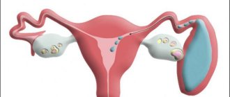
Pregnancy
Getting pregnant and only then finding out that you have health problems is a fear for any woman. Okay, but how will it affect the child? Neoplasms are rare in pregnant women, but they are always a difficult diagnostic and treatment problem. The development could have occurred before conception, but be asymptomatic and detected only on routine ultrasounds. Many tumors during pregnancy manifest as pain in both sides, with an abdominal or colic-like character. Many have hematuria and arterial hypertension. In rare cases, the classic triad of kidney tumors (lateral pain, hematuria, palpable mass) is observed. Angiomyolipomas are sensitive to estrogen and tend to grow in size under the influence of estrogen. As a result, increased tumor growth occurs during pregnancy. How does this affect the baby's health? What are the consequences of the tumor? Basically, a caesarean section is performed and the baby is carefully examined. In 99% of cases, he is born absolutely healthy. The only danger is rupture of the tumor, so the patient is hospitalized for constant monitoring.
Etiology of the tumor process
Renal angiomyolipoma is a neoplasia of unknown etiology. Until now, scientists have not even been able to determine for sure whether it is a congenital or acquired tumor. However, it has been established that the occurrence of neoplasms has some dependence on certain genetic diseases. In particular, we are talking about tuberous sclerosis.
Numerous studies aimed at establishing factors that can provoke tumor growth have made it possible to put forward the following assumptions regarding its origin:
- Angiomyolipoma in the kidney is often combined with inflammatory processes in the parenchyma of the organ. This may be pyelonephritis, glomerulonephritis, urolithiasis, etc. Therefore, some scientists are of the opinion that they are triggers in the formation of angiomyolipoma of both kidneys. Opponents of this theory insist that it is the tumor that provokes inflammation, and not vice versa.
- Since the neoplasm most often develops in women during menopause, experts do not rule out that endocrine disorders can influence the fact of its appearance. There is evidence that during pregnancy angiomyolipoma accelerates its growth. This may be due to hormonal changes in the body. This theory is also controversial, since it is not known for certain whether pregnancy gave rise to the development of a neoplasm, or simply provoked the rapid growth of a tumor that already existed.
- The fact that children inherit angiomyolipoma from parents has also not been properly confirmed. It has been proven that with a genetic disorder such as Bourneville-Pringle disease, a person develops multiple small tumors in both kidneys. However, we are not talking about the large size of the neoplasm and its single appearance, as in the case of angiomyolipoma.
- There is also a viral theory of the origin of the tumor. Its supporters argue that in some cases, tumor growth is observed when the body is damaged by certain viruses. But this theory was never scientifically confirmed.
However, scientists agree that there are certain reasons that are most likely to trigger the appearance of a tumor. These include:
- being female;
- entering menopause;
- period of bearing a child;
- hormonal disorders in the body;
- high level of female sex hormones in a man’s blood.
If these factors are combined with kidney inflammation and metabolic disorders in organs, then the risk of developing angiomyolipoma increases.
Diagnostic methods
Angiomyolipoma of the left kidney, what is it and how to recognize the pathology? The methods of diagnostic research conducted help with this. Since a benign formation is not accessible to palpation, the doctor prescribes:
- Ultrasound (detection of compactions in the kidneys - rounded isolated areas of low echogenicity);
- spiral CT and MRI (identification of low-density areas in the fat layer of the organ);
- ultrasound angiography (determining changes and functions of the renal circulatory system);
- x-ray or excretory urography of the entire urinary system (tissue condition and changes in function);
- biopsy (taking a piece of a tumor to exclude a malignant nature);
- scanning the condition of kidney tissue;
- clinical studies of the bloodstream (biochemistry and OA) - determination of indicators of the functioning of the renal organs - creatinine and urea;
- OA of urine (micro- and macrohematuria is detected);
- multislice computed tomography (MSCT) with contrast (slice examination of the kidney) – assessment of blood flow and supply to the organ.
The therapist and urologist should consult the patient and prescribe individual treatment. The doctor himself chooses the most suitable research methods for the patient, but taking blood, urine and undergoing an ultrasound are mandatory.
Diagnostics
Only with early diagnosis can a complete cure be guaranteed. To determine a neoplasm, use:
- ultrasound scan of the kidneys;
- spiral MRI or computed tomography;
- blood chemistry;
- Ultrasound angiography;
- excretory urography;
- renal tissue biopsy.
Treatment
The obtained diagnostic results give the doctor grounds to prescribe an individual plan of medication for the treatment of angiomyolipoma of the left or right kidney, taking into account the development of the stage and characteristics of the benign formation (detected by biopsy), the individual characteristics of the body, concomitant chronic pathologies and the tendency to allergic reactions. Surgical operations are prescribed only after the tumor has been accurately determined to exceed 5 cm in diameter. It is better to remove such tumors, because this will protect against complications in the future, and will not interfere with the normal functioning of the kidney organ and the body as a whole. In addition, indications for intervention are the following:
- a rapid increase in tumors (this indicates the start of the irreversibility of the process, the patient’s condition will only worsen) or malignancy;
- diagnostic indicators of renal circulatory failure;
- worsening clinical manifestations of the disease;
- detection of blood inclusions in urine;
- resection of part of the kidney;
- embolization (introduction of a chemical foam or metal coil into the artery feeding the tumor);
- enucleation (removal of formation using a special method of enucleation);
- cryoblation (a low-traumatic method that occurs without postoperative complications);
- nephron-sparing method (special sequential tactics aimed at preserving all renal functions).
A large angiolipoma of the right kidney, while the second is normal, is accompanied by severe pain (risk of developing huge hemorrhage, shock, sepsis, death). Surgical interventions used:
Nutrition for illness
When detecting angiomyolipoma of the kidney, it is necessary to follow a diet that will not provoke accelerated tumor growth and will not cause an exacerbation of the disease. The first thing you should do is, if possible, eliminate salt from your diet. You should also stop smoking, drinking alcohol and coffee. You should eat small meals 5-6 times a day and drink about 1.5-2 liters of water. You can eat dairy products, eggs, vegetables, lean meat, vegetable broths, pasta, honey, dried fruits. The list of prohibited foods includes pickles, smoked meats, meat broths, fatty meats, spices and spices, onions, garlic, parsley, and sorrel.
Medicinal prescriptions
Drug therapy includes the use of drugs related to symptomatic therapy. Painkillers, anti-inflammatory, antispasmodic drugs, vitamins, corticosteroids, immunomodulators, etc. are used. In some cases, for possible bacterial complications (threat of sepsis), antibiotics are prescribed. The patient should know: the diagnosis of angiomyolipoma of the left or right kidney indicates that self-medication and herbal medicine will not be beneficial - this will only aggravate the process, since such help will not have any effect. The traditional method of treatment will not help and the person will miss time that he could spend on an adequate medical course. Angiomyolipoma has a multiple and bilateral nature in patients with tuberous sclerosis, in whom pathologies such as mental retardation, epilepsy, polycystic disease, kidney oncology have been identified, but most often, the tumor is an independent disease that occurs in the form of a single formation. Small tumors (less than 4 cm in diameter) do not have a negative effect on the patient’s body, since they do not manifest clinical signs, but require monitoring and medical supervision. The left kidney is affected less frequently. Women get tumors of the kidney organ more often than men. Video: Angiomyolipoma. What to do?