KOENIG'S DISEASE
(F. Konig, German surgeon, 1832-1910; synonym
dissecting osteochondrosis
) - subchondral aseptic necrosis of a small area of the articular surface of the epiphysis of the bone.
The disease was first described by F. Koenig in 1888. K. b. belongs to the group of osteochondropathy (see); It differs from other types of osteochondropathy in that the affected area is always a strictly delimited area of the articular surface of the bone, which is ultimately rejected (“cut off”), and a bone regenerate appears in its place. The favorite localization (at least 85% of all cases of K.) is the area of the knee joint, then the elbow, ankle, hip and shoulder joints. There is evidence that K. b. occurs in 0.8% in relation to other orthopedic diseases in young people (from 15 to 35 years).
General information
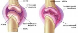
Koenig's disease , or osteochondrosis dissecans of the hip and knee joints , is one of the types of osteochondropathy; the disease is characterized by subchondral necrosis of an area of the articular surface of the bone with its further separation from the cartilage.
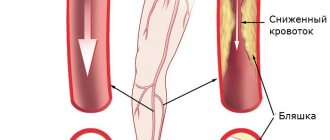
During the separation process, the osteochondral fragment enters the joint cavity, forming a so-called “articular mouse”; its size can vary from a millet grain to the size of a bean. This disease of the knee joint was first described by the German surgeon Franz Koenig , according to his assumption, the cause of the pathology is injury. After a series of studies, a hypothesis was put forward stating a hereditary predisposition to this disease. The exact causes of the development of osteochondrosis dissecans are not yet known, but presumably the disease occurs due to hereditary characteristics in the structure of joints, endocrine disorders, minor repetitive injuries, and deep vein thrombosis . These factors lead to disruption of the blood supply to the bone tissue in the joint, and a focus of non-infectious necrosis appears under the cartilage. Necrosis of an area of osteochondral tissue leads to the formation of blood clots in the vessels, which further impairs blood supply and contributes to the rejection of part of the bone tissue.
Osteochondrosis dissecans most often affects the knee joint . Less commonly affected are the hip, radial, ankle and elbow joints. The disease develops between the ages of 15 and 35, but can occur in both children and the elderly. Men are more often affected than women, which is associated with a more active and traumatic lifestyle.
Treatment
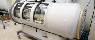
In the first stage of the disease and in atypical forms, conservative treatment is usually indicated. It consists of immobilizing the limb, prescribing thermal procedures in the form of paraffin or ozokerite applications, phonophoresis with hydrocortisone, electrophoresis with calcium chloride, treatment. gymnastics, massage. Among medications, rumalon, etamide, and vitreous injections are recommended. In the second and third stages of the disease, surgical treatment is indicated. The main methods of treatment for K. b. are Beck's operation - the formation of a tunnel towards the lesion using extra-articular access, as well as the removal of an osteochondral fragment with plastic surgery of the defect using shavings from a bone allograft.
Diagnosis and symptoms of Koenig's disease
The disease lasts quite a long time without obvious clinical manifestations. In many cases where the disease develops as a result of trauma, its symptoms are perceived as traumatic injuries. In rare cases, there may be an acute onset of the disease immediately after the injury; more often, symptoms increase gradually some time after the injury.
Based on the progression of symptoms, Koenig's disease has four stages of development. The disease is diagnosed using x-ray studies. If necessary, computer and magnetic resonance imaging and ultrasound examination are performed to clarify the stage of the disease. In difficult cases, arthroscopy is used, in which the inner surface of the joint is examined using an arthroscope through small incisions.
The first stage of the disease is manifested by mild clinical symptoms. Most often, the symptoms of Koenig's disease are a feeling of discomfort and mild pain in the joint area when putting stress on it. When diagnosed on an x-ray, a wedge-shaped or oval-shaped focus of necrosis is observed.
In the second stage, when the joint is loaded, the pain becomes more intense and swelling appears. The pain intensifies when bending the knee joint, walking, or palpating. The radiograph clearly shows the area of necrosis.
At the third stage, rejection of the dead area begins to occur. The osteochondral fragment, which has not yet completely separated, leads to joint contracture. When diagnosing, you can see the shadow of the area that has not separated.
Stage four dissecting osteochondrosis is characterized by increased pain and inflammation. In this phase of the disease, the necrotic area is completely rejected into the joint cavity. Migration of the joint mouse leads to increased symptoms. During diagnosis, the intraarticular body is clearly visible. Musculo-articular dysfunction is often observed , the joint stops moving, and the muscles gradually atrophy.
Symptoms
The main symptom of Koenig's disease is pain, which is localized on the anterior or anterointernal surface of the knee joint. As tissue necrosis spreads, there is an increase in pain and other symptoms:
- discomfort in the area of the injured knee while walking;
- swelling of the joint - the affected area is clearly visible and visible to the naked eye;
- limitation of limb mobility - indicates the beginning of a necrotizing process and separation of cartilage from the bone;
- a significant decrease in performance is caused by complete immobility of the joint. It is quite difficult for a person to walk and withstand minor physical activity.
If the above signs of osteochondritis dissecans are ignored, the person is taken to a medical facility on a stretcher, since he completely loses the ability to move independently. Timely treatment at the initial stages of symptoms makes it possible to save the patient from serious consequences, in particular arthrosis.
Diet, nutrition for Koenig's disease
Diet for sore joints
- Efficacy: therapeutic effect after 2-3 months
- Terms: 2-6 months
- Cost of products: 1700-1800 rubles. in Week
Diet for osteochondrosis
- Efficacy: no data
- Timing: constantly
- Cost of food: 1450-1780 rubles per week
Diet for arthrosis
- Efficacy: healing effect
- Timing: constantly
- Cost of food: 1550-1700 rubles per week
Diagnostics
Diagnosis of Koenig's disease is complicated by the fact that this disorder shares many of the same symptoms with other diseases, such as synovitis or meniscal tears. Studying the patient's medical history has no diagnostic value, because the list of complaints does not always allow the specialist to understand for what specific disease treatment should be prescribed. It is for this reason that preference is given to hardware examinations of the patient, which include:
- radiography - the procedure is informative only in the later stages of the disease;
- CT scan – makes it possible to diagnose this disease at an average and moderate stage;
- MRI of the joint, depending on the location of inflammation, is an informative technique that allows you to identify osteochondritis dissecans at the beginning of its development;
- arthroscopy is the most reliable diagnostic method. It allows not only to evaluate the surfaces of the joint, but also makes it possible to simultaneously carry out therapeutic measures.
After receiving all the results of the patient’s examinations, the specialist prescribes individual methods of treating Koenig’s disease.
List of sources
- Merkulov V.N., Yeltsin A.G., Mininkov D.S., Avakyan A.P. Treatment of osteochondritis dissecans of the femoral condyles in children and adolescents // Journal of the Russian State Medical University No. 4 (63) Special issue. Materials of the first joint scientific and practical forum of children's doctors in Orel 2008 (19-23/V) p. 136
- Shapiro, K.I. Frequency of injuries to large joints in adults / K.I. Shapiro // Diagnosis and treatment of injuries to large joints. – St. Petersburg, 1991 – pp. 3–5.
- Mironov, S.P. Classification and methods of treatment of cartilage defects / S.P. Mironov, N.P. Omelyanenko, E. Kon, A.K. Orletsky, I.N. Karpov, A.P. Kurpyakov // // Bulletin of Traumatology and Orthopedics named after. N.N. Priorova. – 2008. – No. 3. – P.81-85.
Varieties
Depending on what the x-ray shows, there are four stages of this disease:
- initial - characteristic signs are not observed on the x-ray. The pathological process is just beginning to form - softening of the tissue of the knee cartilage occurs;
- medium - the pictures show small compactions separated from the joint. At this time, significant softening and protrusion of the articular joint occurs;
- moderate severity - a partial separation of the affected area of cartilaginous tissue is clearly visible on the x-ray. This is due to its melting and fiberization;
- severe - complete separation of dead cartilage of the knee joint.
Stages of Koenig's disease
Depending on the degree of manifestation of symptoms and the age group of patients, there are several forms of Koenig's disease:
- children's - differs in the expression of characteristic features to a slight extent. The disease has a positive outcome with timely treatment. The childhood form is characterized by the fact that with it there is often a bilateral development of the disease, and there are also frequent cases of spontaneous recovery;
- juvenile - similarly to the previous form, it can be eliminated without problems with the help of drug therapy. This is explained by the fact that the tissues of the knee joint are not fully formed, which makes it possible to influence them with medications;
- adult form - the symptoms of this disorder are expressed depending on its stage. The further the disease progresses, the more painful it becomes. Treatment is only surgical, since taking medications does not give the expected effect.
Types of disease
In medicine, it is customary to distinguish two forms of pathology:
- The juvenile form, which is diagnosed in childhood and adolescence, responds well to therapy.
- An adult pathology that occurs before the age of fifty, it is poorly treated and gives worse results.
Recently, scientists have viewed the juvenile form of the disease, or Koenig's disease in children, as a normal process of bone growth that ends with spontaneous recovery.
Reasons for the development of pathology
At present, the exact causes of the development of the disease have not been established. Presumably, the impetus for the development of necrosis is a malnutrition of the bone and cartilage area, which occurs due to injury or heavy force loads. Usually, it is not possible to establish a connection between injury and Koenig's disease of the knee joint, since necrosis of the area occurs over a long period of time, and the disease progresses gradually. If spontaneous healing does not occur or surgery is not performed, over time the necrotic area of cartilage will separate and be replaced by an area of bare bone.
Doctors also tend to identify the following provoking factors that influence the development of the disease:
- hereditary predisposition;
- vascular pathology;
- repeated microtraumas;
- endocrine system disorders;
- bone growth disorder.
Causes of the disease
- Acute embolism (blockage) of blood vessels supplying a certain area of the bone.
- Repeated microtraumas of the joint, both external (trauma) and internal (chronic instability of the knee joint, habitual dislocation of the patella, chronic damage to the menisci).
- Disruption of ossification processes in a certain area of the bone.
- Hereditary predisposition and endocrine disorders.
Koenig's disease
There are many pathologies that affect various joint joints in the human body, among them Koenig's disease, which is a type of osteochondropathy. In most cases, it is diagnosed in representatives of the teenage age group (from 12 to 15 years). In later or early life, symptoms are almost completely absent. Koenig's disease is characterized by necrosis of the bone structures of the joints. Subsequent progression of the pathology leads to cartilage detachment. If treatment is not started in a timely manner, the patient is likely to become disabled.
External signs of pathology
A feature of the presented disease is damage to the cartilage tissue in the knee joint. Rarely, but still there were clinical cases in which the patient's disease affected the hip joint, elbows, ankles and wrists. Doctors say that there are a lot of factors that provoke tissue necrosis, but there are some reasons that are likely to contribute to the onset of this process.
In the first place is a genetic predisposition to pathology. Quite often, doctors diagnose the disease in children whose parents also suffered from Koenig. In this case, the pathological process can begin even in a completely healthy child.
Inadequate or impaired blood circulation also occupies a leading position. This condition is observed after severe physical strain, as a result of an injury or wound. Most often, the disease is diagnosed in adolescents who play sports professionally.
Various pathologies of the endocrine system are also the impetus for the manifestation of the first symptoms of Koenig's disease. It is also possible that tissue necrosis develops due to improper or insufficient nutrition. The fact is that a person receives all the necessary elements and vitamins with food, and if this does not happen, then the functionality and structure of the joint is disrupted.
If Koenig's disease is detected in adults, the causes may be bad habits (alcoholism, drug addiction, smoking), heavy work requiring constant and strong physical activity, heavy lifting, regular contact with chemicals, which provokes tissue mutation.
There are frequent cases of diagnosing pathology against the background of vein thrombosis and infectious diseases that have spread to internal organs. The disease also occurs in children with diagnosed rickets or hyperactivity.
Causes
The actual origins of Koenig's disease are unknown. Experts put forward several theories about its appearance:
- The most common version is the ischemic origin of the disease. The death of cartilage structures is associated with disruption of blood circulation in the bones. Lack of nutrients and oxygen starvation provokes the death of chondrocytes.
- The traumatic variant reports the development of necrosis under the influence of fractures and microtraumas.
- The version about the hereditary factor is associated with the anatomical features of the design of the joints and disorders of the development of bone tissue.
The disease occurs 50% more often in males.