Filariasis is a very common disease on the tropical continent. About 1.4 billion people in 73 countries are constantly at risk from this disease. And about 40 million have already become disabled as a result.
This disease can cause changes in the lymphatic system and cause abnormal expansion of any part of the body, causing pain, disfigurement and making normal life impossible for a person.
Filariasis - what is it? How can you alleviate the condition of this disease and prevent its occurrence? We will try to answer these questions in this article.
Pathogens of filariasis
Filaria are parasitic worms that have a round and smooth body covered with a special protective membrane. They are thread-like and long. Females are usually much longer than males. Their length reaches 10 cm. Males are half shorter. The body diameter of parasites is usually no more than 0.5 mm. The appearance of worms is not much different from white threads.
Worms settle in the human lymphatic system.
They are long-lived and live up to 17 years. During all this time, the female produces 1 million larvae.
What provokes the development of pathology
The causative agent of filariasis is a roundworm. The family unites 380 species, eight of them are dangerous to humans.
By settling in the lymph nodes, parasites cause an inflammatory process. Favorite places are the inguinal and femoral lymph nodes, which increase to seven centimeters in diameter. Helminths clog blood vessels, preventing lymph from escaping. After complete blockage, the vessels burst and the liquid flows out. Leakage of lymph provokes the development of various complications.
Signs of filariasis
The disease can occur in three stages - early, carrier stage and blockage.
Signs of an early stage
This stage is observed after an insect bite after approximately 90 days and lasts up to six months. It is characterized by the following symptoms:
- Increased body temperature. Thus, the human immune system always responds to the presence of foreign bodies in the body. With the help of elevated temperatures, all the protective properties of an infected person are activated.
- Painful allergic rashes, characterized by swelling of areas of the skin that become thinner if not treated in a timely manner. They take on an aged appearance and change pigmentation.
- Enlarged lymph nodes, and their pain is most often not noticeable.
- Pain in joints affected by parasites.
- Allergic bronchitis and attacks of bronchial asthma. Such symptoms appear from allergens of helminth larvae, causing spasm or suffocation.
- Mastitis, in which the breasts swell, harden, and feel hot. Such symptoms appear when microfilariae enter the mammary glands.
- In men, there is painless swelling of one of the testicles, in most cases without hyperemia of the scrotum. This happens when parasites penetrate the seminal canal.
- Inflammatory lesion of the spermatic cord, often complicated by hydrocele.
Signs of the carrier stage
During the carrier stage, during which the development of parasites until sexual maturity is observed, the following signs appear:
- Lymphadenitis , which occurs due to the entry of larvae or adult filariae into the affected cells, causing severe inflammation. In most cases, this area is affected in the area of the thighs and groin; formations of nodes with a diameter of about 7 cm can be observed there. Typically, the course of lymphadenitis occurs in waves. The attack is characterized by fever, severe weakness, nausea and vomiting.
- Lymphangitis , which appears as a result of allergies and inflammatory processes in the capillaries. This causes blockage of the lumen of blood vessels and the movement of lymph. On palpation, pain and swelling are observed. Over time, connective tissue grows and increases in volume.
- Lymphatic vessels rupture , and their contents leak out or into internal organs. This occurs from a violation of the outflow of lymph, overflow of blood vessels and rupture of their walls. Lymph may leak into the groin or mammary glands. It is observed in the intestines and bladder. The entire process of lymph flow can last more than an hour.
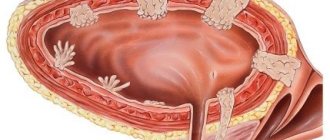
- Filaria , which are elastic nodular compounds, are observed in the eyes. A person suffers from photophobia, lacrimation, and inflammatory processes in the eyes. Their movement and blinking of the eyelids leads to painful symptoms. Cloudiness is observed on the iris, sometimes it becomes completely discolored. A clear liquid with a brown tint accumulates under the conjunctiva. As a result of the parasitic activity of filaria larvae, the optic nerve atrophies and blindness can occur.
- Parasite larvae can accumulate in the subcutaneous tissue , presenting as nodes rolling under the skin. Such knots cover the head, ribs, and legs of a person. Sometimes they become inflamed, fester and open, turning into a non-healing wound, and then into a scar.
Signs of the stage of stagnation
The stage of stagnation is characterized by the following symptoms:
- Elephantiasis , in which some parts of the body become enlarged due to stagnant lymph and inflammatory processes throughout the lymphatic system. The increase is significant, sometimes exceeding the normal, usual size by ten times. This happens due to the growth of fatty tissue under the skin, which is subsequently replaced by connective tissue. Papillomas, warts and ulcers form on the skin. It becomes not as elastic as before, but thin and wrinkled, its pigmentation changes. Most often, the lower extremities are affected, and less often the genitals, arms and eyelids. It happens that fever is added to these symptoms.
- Hyluria , in which lymph is found in the urine. Lymph appears in the bladder due to dilation and damage to blood vessels. The color of the urine becomes milky, and in the evening it turns red from blood impurities.
- Ascites , manifested by the accumulation of lymph in the abdominal cavity. This causes the stomach to increase significantly in volume. Often this condition is accompanied by diarrhea with lymph.
- Abscess , characterized by a cavity filled with pus. Due to illness, the skin loses its protective functions, blood circulation in it is disrupted, which leads to the proliferation of pathogenic bacteria. On the skin there is the appearance of areas with a purulent inflammatory process, the opening of which leads to the formation of rough scars.
- Pneumonia can begin if filariae enter the lungs. They easily manage to destroy the pulmonary alveoli and disrupt the outflow of blood and lymph.
Activities aimed at identifying the disease
Mosquitoes:
- Malaria
- Dengue fever
- West Nile fever
- Dirofilariasis
- Yellow fever
- Lymphatic filariasis or elephantiasis (vucheriosis, brugiosis)
- Chikungunya
- Zika disease
- Japanese encephalitis
- Rift Valley Fever
- Setariasis
Mosquitoes:
- Leishmaniasis
- Mosquito fever (phlebotomy fever)
They carry more than 15 infectious diseases - see their full list here
- Chagas disease (American trypanosomiasis)
Tsetse flies:
- Sleeping sickness (African trypanosomiasis)
Fleas:
- Plague
- Endemic or flea (rat) typhus
- Trench (Volyn trench) or five-day fever
- Onchocerciasis (river blindness)
Midlings:
- Mansonellosis
- Streptocerciasis
- Acanthocheilonematosis (syn. dipetalonematosis)
- Typhus
- Epidemic relapsing fever
Horseflies:
- Loiasis (another human filariasis, but not lymphatic, but bloody)
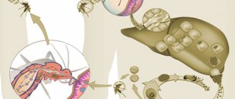
The source of infection is vertebrates, in whose bodies microfilariaemia occurs (circulation of helminth larvae in the bloodstream).
Need advice from a beauty expert?
Get advice from a beauty expert online. Ask your question right now.
ask a free question
The disease is transmitted through an insect bite (mosquito, horsefly). Mosquitoes of the genus Anopheles (which transmit malaria) and Aedes (which transmit the viral infection that causes Dengue and Zika) are the main vectors of filariasis.
Mosquitoes are intermediate hosts. When an insect bites an infected person, it becomes infected with immature larvae of the parasite – microfilariae. Over the course of 1-2 weeks, the larvae undergo a transformation stage inside the mosquito. Afterwards they become able to penetrate the human body through a bite.
In the body of an infected person, microfilariae mature into adult parasites (macrofilariae), which accumulate in the lymphatic system. Then they begin to multiply, leading to filariasis.
The favorite place of settlement in the body of the helminth is the lymphatic system, a complex drainage network of vessels and organs, through which the fluid that enters the tissue returns back to the bloodstream.
Swelling of the lymph nodes in lymphatic filariasis is the result of a reaction by the body's immune system, which is trying to get rid of the worm. The inflammatory reaction is caused by the addition of a bacterial infection. Constant relapses of secondary infection are the main cause of elephantiasis, hardening of the skin and tissues.
Three periods of illness:
- Early – after a mosquito bite, microfilariae appear in the blood within 3 months. Lasts about six months.
- Carriage (from 2-7 years) – maturation of the parasite to sexually mature forms.
- Blockage – chronic inflammation and impaired lymph flow leads to elephantiasis.
Filariasis is most often detected by the following diagnostic methods:
- The Masotti test is carried out by provocation.
- Ultrasound of nodes that are localized under the skin. This method makes it possible to carry out differential diagnosis of the accumulation of helminths, abscesses and neoplasms.
A Bancroft's filamentous lesion will be indicated by clear edges of the node, its heterogeneity and movement within it.
- Examination of the patient's blood using the microcapillary method, negative drop method, sedimentation and filtration.
- Examination of skin sections.
- Microscopy of material collected by puncture of nodes that are localized under the skin.
- Ophthalmoscopic examination of the eyeballs.
As already mentioned, the disease occurs in three stages. At the initial stage, a painful rash is identified on the skin, which is the body’s response to the waste products of helminths. Over time, the rash goes away, but swelling remains.
The characteristic symptoms of filariasis in the first stage are inflammation of the lymph nodes, as well as stiffness of movement and pain in the joints. In men, inflammation of the spermatic cord may occur; in women, mastitis. The initial stage is characterized by asthmatic attacks and bronchitis.
During exacerbation, fever is noted. Body temperature rises in response to the penetration of a foreign body into the body.
At the second stage, enlarged lymph nodes are accompanied by headaches, lethargy, and hyperthermia. Lymphatic capillaries are destroyed. At this stage, parasites accumulate under the skin, which looks like a dense elastic knot. Sometimes the nodes begin to fester and open on their own.
At the third stage, elephantiasis is added. An enlargement of a certain part of the body occurs. This pathology is caused by blockage of blood vessels and accumulation of lymph inside. When lymph penetrates the joint tissue, inflammation of the synovial membrane begins, accompanied by severe pain and limited joint mobility.
At the last stage of filariasis, symptoms such as pneumonia, abscesses, and diarrhea accompanied by ascites may appear.
Tourists visiting the tropics do not have the same immunity as local residents, so they develop elephantiasis within six months to a year. Microfilariae are not present in the blood of such patients, since filariasis is the body’s immune response to the pathogen. The more expressive the symptoms, the fewer parasites in the body.
What do filariae look like under the skin?
Approximately 2-7 years after infection, an accumulation of adult filariae under the skin in the form of mobile nodes is observed. They are the size of a pea or a quail egg.
Basically, these nodes do not hurt; the skin in their area turns slightly red. Such nodes can form on the knee or elbow joints, under the arms, in the ribs or on the head. When pathogenic bacteria enter the nodes, a purulent inflammatory process occurs with pain, tissue swelling and fever.
In places where larvae accumulate under the skin, a change in its pigmentation occurs, the appearance of light or dark spots, accompanied by itching.
Treatment and prevention
Diagnosis of filariasis can be difficult due to the fact that this disease is atypical for post-Soviet countries. However, if helminths are suspected, a comprehensive diagnosis of the body is carried out based on all existing symptoms. It is extremely important to draw up a medical history of the patient , taking into account his movements around the world over the past 5-7 years.
Therapy for the disease includes special anthelmintic treatment , based on taking medications that kill adult nematodes, as well as eliminating the effects of infection by larvae from the body. In addition to Albendazole-based drugs, your doctor may prescribe anti-inflammatory and antihistamines.
In order to prevent this dangerous disease, you should pay due attention to the use of insecticides when traveling to countries with warm and humid climates. Also, to be on the safe side, it would not be superfluous to carry out a thorough analysis of the organs and systems of the body upon return for helminth infestation.
Type of filaria under the conjunctiva
When filariae enter the eye, the thin and transparent tissue of the conjunctiva becomes cloudy. Under this tissue, gray-brown spots, dilated and blood-filled vessels are clearly visible.
When examining the conjunctiva, you can notice an uneven surface on it, and sometimes the curled filariae themselves. A person is haunted by a constant sensation of the presence of a foreign body in the eyes. Sometimes there is a sensation of floaters, and the eyeballs hurt. Involuntary closure of the eye occurs, which is called blepharospasm. With a retracted eyelid, a ridge 2-3 mm in height is clearly visible, which is a sign of thickened conjunctiva.
It should be remembered that any disease is easier to prevent than to cure. Filariasis is no exception.
It is much easier to expel parasites from the body in the initial stage of the disease, even before the onset of serious disorders in the lymphatic system and the appearance of elephantiasis. In such cases, sometimes doctors may be powerless.
When visiting tropical countries, you need to make every effort to combat insects and treat the skin with insecticidal agents, which will help protect against infection with filariasis.
| Filariasis | |
| ICD-10 | B 74 74. |
| ICD-10-CM | B74, B74.9, B74.8, B74.1, B74.3, B74.4, B74.0 and B74.2 |
| ICD-9 | 125.0 125.0 — 125.9 125.9 |
| ICD-9-CM | 125.9 [1] [2] |
| MeSH | D005368 |
Filariasis
(filariatoses, filariasis) - nematodes, helminthic infestations of humans and animals caused by filariae (filarias) - nematodes from the family Onchocercidae (order Spirurida).
Prevention of filariasis
If you find yourself in the tropics, the main thing is to avoid being bitten by blood-sucking insects both indoors and outdoors. They are removed from the room using fumigators, bed and window nets, and air conditioners. When going outside, you need to wear long sleeves and apply repellent to unprotected areas of the body.
In areas with severe spread of the disease, doctors recommend taking drugs with diethylcarbamazine at the rate of 10 mg per kilogram of a person’s weight for 2 days every month.
Thanks to these measures, you will not be at risk of filariasis. The symptoms, photos of patients and other unpleasant moments that were presented in this article will not be able to poison your memories of a trip to the tropics. Be healthy!
Development [edit | edit code]
Cycle [edit | edit code]
The development cycle of parasites is carried out with a change of two hosts - the final (vertebrate) and the intermediate (blood-sucking insect from the order Diptera). Sexually mature females, localized in the internal organs (including lymphatic vessels) of the definitive host, give birth to live larvae - microfilariae, which exit into the peripheral blood vessels or skin. When a blood-sucking insect feeds, they penetrate through the bloodstream into its intestines. and then through the intestinal wall they exit into the body cavity and muscles. After moulting twice or three times, the larvae become invasive and penetrate the insect’s mouthparts. When sucking blood, they break through the insect's proboscis, emerge onto the skin of the final host, penetrate the blood vessels through wounds and cracks in the skin, then undergo the final molt and reach maturity. Sexually mature worms live up to 15-17 years.
Distribution[edit | edit code ]
Filariasis is widespread throughout the tropical zone - Africa, Asia, South America, and the Pacific Islands.
Classification [edit | edit code ]
Filariae, or filaments, are nematodes from the order Spirurida.
, suborder
Filariata
, family
Filanidae
, viviparous helminths.
Routes of transmission [edit | edit code]
Humans and vertebrates are the definitive hosts; blood-sucking dipteran insects of various species are intermediate hosts, and they are also carriers of the parasite. In the human body, mature helminths parasitize in the lymphatic vessels and nodes, in the mesentery, retroperitoneal tissue, in various body cavities, in the skin and subcutaneous tissue. When an insect sucks blood, microfilariae enter the stomach with the blood, then migrate to the muscles, where they transform into infective larvae. They are carried by a current of hemolymph into the piercing proboscis of the insect and during the next blood sucking, the larvae enter the body of the final host through a wound in the skin. Migrating, the larvae reach the habitat where they transform into adult filariae. The concentration of larvae of some types of filariae circulating in the blood may change during the day in peripheral vessels.
Course of invasion [edit | edit code]
In this regard, three types of invasion are distinguished: periodic - a pronounced peak in numbers occurs at a certain time of the day - day or night, subperiodic - the larvae are constantly in the blood, but at some time of the day their concentration will increase; non-periodic (constant) - microfilariae are found in the blood at any time in the same quantity. The frequency of microfilariaemia is determined by the time of maximum activity of the insect vector.
Symptoms at various stages of the disease
As already mentioned, the disease occurs in three stages. At the initial stage, a painful rash is identified on the skin, which is the body’s response to the waste products of helminths. Over time, the rash goes away, but swelling remains.
The characteristic symptoms of filariasis in the first stage are inflammation of the lymph nodes, as well as stiffness of movement and pain in the joints. In men, inflammation of the spermatic cord may occur; in women, mastitis. The initial stage is characterized by asthmatic attacks and bronchitis.
During exacerbation, fever is noted. Body temperature rises in response to the penetration of a foreign body into the body.
At the second stage, enlarged lymph nodes are accompanied by headaches, lethargy, and hyperthermia. Lymphatic capillaries are destroyed. At this stage, parasites accumulate under the skin, which looks like a dense elastic knot. Sometimes the nodes begin to fester and open on their own. Eye damage is often identified, with associated symptoms. A characteristic feature is the wavy course of lymphadenitis. During an exacerbation, the patient feels severe fatigue, headaches, and nausea.
At the third stage, elephantiasis is added. An enlargement of a certain part of the body occurs. This pathology is caused by blockage of blood vessels and accumulation of lymph inside. When lymph penetrates the joint tissue, inflammation of the synovial membrane begins, accompanied by severe pain and limited joint mobility. The subcutaneous tissue grows and is replaced by connective tissue. Various growths, warts, and ulcers begin to appear in large numbers on the surface of the dermis. The upper and lower extremities, eyelid area, and genitals are more susceptible to elephantiasis. In the advanced stage, lymph appears in the urine.
At the last stage of filariasis, symptoms such as pneumonia, abscesses, and diarrhea accompanied by ascites may appear.
Tourists visiting the tropics do not have the same immunity as local residents, so they develop elephantiasis within six months to a year. Microfilariae are not present in the blood of such patients, since filariasis is the body’s immune response to the pathogen. The more expressive the symptoms, the fewer parasites in the body.
Life cycle and routes of infection
Filariasis is not one disease, but a whole series of pathologies (onchocerciasis, mansonellosis, etc.).
Filaria enter the human body after the bite of a winged carrier (horsefly, midge, mosquito). It is easy to catch filariasis from mosquitoes. By the way, the larvae enter the body of these insects in the same way. A mosquito or midge bites an infected person, in whose body the filaria larvae are concentrated under the skin, and become carriers of life-threatening helminth parasites.
Treatment of the disease
For a complete recovery, an integrated approach to therapy is required, most often carried out under the inpatient supervision of doctors. It is important to understand that treatment of filariasis takes a long time and may require not only the use of potent drugs, but also surgical intervention. Most often, surgery is indicated for the development of “balls” of parasites under the skin or in the eye, inflammation of the genital organs, or severe disruption of lymphatic circulation. Your doctor may prescribe the following medications:
- anthelmintics - Albendazole, Diethylcarbamazine citrate, Ivermectin;
- antihistamines - Zodak, Loratadine, Citrine;
- antibiotics - Doxycycline;
- glucocorticosteroids - Prednisolone.
When elephantiasis develops, doctors can only stop the negative symptoms, but complete recovery at an advanced stage is impossible. Folk remedies are ineffective in treatment, especially if lymphatic filariasis is diagnosed, which must be promptly treated under medical supervision. However, in the early stages, if pathogens have recently entered the body, eating garlic, cinnamon, ginger and citrus fruits may help.
More details on the website: infoparazit.ru/
Stages of the disease and symptoms
When an insect bites a person, the larvae enter the human bloodstream and experience:
- Maturation period. It differs from person to person. In some cases (when a person has a weak immune system), the parasites “man up” for several months, in others – more than six months.
- When filaria worms become adults, the vector stage begins. Filaria only lay larvae that enter the human bloodstream.
- Blockages develop after 3-5 years when adult filariae:
- penetrate into the organs of the reproductive system;
- make their way into the lymph nodes and lymphatic vessels (most often the lower extremities, but also the mammary glands or scrotum);
- settle in the eyeball, eyelid and various cavities (including the abdominal).
Filariasis begins after adult filariae completely occupy a lymphatic vessel. It becomes blocked.
Symptoms of filariasis vary depending on how far the disease has progressed and its location:
- At the initial stage (when the larvae just entered the bloodstream and began to gradually “settle” in one or another area of the body):
- the skin becomes covered with ulcers and swelling, which are formed due to problems with lymphatic drainage. This is an allergic reaction to the invasion of “small” microfilariae into the subcutaneous area, an attempt to get rid of them and the toxins formed during their life activity. If treatment is not started in time, atrophy and premature aging of the affected skin areas may occur;
- fever begins, body temperature rises slightly. With the help of elevated temperature, the immune system tries to destroy uninvited guests and destroy individuals;
- the allergic form of bronchitis begins to bother you, cough and spasms appear;
- the volume of lymph nodes increases. This does not cause painful sensations at the onset of the disease;
- Men have problems with the genitourinary system. Funiculitis develops (inflammatory processes in the area of the spermatic cord), as well as hydrocele (dropsy of the testicle) and orchiepididymitis (inflammation of the epididymis and itself). Filariasis of the genitourinary organs occurs most often in men;
- women develop mastitis (inflammatory pathology of the mammary glands);
- the synovial membrane in the joint becomes inflamed (synovitis). The joint becomes difficult to move.
- In the second stage (carrier):
- the lymph nodes become very inflamed (especially in the groin and thighs). Lymphadenitis begins, leading to compression of the lymphatic vessels. It is accompanied by vomiting, migraine and lack of strength;
- lymphangitis develops (lymphatic vessels become inflamed). Lymph ceases to flow normally, and pathological growth of connective tissue occurs in the area of inflammation. If the pathology is neglected, the lymphatic capillaries may rupture, and the lymph will end up in the cavity of the inflamed organ;
- Whole colonies of filariae accumulate under the skin. They have the shape of flexible nodules that roll like waves. The accumulations may begin to fester. They form a difficult-to-treat ulcer;
- the eyes are affected. Parasites may appear in the eyelid area, in the middle of the eyeball, or under the conjunctiva. Symptoms of ocular filariasis include severe lacrimation, fear of light, and conjunctivitis. Pain appears when a person moves his eyes, opens them and closes them. Uveitis in filariasis, inflammation of the blood vessels in the eyes, is a common occurrence. Cloudy areas form in the iris, and clear fluid accumulates under the conjunctiva. Without treatment, eye damage from filariasis can result in blindness.
- At the last third (clogging) stage:
- the so-called elephantiasis (elephantiasis) develops. The limbs (almost always the lower ones) or certain areas of the body (most often the genitals or eyelids) become many times larger due to the fact that lymph stagnates and the lymphatic vessels are inflamed. Fatty tissue begins to grow uncontrollably, gradually turning into connective tissue. A large number of papillomas and ulcers appear on the skin, it becomes thinner, covered with wrinkles and age spots. A person is constantly tormented by fever, sometimes there is weight loss in the upper extremities;
- lymph appears in the urine (chyluria);
- ascites develops (lymph appears in large quantities in the abdominal cavity);
- if the pathology continues untreated, pneumonia will begin;
- Bacteria multiply in cavities filled with pus (lymph) and inflammation develops. It ends in an abscess through which lymph flows out. Scar-like scars remain on the skin.
If the disease is not eliminated, it can cause purulent-septic complications and lead to death. Filariasis can also make someone disabled (deprived of vision or disfigured limbs).
Filariasis: symptoms of the disease at different stages
Lymphatic filariasis can have asymptomatic, acute and chronic forms. And most often the disease proceeds unnoticed by the patient, without manifesting itself in any way. The incubation period can last from 4 months to 2 years. But even at this time, parasites injure the lymph nodes and blood vessels, and their waste products gradually poison the body of the infected person.
- In the first stage of a disease called filariasis, symptoms include fever and an allergic reaction, most often in the form of a rash on the hands. Enlargement of the lymph nodes is observed (they change their shape and become painful), mastitis and bronchopneumonia occur.
- At the second stage of the disease (it occurs between 2 and 7 years from the onset of infection), the patient develops varicose veins and inflammation of the lymphatic vessels. Sometimes their ruptures are observed, accompanied by the appearance of signs of chyluria (milky color and gelatinous consistency of urine), chylous ascites (lymphatic fluid accumulates in the abdominal cavity) and chylocele (the appearance of a tumor containing lymph).
- At the third (obstructive) stage of the disease, elephantiasis develops in the lower limbs and sometimes in the genitals.
Diagnostics
To diagnose filariasis, the doctor performs a thorough examination of the patient. He also finds out whether the patient has visited countries with hot climates in the last few years, and whether he has been bitten by insects that carry the infection. If the doctor detects external symptoms of the disease (pigmentation, swelling, inflammation), then to make sure that it is elephantiasis and not any other, he prescribes the following diagnosis:
- Mazzotti test (challenge with diethylcarbamazine). For filariae to be detected, the patient takes (once) 50-70 mg of diethylcarbamazine. This remedy “expels” parasites to the area of surface capillaries, where they are much easier to identify. Approximately one to one and a half hours after taking the drug, the patient donates blood for analysis;
- Ultrasound. An ultrasound can show whether there are filariae in the subcutaneous area if it has become denser;
- puncture (in the area of subcutaneous nodes). The sample is taken using a special needle. If a person is sick with filariasis, histiocytes, erythrocytes and granular matter will be found in it;
- unchanged drop. This is a special blood test. The doctor takes 0.3 ml of blood from the patient’s finger. If a person's disease is already quite advanced, this test will detect it;
- microcapillary method. The blood is placed into thin tubes called “capillaries”. They “drive” it through a centrifuge to separate the plasma, in which parasites can then be easily identified;
- sedimentation and membrane filtration. Helps to detect filariae at the very beginning of the pathology;
- study of unfortified and fortified drugs. A small piece of skin is cut off with a scalpel and looked at under a microscope.
Diagnostic methods for detecting filariasis
Therapy includes the use of ivermectin, diethylcarbamazine, and less commonly, albendazole. Prescribing diethylcarbamazine at a dose of 6 mg/kg body weight every six months or a year is sufficient to kill microfilariae and adult helminths. The combination of some drugs (diethylcarbamazine and albendazole) is more effective than use alone.
When a secondary infection occurs, various antibiotics are used.
In case of severe cosmetic defects, cosmetic surgery must be performed, where the extent of the surgical intervention determines the extent of the lesion. Sometimes it is necessary to amputate a limb.
Filariasis cannot be treated with folk remedies; if you do this, there is a danger of developing serious complications. Antifilariasis therapy is considered quite effective if treatment is started in a timely manner.
- rashes;
- pigmentation of the dermis;
- elastic round seals;
- obstruction in the bronchi;
- severe swelling.
Laboratory diagnosis of filariasis includes the Masotti test - the patient is given 50 mg of diethylcarbamazine to drink, and an hour later blood is taken for testing. If a severe exacerbation of allergies occurs during the day, this may be a symptom of infection with worms.
A puncture is taken from the nodes on the skin, which makes it possible to see the presence of non-standard cells.
At the carrier stage, they use the constant drop method - for the study you need a small amount of blood from a finger, which is distributed over the glass. At the second and third stages of filariasis, mobile larvae can be seen.
At the first stage, the presence of larvae is detected by membrane filtration and sedimentation.
An instrumental diagnostic method to exclude or confirm abscesses and tumors is ultrasound.
Treatment of pathology
For filariasis, the doctor prescribes:
- Antihelminthic therapy. Anti-parasite medications usually work after the first dose. Most often, the doctor prescribes Albendazole, Ditrazine Citrate or Invermectin. Treatment of filariasis with Piperazine also gives results.
- Medicines for the treatment of concomitant infections. Antibiotics are prescribed to not only remove bacteria, but also to slow down the proliferation of filaria parasites. Doxycycline is usually prescribed for this. You need to take the product for 2 months.
Anti-inflammatory drugs. They are used if the patient’s disease has caused a severe allergic reaction, as well as if trophic changes occur in the tissues. To eliminate the problem, hormonal drugs are prescribed: Dexamethasone, Hydrocortisone, Loratadine. The dosage of the drugs and the duration of their use are determined by the doctor. During pregnancy, they should be used with extreme caution.
- If the patient's allergies are very severe, glucocorticosteroids are recommended. Most often, a doctor prescribes Prednisolone.
- Desensitizing therapy. Desensitization agents are used to eliminate painful burning and itching on the surface of the skin. For this purpose, antihistamine tablets are used - Loratadine, Cetrin, Suprastin and some others.
If the pathology is in late stages and cannot be removed with medications, the specialist will prescribe surgical intervention.
This is how doctors eliminate abscesses, achieve lymph drainage and return the affected area of the body to its normal appearance:
- With the help of a surgical operation, it is possible to remove filariasis, which is located in the muscle and subcutaneous area. To restore normal lymph flow, the doctor removes the affected part of the lymphatic vessel and stitches what remains with a vein;
- To eliminate the source of the disease in the eye, vitrectomy (removal of the vitreous humor) is performed. It is replaced with a kind of prosthesis - silicone oil. This is how the patient manages to save his vision;
- to remove the focus of filariasis in the abdominal cavity, it is punctured and the accumulated pus is pumped out;
- to remove hydrocele of the testicles, a puncture or incision is made;
- To remove an abscess, an incision is made, and then the resulting cavity is washed out with antibiotics. The abscess is also excised.
with folk remedies . They can help slightly only when the eyes are affected. Lotions and compresses made from string, tea, and chamomile will bring short-term relief.
Treatment of filariasis
It should be noted right away that elephantiasis is not easy to treat. Even if all parasites are eliminated, irreversible changes remain in the body.
In the genital form of the disease, significant improvement is observed after special operations. But returning your legs to their previous shape is very problematic. Patients receive some relief after courses of massage that activate the lymph flow, or by wearing compression tights.
If filariasis is diagnosed, treatment is carried out using diethylcarbamazine drugs (Ditrazin, Banotsid, etc.), which cause the death of parasites. They are taken for a week or 10 days, but these medications can cause a severe allergic reaction, making them difficult to use.
The massive death of helminths also leads to a deterioration in the patient’s condition, so treatment is carried out according to a special scheme: they combine the above-mentioned drugs with steroids, other antiparasitic drugs, as well as with the antibiotic Doxycycline, which gives a good effect.
Identification (diagnosis) of the disease
Lymphatic filariasis is a disease characteristic mainly of countries with tropical climates. Therefore, diagnosing it is not always easy. It is necessary to mention the visits to exotic countries over the past seven years. In order to distinguish filariasis from a number of other diseases with similar symptoms, after the main examination, the doctor prescribes additional tests. The following indicators will indicate the possible presence of the disease:
- skin disorders, rashes, itching, skin pigmentation disorders;
- severe cough that produces phlegm;
- development of lymphostasis (elephantiasis);
- inflammation of the mucous membrane of the eye and eyelid, purulent discharge;
- enlarged lymph nodes, accompanied by painful sensations;
- the occurrence of swelling in some parts of the body.
General information
Elephantiasis disease or filariasis is an invasion common in tropical areas, caused by filariae. Filaria worms belong to the family of roundworms. There are more than 400 species of filariae in the world. Despite their large number, only a few of them are dangerous to humans. Specific types of filariae provoke a certain variant of the disease (onchocerciasis, mansonellosis, brugioz, etc.), which differ only in some symptoms:
- onchocerciasis - most often the targets are the outer integument of the body and the eyes;
- mansonellosis usually occurs with fever, severe pain in the joints, and rashes on the surface of the skin;
- wuchereriosis and brugiosis manifests itself through inflammation of the nodes of the lymphatic and reproductive systems;
- Loiasis is characterized by the migration of filaria through the tissues of the body.
Routes of infection
Formed individuals of this worm parasitize directly in the body of a person or animal. Carriers of helminths are blood-sucking insects such as midges, horseflies, and mosquitoes. The life cycle of filaria development begins with a bite from a person or animal that has already been infected. The larvae of worms, along with the blood, are transferred to the stomach of the insect, where there are ideal conditions for their final maturation. With a subsequent bite, the parasite again enters the body of a person or animal.
In the body, filariae usually form massive subcutaneous formations in which individuals of both sexes coexist. Sometimes movement in them can be detected with the naked eye without additional palpation. The larvae born after the mating process are called microfilariae. They circulate throughout the body in huge quantities and collect in capillaries under the skin. Microfilariae cause allergies, which are associated with the main symptoms of the disease. Cardiac filariasis often occurs. According to statistics, the right ventricle suffers much more often.
Clinical signs
Filaria parasitize all human tissues and organs, since viviparous females give birth to microscopic larvae capable of actively moving. Microfilariae are carried by the bloodstream to all parts of the vertebrate body.
In humans, filariae can live in subcutaneous tissues, the lymphatic and circulatory systems, cavities and organ tissues. Therefore, the signs of the disease vary depending on in which part of the body the parasites are localized.
The spread of parasites occurs transmissibly, that is, as a result of the bites of infected blood-sucking insects. Since the size of the insect population depends on the climate, the disease is typical for climatic zones with high temperature and humidity.
General signs of filariasis are expressed as:
- febrile phenomena;
- itching in places where microfilariae are localized;
- skin manifestations of an allergic reaction.
As the population grows, regional lymph nodes enlarge and inflammatory processes develop in tissues affected by helminths.
Types of filariasis depending on the location of the parasites:
- genital;
- lymphatic;
- ophthalmic;
- skin, etc.
A person is the host of about 8 types of filariae, which cause their own type of pathology; they mainly parasitize in some organ or tissue and, accordingly, each disease has its own symptoms.
Symptoms of onchocerciasis
Onchocerci are a type of filariae that parasitize the muscles, eyes, lymph nodes and skin. The initial stage of the disease causes symptoms such as fever and general deterioration. The skin becomes dry and rough to the touch.
Nodules form that turn into papules. If the papules open, the skin becomes covered with ulcers. The appearance of the skin becomes similar to an orange peel. It becomes rougher, covered with pits and folds. The color of the skin becomes uneven due to the formation of areas without pigment.
These symptoms are a sign of onchocerciasis. Often the symptoms are similar to erysipelas. Dark red lesions appear, hot to the touch and protruding above the level of the epidermis. Body temperature rises to fibril levels.
Dense, mobile fibrous nodes form under the skin. There is swelling in the ears and lips. As a result, the ears become smaller and bend forward. There is a loss of fatty tissue and atrophy of muscle tissue.
In some cases, perforation of the skull bones, penetration of filariae into the brain and the appearance of symptoms of epilepsy are noted. Sometimes skin cavities are formed in which enlarged and sclerotic lymph nodes are located. Most often, nodes in the groin undergo such reduction.
Parasitism of helminths in the periosteum and joints is noted. One of the most dangerous manifestations of filariasis is damage to eye tissue by worms. At the initial stage, white spots are noted on the periphery of the cornea, which is accompanied by symptoms such as lacrimation, fear and intolerance of light, persistent closure of the eyelids as a result of muscle spasm.
Then the lesion spreads to the center of the eye, causing clouding of the cornea, decreased visual acuity, and even complete loss of vision. The iris loses color, atrophies, and brown exudate accumulates in the front of the eyeball. At the junction of the cornea and the sclera, a thickening appears and a ridge gradually forms.