Sycosis is a chronic pustular lesion of the skin caused by Staphylococcus aureus or streptococci. In the pathogenesis of the disease, disruption of the nervous and endocrinological systems plays a key role.
The disease is predominantly diagnosed in men.
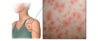
Pathological foci are localized in the area of the beard, cheeks and upper lip.
The onset of sycosis resembles the clinical course of follicles. Over time, the foci of suppuration merge and become recurrent. Diagnosis of pathology is carried out by a dermatologist. Successful treatment of the disease is possible only after establishing a reliable cause of skin suppuration.
Etiology of the disease
The causative agents of sycosis are coccal and fungal infections. Experts distinguish the following risk factors:
- cuts and microtraumas that occur during shaving;
- genetic predisposition;
- general decrease in the level of immunity;
- chronic rhinitis, in which the skin in the upper lip area is constantly infected with streptococci;
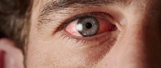
- frequent conjunctivitis also provokes sycosis of the upper and lower eyelids;
- Removal of hair from the nasal passages is also sometimes a trigger for the development of pyoderma on the inner surface of the nose.
Sycosis - what is it, causes, classification, symptoms, treatment
Sycosis is an inflammatory skin disease. The name of the disease comes from the Greek sykosis, which means ulceration. The causes of sycosis have not yet been fully elucidated.
However, it is reliably known that most often the pathology affects men. Inflammation is localized in the mustache area, and sometimes in the beard area. It is very rare, but sycosis occurs on the scalp. It is believed that the main risks for the disease are poor hygiene when shaving and resulting microtrauma to the skin.
However, evidence has been obtained that nervous and endocrine problems can also contribute to the development of sycosis.
The main causative agent of the disease is Staphylococcus aureus (staphylococcal sycosis) and partially (parasitic sycosis) Trichophyton fungi.
Sycosis - what is it
Sycosis is a chronic disease prone to frequent relapses that affects hair follicles in areas of the skin where hard, bristly hair grows (hair follicles on the face (eyebrows, beard and mustache in men), pubic area, armpits, less often on the legs).
For reference. In isolated cases, the scalp may be affected.
The main causative agent of the disease is Staphylococcus aureus (staphylococcal sycosis).
The ICD10 sycosis code is L73.8.0 (the disease is included in the class of other specified diseases of the hair follicles).
Reasons for the development of sycosis
Non-parasitic, staphylococcal sycosis is caused by S. aureus (Staphylococcus aureus). Parasitic sycosis is part of the group of trichophytosis caused by fungi of the genus Trichophyton.
For reference. A rare and difficult to treat form of sycosis, lupoid sycosis, is also caused by Staphylococcus aureus, but the clinical picture of the disease differs from the symptoms of ordinary staphylococcal sycosis.
Sycosis is much more common in men than in women. The favorite localization of rashes with sycosis is the skin in the area and above the upper lip.
Beard sycosis in men is often associated with skin irritation during shaving.
Sycosis in children is rare and is usually associated with changes in hormonal levels due to puberty and improper skin care.
Factors contributing to the development of inflammation:
- insufficient skin hydration;
- use of cheap, low-quality blades;
- rare change of shaving blades;
- the presence of abrasions, scratches, cuts, irritations on the skin;
- frequent skin damage while shaving;
- presence of ingrown hairs;
- using low-quality shaving foam or lotion;
- the presence of allergic dermatitis, psoriatic skin lesions, eczematous rashes, neurodermatitis and other dermatoses;
- chronic, acute rhinitis or sinusitis (such patients often experience nasal sycosis);
- the presence of concomitant fungal skin lesions.
For reference. The exact causes of the development of lupoid sycosis are not known, however, this form of the disease most often occurs in weakened patients, people with decompensated forms of diabetes mellitus, immune pathologies, autoimmune diseases, etc.
According to the duration of the disease, sycosis is divided into:
- acute (onset of the disease);
- chronic (relapsing form).
Depending on the depth of the inflammatory process, sycosis can be superficial and deep (lupoid sycosis).
Depending on the location of most of the rashes, there are:
- sycosis of the beard and mustache;
- sycosis of the nose (sycosis of the vestibule of the nose and sycosis of the nostrils);
- sycosis above the upper lip;
- pubic sycosis;
- sycosis of smooth skin (damage to vellus hair follicles).
Attention. In isolated cases, facial sycosis may be accompanied by damage to the eyebrows and eyelids.
This form of the disease is the most common. Staphylococcal sycosis is characterized by superficial damage to the skin and, as a rule, is not accompanied by the formation of scars.
What is typhoid fever, symptoms and treatment
The disease is manifested by the appearance of pustular rashes on the skin, occurring as ostiofolliculitis or folliculitis. The number of rashes depends on the stage of the disease and its severity.
For reference. Vulgar sycosis primarily affects the skin of the face, but can also be located on the pubis, armpits and legs.
In the first stages, small, single ostiofolliculitis may be observed. Subsequently, as the disease progresses, the area of pustular lesions of the skin increases.
With multiple rashes, the skin is swollen, inflammatory-infiltrated, hyperemic (inflammatory redness).
Pustules are painful when pressed. There is also itching, burning, and a feeling of tightness of the skin.
Due to the dense and tense covering of the pustules, their spontaneous opening does not occur. Over the course of several days (depending on the size of the pustule), its contents gradually dry out with the formation of yellowish crusts.
In the future, the crust disappears on its own without the formation of erosions and scars.
For reference. At the site of inflammation, a focus of congestive hyperpigmentation remains, which disappears within a few weeks. Moderate peeling of the skin is also possible.
With a large accumulation of pustular rashes, the formation of foci of weeping skin covered with a large number of yellowish crusts is possible.
When severe sycosis of the nasal vestibule occurs, large accumulations of dense crusts can make nasal breathing difficult.
When pustules or unformed crusts are injured, pus is released and moderately bleeding erosive surfaces are exposed.
Peeling off crusts or pustules leads to the spread of infection and the appearance of new rashes.
The general health of patients with vulgar sycosis is, as a rule, not impaired. They do not have general intoxication symptoms, fever, etc.
Attention. Due to the pronounced cosmetic defect, sycosis significantly complicates the patient’s social activity and is often accompanied by depressive disorders, psychoses, etc.
A specific form of staphylococcal sycosis is lupoid sycosis. The exact cause of the development of this form of the disease is still not known.
The disease most often occurs in men after forty years of age. Risk factors for the development of the disease are immunodeficiency states, systemic connective tissue diseases, severe endocrine pathologies, etc.
Rashes with lupoid sycosis primarily affect the skin in the growth area of the mustache and beard, as well as the scalp (parietal region and temples).
Lupoid sycosis has a chronic course and is difficult to treat.
For reference. The main difference from vulgar staphylococcal sycosis is the small number of pustular rashes. The lesions appear mainly on the periphery of the inflammatory focus; new pustules appear extremely rarely in its center.
In the center of the inflammatory focus, cicatricial atrophy of the skin is observed (in rare cases, keloid scars may form), hair loss, and the appearance of a smooth and shiny plaque with a diameter of up to three centimeters.
New rashes never appear on the surface of the plaque, but it may be surrounded by a specific purulent ridge of fresh pustules.
Also, a small zone of inflammatory infiltration (a focus of edema and hyperemia up to 1-2 centimeters) may appear around the plaque.
The rashes along the periphery of the plaque are not symmetrical, due to which its outlines often acquire an irregular, asymmetrical character.
Attention. Rashes on the scalp can be quite painful. The general condition of the patient is not impaired.
The disease can last for years, accompanied by alternating periods of subsidence of the inflammatory process and exacerbations, leading to the appearance of areas of alopecia (baldness). The hair around the alopecia area is dull, thin, and split.
What is folliculitis, classification, symptoms and treatment
Sycosis - treatment
For reference. Treatment of sycosis is selected by a dermatologist individually for each patient and depends on the form of the disease and its severity.
General therapy for sycosis consists of:
- prescribing antibacterial therapy, taking into account the sensitivity of the pathogen (systemic antibacterial therapy is prescribed for a large number of pustular rashes);
- the use of specific immunobiological drugs (staphylococcal toxoids);
- selection of proper hygienic skin care;
- prescribing local disinfectant and antimicrobial therapy;
- the use of drugs that soften crusts and accelerate the healing of the skin;
- carrying out immunostimulating therapy (according to indications);
- manual hair removal at the site of inflammation (treatment of nasal sycosis can begin with X-ray irradiation of the affected area in an epilation dose).
It is also recommended to follow a diet limiting gastrointestinal irritating foods, sweets and alcohol.
Systemic antibiotics for sycosis are prescribed doxycycline, penicillin group, and macrolides.
Treatment of affected areas of the skin with solutions of chlorhexidine or salicylic acid is effective. At the stage of crust formation in sycosis, salicylic ointment can be used.
For reference. To speed up skin regeneration, ointments with panthenol, vitamins A and E can be used.
Tar ointment can be used to resolve large infiltrates.
According to indications, UV irradiation is carried out in an erythemal dose.
Source: https://klinikanz.ru/sikoz/
Clinical picture
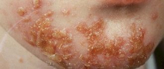
Sycosis of the mental area
Staphylococcal and streptococcal sycosis in men is concentrated on the scalp, mustache and beard area.
Typical places for the development of purulent foci for women are the eyebrows, eyelids and wings of the nose.
The onset of the disease is characterized by the formation of single round follicles. Gradually, the number of purulent foci increases, the lesion spreads to the deep layers of the skin and the pathological area increases. After a few days, purulent changes in the skin disappear spontaneously.
After a short period of time, the patient again forms follicles, which already have a protracted course. In this case, a typical clinical picture of sycosis is formed in the following form:
- inflammation and swelling of a limited area of skin;
- severe hyperemia of the skin;
- increased sensitivity and pain in the affected area of the body.
- multiple formation of pustules.
As the disease develops, pus is released from the inflammatory focus, which covers the entire pathological area of the skin. As a result, the affected epidermis becomes covered with yellowish or green crusts. After accidentally removing such deposits, a weeping and inflamed surface of the mucous membrane is discovered.
Multiple ulcers form along the periphery of the focus of sycosis. Such pyoderma gradually merges with purulent crusts. In this case, the disease does not cause enlargement of regional lymph nodes, and a rise in body temperature is observed in exceptional cases.
In addition to the local manifestation of sycosis, the disease is often accompanied by depression. This is due to a significant distortion of the patient's appearance.
Other types of pyoderma: folliculitis, boil, carbuncle, hidradenitis, impetigo.
Primary signs
The primary symptoms of staphylococcal sycosis in the form of shallow folliculitis appear suddenly and disappear just as quickly. However, after a short period of time, folliculitis resumes, its course becomes more protracted, deep lesions develop, and a typical clinical picture can be observed. Staphylococcal sycosis on the face develops very often.
The skin affected by this pathological process is hyperemic, inflamed and swollen. Pain during touch and increased sensitivity of the affected area increases. With staphylococcal sycosis, the skin becomes covered with many pustules, which are located very close to each other, their base has a bright red color and compactions. Purulent pustules dry out quite quickly, as a result of which the entire surface is covered with their contents.
As a result, the skin affected by staphylococcal sycosis becomes covered with greenish or dark yellow crusts. Over time, such crusts disappear, but due to a long purulent process, ulcers reappear. After removing the crusts, the inflamed, moist surface is exposed. Often with this pathology, swelling and redness extend beyond the main focus of infection.
Classification of sycosis
Depending on the clinical course of the disease, dermatologists distinguish the following forms of sycosis:
- Common or vulgar . The pathology is manifested by the formation of multiple foci of suppuration and is characterized by a chronic course.
- Parasitic species . In the patient, the disease is acute, causing significant purulent-necrotic changes in the skin. At the same time, parasitic sycosis does not recur and can spontaneously disappear when the general immune system is stimulated.
- Lipoid sycosis. This is a fairly rare form of purulent lesions of hair follicles of staphylococcal origin. The disease has a sluggish, slowly progressive treatment. Lipoid sycosis is mainly diagnosed in middle-aged and older men. The onset of the disease is manifested by persistent erythema of the scalp. Over time, the affected area becomes covered with purulent plaques and crusts, which merge and form a rounded focus of inflammation. In the central part of such a neoplasm, a zone of epidermal atrophy is noticeable.
Lipoid sycosis can bother a person for many years and occur with long periods of remission. In most cases, such changes do not cause pain or increase in body temperature. The main complaint of patients is a cosmetic defect of the scalp.
Parasitic form of sycosis Lipoid sycosis of lipoid form Vulgar sycosis of the scalp in the form of single pustules
Impetigo
At the periphery of the area affected by sycosis, scattered isolated phenomena of impetigo are observed, which, as the main focus grows, merge with it. Since follicular pustules form one after another, without adequate treatment, the inflammatory infiltrate begins to gradually increase.
The area affected by this staphylococcal skin disease is painful only when touched, but otherwise patients very rarely complain of burning and itching. Sycosis disfigures the human face, therefore, in addition to the main purulent process, patients experience depression, people become withdrawn and refuse to lead a normal lifestyle. The general condition does not suffer during the development of this disease, hyperthermia is extremely rare, and the lymph nodes do not enlarge.
Despite the fact that the main cause of the disease is Staphylococcus aureus, the mechanism of its development and etiology are not fully understood, since the microbial factor is considered only one of the links in pathogenesis. With the development of the pathological process, parallel colonization of the follicular apparatus by other gram-negative microflora is possible. Often staphylococcal sycosis occurs against the background of diabetes mellitus, seborrhea and focal infection of a chronic nature.
Most often, the pathological phenomenon is observed in elderly and middle-aged men; the mustache and beard area, the parietal and temporal parts of the head are affected. The disease is chronic and begins with congestive erythema, against which grouped pustules, follicular nodules and light yellow crusts appear. Grayish scales are located near the affected follicles and are removed with light scraping.
Over time, the pustules merge, forming a round plaque with a diameter of up to 3 cm, clearly demarcated from the healthy skin. As a result of infiltration, it is red in color and localized on a flat, compacted, painless base. Subsequently, the plaque gradually turns pale from the center to the periphery, the surface of the skin above it begins to thin out and becomes smooth. In this case, retraction of the element develops with skin atrophy in the center.
Diagnosis of sycosis
The pronounced clinical picture of the disease, as a rule, does not create any particular difficulties for diagnosis. An experienced dermatologist, after examining the patient, determines a preliminary diagnosis.
To reliably identify the causative agent of the disease, doctors recommend conducting a microscopic examination. To do this, specialists culture the purulent contents and identify the type of sycosis in the laboratory. Thus, the parasitic form is provoked by a fungal infection, which is gradually joined by a coccal infection. But lipoid sycosis is caused by Staphylococcus aureus.
Diagnostics
Diagnosis of sycosis is quite easy. Already by external signs you can identify a disease that is not similar to other inflammatory processes. You should contact a dermatologist, as well as an infectious disease specialist and mycologist. They will conduct a general examination, as well as additional tests:
- Sowing crusts and purulent discharge.
- Skin microscopy.
- Blood analysis.
- Cultural examination of crusts and purulent discharge.
go to top
The diagnosis of “sycosis” is established by a doctor based on the patient’s complaints, the history of his illness, examination data and objective examination. Typically, diagnosing this pathology does not cause difficulties for a doctor. No special examinations or tests are required for this.
However, sometimes the disease is limited in nature, when the skin of only the upper corner of the tip of the nose is affected. This location of the pathological focus can cause some difficulties in the diagnostic process, since this place is more difficult for the doctor to examine.
Basic methods of treating sycosis
The choice of treatment method depends on the clinical stage of the pathology and is carried out by a dermatologist. This treatment is usually very long-term.
During acute periods, experts recommend using disinfectant lotions based on boric acid and potassium permanganate. These measures are aimed at preventing relapses and softening pathological compactions. After the acute period has subsided, doctors begin the spot application of aniline dyes (methylene blue and brilliant green). It is advisable to apply an iodine solution along the periphery of the ulcers.
Treatment of the disease necessarily includes systemic administration of tetracycline antibiotics. These medications can be in the form of injections, tablets, or topical ointments. Experts recommend the following medications:
- Tetracycline;
- Chlortetracycline;
- Oxytetracycline;
- Syntomycin ointment;
- Gentamicin ointment.
In some cases, with severe sycosis, dermatologists resort to injections of staphylococcal vaccine.
General principles of therapy
The course of treatment for the disease is carried out according to the following scheme:
- prevention of further disorders in the functioning of the nervous system is achieved through iron supplements;
- local treatment of the purulent focus with brilliant green;
- during an exacerbation, lotions with antiseptic medications;
- injections of broad-spectrum antibacterial agents;
- vitamin therapy and physical therapy during remission;
- diet therapy, according to which the patient is prohibited from consuming salty, peppery and sweet foods;
- patient’s refusal of bad habits (alcohol abuse and smoking).
Many dermatologists believe that streptococcal lesions require exclusively antibiotic therapy.
High effectiveness in the treatment of sycosis is demonstrated by:
- Ural Federal District. Ultrafiltration irradiation of pathological tissues has anti-inflammatory, wound-healing, anti-edematous and analgesic effects.
- Immunotherapy. Stimulation of the body's defenses with the help of immunostimulants and immunomodulators directly affects one of the causes of sycosis. As a result, the patient recovers in the shortest possible time, and the symptoms of the disease become less intense.
- Autohemotherapy. Subcutaneous administration of one's own venous blood reduces the level of inflammation of the epidermal tissue.
- Laser therapy. Treatment of purulent lesions with a laser beam disinfects and desensitizes the surface layer of the skin.
Before carrying out the therapeutic method, doctors recommend removing hairs from the affected area of the body using tweezers.
Symptoms and signs
First of all, sycosis forms on the hairy side of the face, affecting the beard and mustache. It should be said that sycosis is diagnosed much less frequently in women. In women, sycosis is usually localized on the inside of the wings of the nose, as well as on the surface of the eyelids and eyebrows. On the skin in the armpit area, on that part of the body that is covered with thick and long hair, as well as on the pubis, sycosis can occur in isolated cases.
During the development of sycosis of the beard, a process of shallow folliculitis occurs (exclusively the upper part of the follicle is involved).
When folliculitis occurs, the inflammatory process can spread along the entire length of the follicle. When removing the affected hair with tweezers, a frame in the form of a purulent muff is found on its root.
After a certain period of time, lesions form with the help of follicular pustules.
The vestibule of the nose, beard, eyelids, eyebrows, and the circumference of the oral cavity are all places in the human body where sycosis occurs most often. Being in close proximity to the mucous membranes allows staphylococcus to receive a stable supply of moisture and actively multiply.
Much less often, the disease can be found on the pubis and other areas of the skin with hair. This dermatological disease always begins suddenly and occurs in the acute phase in the first 2 days.
Signs of a dermatological problem develop in several stages, and its symptoms are as follows.
- A disc-shaped rash of pale red color appears on the painful area of the epidermal layer. If blood vessels are located close to the surface, it may have a brown tint. The rash is localized and always has an irregular shape.
- As the inflamed formations grow, they fill with purulent fluid and may acquire a yellowish tint.
- Such unpleasant sensations as burning, itching, a feeling of swelling and heat in the affected area are added.
- The rash, filled with pus and ichor, gradually dries out, cracks, and the liquid it contains flows out.
- A dry crust of bloody secretions forms on the inflamed skin, which periodically becomes wet and cracks. If the patient does not receive adequate drug therapy, the wound surface increases and the disease progresses. This is how the chronic form of sycosis develops.
The listed symptoms of the disease are characteristic of the staphylococcal type of sycosis. With a parasitic type of disease, the signs of dermatological pathology are not so pronounced.
The accumulation of pus is less voluminous, and due to the absence of staphylococcus in the wound, the patient’s immune system copes with the disease much faster (6-10 days).
Sometimes the use of medications is not required at all.
Sycosis is usually localized on the scalp of the face, in the area of the mustache and beard, less often on the inner surface of the wings of the nose, eyebrows, edge of the eyelids, much less often on other areas of the skin covered with long hair (in the armpits, pubic area, etc.).
Complications of the disease
The main danger of sycosis is considered to be the transition of the disease to the stage of eczematization, which occurs quite often. The clinical picture of this complication includes signs of weeping and intense itching of the skin.
In some cases, the pathology is accompanied by the development of such lesions:
- impetigo in the form of pustular processes on the surface layers of the skin;
- furunculosis, which is an acute purulent-inflammatory damage to the hair and sebaceous follicles.
Causes of the disease
Since sycosis is classified as pyoderma, it can even be provoked by microtrauma or accidental cuts during shaving. Interestingly, chronic rhinitis also provokes sycosis. Since the skin located above the upper lip is usually loose, and at the moment of blowing the nose there is rubbing in of mucus containing a significant number of staphylococci, this can become a provoking factor for sycosis.
It should be separately noted that nasal sycosis is often confused with streptoderma and lip abscess. This can be explained by similar symptoms: lesions begin to appear in the corners of the lips and shift to the nose area.
Only a qualified dermatologist can make an accurate diagnosis and select the necessary course of treatment.
Any acute or chronic purulent disease of the nose or its paranasal sinuses (rhinitis, sinusitis, and so on) can cause the development of sycosis.
Beard sycosis is characterized by the formation of an inflammatory process in the hair follicle and is provoked by the staphylococcus bacterium.
sycosis in the photo
Sycosis is a skin disease that most often affects areas of the nose, beard, and the circumference of the oral cavity. It is impossible to single out only one cause of the disease in the form of an infectious pathogen.
Classification of the disease
The classification of the disease is based on the principle of the origin of the pathology and the type of skin lesion. There are three forms of the disease. The features of their clinical picture are shown in the table.
| Form name | Pathogen | Duration | Clinical manifestations |
| Vulgar sycosis | Staphylococcus | Can last for years, constantly recur, worsening quality of life, causing mental disorders | Develops gradually. First, one pustule appears on the affected area. It is formed in the zone of penetration of pathogenic bacteria. Then other elements appear around it, pustular ostiofolliculitis develops, which leads to the formation of inflammatory infiltration of the dermis. Purulent masses, moving deeper, reach the ducts of the sebaceous glands. The infection process is long. Therefore, at the same time, over time, on the affected skin you can see drying out pustules, covered with a purulent-serous crust, and pustules beginning to form. A distinctive feature of vulgar sycosis is renewal. During the next exacerbation, the pathology develops in the follicles that have already become scarred, and at the same time new areas of healthy skin are captured. If you pull out a hair from an inflamed bulb using tweezers, you can see a purulent sheath at its end |
| Lupoid | Staphylococcus | Develops slowly and takes a long time | Follicles located on the face and scalp are affected. They become inflamed and filled with pus, but the pustule does not open. The hair follicle dies and a new abscess forms next to it. An atrophic scar forms in place of the dead follicle. The development of the disease leads to baldness |
| Parasitic (a type of ringworm) | Zoophilic and anthropophilic fungi | The pathology often goes away on its own; the duration of exacerbation depends on the state of the immune system. | Sharply limited nodes of a red-bluish color appear on the skin of the chin, neck, and upper lip. Usually they are located asymmetrically. Perifollicular pustules gradually form on the surface of the nodes. They merge and provoke the development of abscesses. After each opening, the pus comes out partially, and the abscess becomes covered with a crust. Purulent passages, or cavities, form underneath it. Hair growing in the affected area becomes thin and dull and falls out. Despite the active development of the inflammatory process, skin rashes do not cause pain. The patient may only complain of symptoms of skin irritation |