Basic treatment methods
Treatment of hemangioma is most often surgical.
Regardless of which method of getting rid of the problem is chosen, it is worth considering that the tumor can recur. Medications are used less frequently. Timolol or propranolol-based products are suitable for treatment. Apply a pressure bandage to the affected area and leave it for some time.
- surgical intervention;
- laser exposure;
- electrocoagulation;
- cryodestruction;
- sclerosing therapy.
Surgical removal is performed under general anesthesia. The technique is traumatic and ineffective, since in more than half of all cases the tumor returns again.
Electrocoagulation allows using electric currents to destroy tumor tissue, and healthy skin appears in its place. The method is not very suitable for getting rid of cavernous hemangiomas due to the appearance of deep scars.
Cryodestruction, or cauterization with nitrogen, is used most often and regardless of the stage of the disease. Complete disappearance of the formation occurs after 3 sessions. Ideal for the treatment of capillary hemangiomas.
Sclerotherapy of hemangiomas is carried out by introducing special mixtures into the tumor, which stop feeding the tumor and its destruction occurs.
With timely treatment using the right methods, getting rid of hemangioma is quite possible. Much depends on the initial location and extent of the lesion. In most cases of the disease, the prognosis is favorable.
Hemangioma is a tumor of vascular origin, which is benign in nature, and is found mainly in children...
Hemangioma is a tumor of vascular origin, which is benign in nature, and occurs mainly in young children. At a certain stage of development, even in the prenatal period, an area of accumulation of vessels is formed, which can have different calibers and are located in the soft and hard tissues of the body.
When a hemangioma tends to rapidly grow to the sides and deep into the tissue, this can disrupt the function of the main tissue of the organ and harm the physical development of the child.
Therefore, once a hemangioma is diagnosed, it should be treated, or monitored as directed by a doctor. The main method of treatment is surgical; the most effective is the use of laser.
Causes of hemangioma. Hemangioma develops as a result of a hereditary tendency. The chances of this happening are especially high in children whose one or both parents suffer from hemangioma.
The probability of manifestation of unfavorable genetic information increases when the germ cells are affected by alcohol, tobacco poisons, and other toxic substances (for example, some drugs).
Symptoms of hemangioma. Flat hemangiomas are most often located on the face in the area of innervation of the trigeminal nerve, or along the nerve fibers of other plexuses of the head.
They have the characteristic appearance of spilled wine and are called wine stains. The color of the spots ranges from bright red to bluish; the skin over the spot is very thin, and subsequently becomes even thinner and may become covered with bleeding cracks.
Hemangiomas that begin as small red bumps above or within the skin tend to increase in volume and are most effectively treated early in their appearance.
Formations can be located on any part of the body, in internal organs, bones. On the skin, such hemangiomas are surrounded, first by a small, and then by a more pronounced vascular network.
Diagnosis of hemangioma. In the fetus, hemangioma can be diagnosed using ultrasound in the 2nd – 3rd trimester of pregnancy. After birth and in adult patients, selective angiography, radiography, computed tomography, magnetic resonance imaging, and scintigraphy are used.
The most effective method of treatment is radical removal of hemangioma using one of the methods existing today. As a concomitant therapy, as well as to prevent relapse of hemangioma, folk remedies should be used.
Antitumor collection for complex treatment of hemangioma. Make a collection of the following components: Plantain - 50 g, yarrow - 50 g, burdock leaf - 50 g, St. John's wort - 50 g, sweet clover - 50 g, knotweed - 30 g, cinquefoil - 30 g, coltsfoot - 30 g, calendula flowers – 30 g, hazel leaves – 10 g, eranium – 10 g, white willow bark – 10 g, dried grass – 10 g.
Brew a tablespoon of the resulting mixture with half a liter of just boiled water, and leave in a water bath for five minutes. After this, leave for an hour.
Ginseng tincture as an antitumor agent. Grind the ginseng root, fill a half-liter jar 1/3 full and fill it with vodka up to the neck. Close the jar tightly and let the medicine brew in a dark place for 20 days.
Take the tincture twice a day, one teaspoon. After 10 days, continue drinking the tincture one teaspoon at a time for another month. If necessary, after a twenty-day break, repeat a similar course.
Collection for the treatment of cavernous and bleeding hemangiomas. Combine equal parts of knotweed, lingonberry leaf, horsetail, yarrow, birch leaves, sweet clover, and calendula flowers.
Prepare and use the drug in the same way as antitumor collection according to prescription 1. It is also recommended to alternate both collections for two weeks (recipe 1 and recipe 3).
Celandine for external treatment of hemangioma. Grind fresh celandine herb and fill it with warm water 1:1, leave for two hours. Strain the infusion through cheesecloth and squeeze out the herb well.
Soak a gauze pad in the infusion and place it on the tumor. After 40 minutes, change the compress and leave for another 40 minutes. Carry out this procedure in the morning and evening. The duration of treatment is determined by the doctor.
Oak bark and duckweed for hemangioma. Brew crushed oak bark with boiling water 1:5, simmer over low heat for 20 minutes. Add duckweed to the still boiling broth, in a volume equal to the volume of oak bark, remove from heat, leave on the table for two hours.
Treatment of hemangioma with viburnum ice. Crush fresh viburnum fruits and fill them with cool water 1:1. Mix the viburnum well, remember it so that more juice comes out into the water.
apply ice to the hemangioma and do not let go until the ice melts. The product is suitable for the treatment of small tumors. Use under medical supervision.
Icelandic moss and plantain as a remedy for hemangioma. Combine the dried raw materials equally: a tablespoon of Icelandic moss and the same amount of plantain. Pour the raw materials into a thermos, pour a liter of boiling water, leave for two hours.
Sushenitsa and tansy for vascular tumors. Combine two parts of dried cucumber and one part of tansy as a medicinal raw material. Brew two tablespoons of herbs with 200 ml of boiling water, leave for two hours.
Hemlock to combat hemangioma on the skin. Grind the hemlock leaves, pound them in a mortar, put them in a plastic bag and put them in the freezer for two days.
Separate small parts from the frozen mass and apply to the hemangioma for 45 minutes: first it will be an ice compress, and then a regular one, with the active substances of hemlock. Apply once a day until satisfactory results are achieved.
The use of a complex tincture for the treatment of hemangioma. Combine 50 g of crushed calamus roots, green walnuts, elecampane roots, kermek roots, St. John's wort, and celandine.
Add 2 g of Spanish fly powder to the composition, mix everything, and pour one liter of 40° vodka. Close the vessel and leave for two weeks in a dark place. Apply the tincture as a compress to the hemangioma area for 40 minutes every day. Do not use for treating children!
Tincture for the treatment of liver hemangioma as part of complex therapy. Horse chestnut flowers, celandine, mistletoe leaves, eleutherococcus, knotweed, thyme, green tea, peppermint - pour all the ingredients into a liter jar, two heaped tablespoons at a time.
Fill the jar with vodka to the neck, close tightly, leave for 21 days in a dark place. Take two teaspoons of tincture twice a day with warm water.
After three weeks of taking it, stop and start taking ginseng tincture (recipe 2) and plantain infusion with Icelandic moss (recipe 7) at this time. After 21 days, resume the course of treatment with a complex tincture (recipe 11).
St. John's wort juice as a remedy for hemangioma. If we are talking about spinal hemangioma, a mixture of St. John's wort and celandine juice with medical alcohol 1:1 is prepared for topical use.
A compress of multilayer gauze soaked in a solution is placed on the area of the hemangioma and secured with a band-aid. After three hours, the compress is removed and the procedure is repeated the next day. Use for 10 days. If necessary, repeat the course in a week.
How to quickly lower cholesterol at home?
Hemlock
Hemlock herb can treat benign and malignant tumors. To remove hemangioma on the skin, the following healing remedy is prepared from hemlock:
- fresh plant materials are taken;
- crushed and sent to the freezer for 2 days;
- frozen leaves are applied to the problem area;
- stand for 30–40 minutes;
- The procedure is carried out once a day for 2 weeks.
This is a type of benign tumor. Although it does not cause direct harm, it is still dangerous, since its growth is not controlled. Therefore, hemangioma removal is most often performed.
For this purpose, a full range of modern surgical interventions is used. You can try to get rid of hemangioma using folk remedies when treating at home.
There is an opinion that the cause of the appearance of hemangiomas is due to a viral process that occurs in the expectant mother during the week of pregnancy. During this period, the laying of the vascular wall occurs.
Compresses according to traditional recipes for hemangioma
Therapy with such remedies begins with the use of walnuts. Its juice is used, squeezed out when the nut is still green. With the help of compresses, the juice is left attached for a long time.
To carry out treatment with folk remedies for hemangioma, you need to take one tablespoon of copper sulfate and dilute it in half a glass of water. Dip a rolled-up cotton swab into the solution and thoroughly wipe the sore spot.
An onion compress according to a folk recipe for hemangioma is made as follows. You need to grate the onion and apply the resulting pulp to the sore spot, wrap it with a bandage and keep it for twelve hours.
It should be noted that jellyfish (commonly known as kombucha) helps well in the folk treatment of hemangioma. It is necessary to tear off a piece from the so-called jellyfish and apply it to the sore spot.
This compress must be changed once a day. The remaining pieces of kombucha should be stored in a jar of water and, if necessary, take out the next piece.
Before going to the doctor, you need to imagine the causes of hemangioma; you cannot choose a compress for traditional treatment of hemangioma, a decoction or other remedy, relying on your own conjectures.
And specifically at the appointment, they will tell you about the specific features of the problem and advise the most convenient method of treatment. Only then will it be possible to select folk remedies at home to treat hemangioma and its root causes - liver diseases.
In this matter, it is better to trust the doctor and his technique, because such an education may be the first in a series of several. Elimination of one tumor will stop the possible growth of others.
Hemangioma is a layering of blood vessels that form a superficial defect on the skin. Most often it occurs in the ears and neck. Often such pathology is found on the internal or genital organs.
A multiple variant of such a tumor cannot be ruled out. In some cases, spontaneous expression of the neoplasm is possible. Despite the fact that this tumor is benign, it is quite dangerous.
Even minor damage to it leads to serious bleeding, which is quite difficult to stop. According to statistics, hemangioma occurs somewhat more often in girls than in boys. It can be detected immediately after birth or in the first two months of life.
Hemangioma is a benign tumor formation consisting of small vessels. It occurs at any age: from infants to the elderly. In adult patients, it appears as red round spots of regular shape.
It can be convex, rise above the skin or be located in its thickness, having irregular outlines, occupying a large surface area. In old and mature age, the incidence of these tumors is higher than in young people. Is it possible to get rid of hemangioma without seeking the help of a doctor?
Despite the fact that hemangiomas do not tend to transform into cancerous tumors, there is a high risk of rapid uncontrolled growth and re-formation after removal.
Typically, such tumors form within several years after the birth of a child, affecting the skin and sometimes growing into the fatty subcutaneous layer. Less commonly, such a neoplasm appears in internal organs - the liver or kidneys, and even bone tissue is no exception.
Hemlock
Hemangioma is a type of benign tumor. Although it does not cause direct harm, it is still dangerous, since its growth is not controlled. Therefore, hemangioma removal is most often performed.
For this purpose, a full range of modern surgical interventions is used. You can try to get rid of hemangioma using folk remedies. There is an opinion that the cause of the appearance of hemangiomas is due to a viral process that occurs in the expectant mother during the week of pregnancy. During this period, the laying of the vascular wall occurs.
Compresses according to traditional recipes for the treatment of hemangioma
Treatment of hemangioma using folk remedies begins with the use of walnuts. Its juice is used, squeezed out when the nut is still green. With the help of compresses, the juice is left attached for a long time.
To cure hemangioma, you need to take one tablespoon of copper sulfate and dilute it in half a glass of water. Dip a rolled-up cotton swab into the solution and thoroughly wipe the sore spot.
An onion compress for the treatment of hemangioma is done as follows. You need to grate the onion and apply the resulting pulp to the sore spot, wrap it with a bandage and keep it there for twelve hours.
Hemlock
Tip 1: Treat hemangioma with folk remedies
Hemangioma is a benign vascular tumor that appears as red spots. Its localization is possible on any part of the body, including the face. The progression of the disease is most often unpredictable.
A pinpoint formation can grow instantly, while a large one can shrink significantly.
It can be quite difficult to differentiate hemangioma from warts or moles. Unlike other formations, it does not degenerate into a malignant tumor.
Vascular tumors can be either single or multiple, and in girls they occur 2.5 times more often than in boys.
It is advisable to remove hemangioma immediately after it appears on the skin. Treatment of hemangioma with folk remedies is one of the simplest options for removing a benign tumor.
It appears in a child in the first month of life and often does not bring significant discomfort to the baby. But its growth cannot be controlled, and this fact contributes to the decision to remove the skin defect.
There are several types of tumors on various organs and parts of the body:
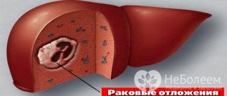
- Hemangioma of the liver. Officially, it is only subject to surgery. Although not everyone agrees with this. It can be found in any part of the organ. A complication of the pathology is possible when the tumor is not limited to the liver, affecting adjacent organs, causing them serious harm.
- Kidney hemangioma. It is quite rare and is benign in nature.
- Spinal hemangioma. Quite a common disease. Mostly found on the lower and middle thoracic spine. The pathological process can be a source of severe pain. The danger lies in the risk of destruction of bone beams that have a supporting function.
- Hemangioma of the lip. This is a benign tumor of the mucous and vascular membranes. Occurs mainly in children.
Many parents, having discovered a suspicious tumor in their child, ask the question: “Is this dangerous for the baby’s life?” and “How to get rid of it?”
To answer these and many other questions, you need to understand what a hemangioma is, diagnose the tumor and then choose the right treatment method.
Hemangioma is a benign tumor of blood vessels. Appears from the first days of a baby’s life. Usually the disease occurs in several stages: the appearance of the spot (the first days of life), the growth of the spot (until the child reaches six months), the reduction of the spot and its disappearance.
A simple tumor forms on the surface of the skin. It appears as spots of a reddish or pinkish color, soft and warm to the touch. Typically, such hemangiomas do not cause any inconvenience other than aesthetic ones.
The problem in preventing this disease today is that the causes of its occurrence have not yet been studied. What is clear is that during the development of the embryo there is a failure during the process of formation of the vascular system.
What caused this failure is not known. Among the assumptions, experts name a mother's cold during pregnancy, injuries or infectious diseases suffered during this period, and poor environmental conditions.
Listed above are the main signs of hemangioma that can be observed at home. But to understand how to treat hemangioma, you need to see a doctor for further examination and treatment.
Surgical treatment is based on complete or partial removal of the tumor using surgical instruments. The operation is performed under anesthesia. The disadvantages of this method are that there is a high risk of severe bleeding. Also, after the operation, scars remain at the site of the hemangioma.
What does hemangioma look like in adults, its causes and treatment?
Everyone knows that tumor diseases can be benign and malignant. One type of benign vascular tumor is hemangioma. It is more common in newborns than in adults.
It is found in ten out of a hundred babies, but after twelve months it stops growing and by the age of five the spot successfully resolves. In adults, hemangioma will not lead to any serious disturbances in the functioning of the body. But it all depends on the size and location.
What is hemangioma?
Hemangioma is a benign tumor of vascular tissue. It affects both external tissues and internal organs. It has a slow growth rate and does not penetrate into neighboring tissues of the body.
It has a clear localization and practically does not bother the patient. It looks crimson or red, which can rise above the skin (the surface is lumpy and uneven), or be a red spot under the skin.
Locations
There are four types of hemangiomas depending on their location:
- cutaneous;
- mucous membrane;
- musculoskeletal system;
- internal.
A cutaneous vascular tumor is located on the surface of the skin. It is less dangerous, does not lead to complications, and does not require timely therapy.
With the exception of formations that are located in the area of the eyes, ears, and genitals, they must be removed, since the likelihood of damage is high. It is possible for many small vascular tumors to develop in different parts of the body.
- Mucosal hemangioma appears on the lips, tongue and genitals.
- Hemangioma of the musculoskeletal system is the least dangerous for an adult, but can lead to skeletal deformation in newborns,
- Internal - affects internal organs: spleen, brain, and even all types of glands. If it is small, the doctor will prescribe treatment that will stop its further development.
Causes
The main reason for the appearance is associated with a violation of the intrauterine development of the vascular system, in particular, with abnormal growth of vascular tissue.
In adults, the presence of hemangiomas growth is provoked by the following factors:
- diseases that cause disruption of blood flow and vascular function;
- exposure to ultraviolet radiation: sun, solarium;
- hypothermia;
- poor environmental conditions: radiation, living in a contaminated area;
- experiencing severe anxiety or a stressful situation.
Note! If an adult has never had a formation that looks similar to a hemangioma, then it is necessary to undergo a thorough examination and diagnosis.
Symptoms and signs
This tumor can be recognized due to the following signs:
- visually similar to a mole, only red;
- its boundaries are clearly defined or blurred;
- does not cause any discomfort or pain;
- under unfavorable factors, it begins its development and growth;
- The main places of localization are the head and neck, but can occur on other parts of the body.
Note! In the initial stages, it is difficult to recognize, since at first the skin turns red little by little, and only small spots of disproportionate shape appear. Then, over time, the spots combine into one large formation, which contains small blood capillaries.
Classification of hemangiomas
- By location: integumentary, mucous, internal organs, skeletal.
- According to the type of cellular structure: simple, cavernous, combined and racemic.
- Simple - red or burgundy with a bluish tint, with defined edges, which is located on the surface of the skin. When you press it, it turns white and then turns red again.
- Cavernous - similar in size and color to capillary, differs only in its structure. Consists of parts that are formed as a result of blood clotting. This type is localized on the skin of the neck or scalp.
- Combined - consists of capillary and cavernous.
- Racemic - characterized by imprecise outlines of shapes and boundaries, the basis is large twisted vessels. Develops preferentially on the head or neck.
Are hemangiomas dangerous?
This is a benign tumor, it does not metastasize to surrounding tissues, but this is not a reason to refuse its therapy.
In turn, its harmless growth can lead to the following consequences:
- hemangiomas located in the oral cavity, on the neck (with germination deep into the tissues) can lead to breathing problems. Also, their localization on the walls of the vessel can interfere with normal blood flow. A formation on the temple, forehead or eyelids threatens the development of glaucoma and a gradual decrease in vision, and in the vessels of the brain it can develop epilepsy;
- damage or frequent trauma to it leads to bleeding, and there is a risk of infection in the wound;
- Aesthetic discomfort occurs in patients whose hemangioma is located in a visible place: face, neck, hands.
Note! Often simple hemangiomas tend to regress, so some of them simply disappear on their own without any treatment. But if it disrupts the functioning of organs, then only the selection of the optimal method of its treatment will help.
Diagnostic methods
It is not difficult to notice a vascular tumor on the surface; they are diagnosed during a visual examination of the patient.
To diagnose internal hemangioma, one of the following methods is used:
- Laboratory research;
- Instrumental – X-ray, ultrasound, MRI;
- Puncture is a puncture of the walls of blood vessels, with further morphological examination of the selected material.
Which doctor should I contact?
It is impossible to single out one specialist who would deal only with this problem. So, upon examination, the therapist diagnoses hemangioma. A dermatologist treats only those spots that are located on the surface of the skin, and the surgeon treats internal ones.
Let's celebrate! only the specialists presented above will be able to confirm the diagnosis and prescribe the necessary physical therapy so that the spot goes away without a trace and painlessly.
hemangioma treatment
For spots that do not grow and do not cause complications, a wait-and-see approach is often used. Hemangiomas with progressive growth, infected and bleeding are subject to treatment or removal.
Removal methods
The following removal methods are available:
- radiotherapy - used in hard-to-reach places;
- laser coagulation of blood vessels;
- dithermoelectrocoagulation – removal of small formations;
- cryodestruction – exposure to liquid nitrogen (freezing);
- sclerodestruction - administration of a sclerosing drug;
- hormone therapy to stop the growth.
- surgical removal.
Let's celebrate! In practice, combined methods are used: surgical removal with radiation therapy, cryodestruction with hormone therapy.
Surgery
Hemangioma, which grows and destroys the body of the spine, puts pressure on neighboring organs, actively grows, is localized near the eyes, ears, and genitals and requires surgical intervention.
But now more and more preference is given to minimally invasive treatment methods, since surgical operations cannot be performed due to a possible cosmetic defect or for other reasons.
Note! In such situations, close-focus radiotherapy is used - radiation exposure. Targeted gamma rays destroy tumor cells, having minimal effect on nearby organs.
Traditional methods
Basic recipes for folk remedies to combat hemangioma:
- Kombucha compress. The compress is applied to the area of the growing vascular tumor for the whole day. the course of treatment consists of three weeks.
- Treatment with copper sulfate. Add a tablespoon of vitriol to half a glass of water, and wipe the stain with this solution. Treatment lasts for 10 days. In parallel with this, you should take a hot bath with baking soda at night (a pack of baking soda per bath).
- Fresh celandine juice is also used in various infusions: from wormwood, coltsfoot, St. John's wort, yarrow, calendula and so on.
Differences between childhood and adult hemangioma
The structure of an adult hemangioma is no different from that of a child. Everything also consists of vascular tissue and is localized in various places in the body. All its types are found in both adults and children. Moreover, it occurs 7 times more often in women than in men.
But the main difference is that in children under seven years of age it can go away on its own, without any type of treatment. As a rule, it does not require an increased degree of attention, however, it should be constantly monitored.
Let's celebrate! If the spot is actively growing and increasing in size, changing its color, then you should immediately contact your pediatrician to determine the course of therapy. In addition, the hemangioma should be removed if it is constantly injured, as this is fraught with the development of bleeding.
Prevention measures
Special preventive measures have not yet been developed.
In order to minimize the risk of developing this tumor, the causes of its occurrence should be controlled:
- environmental influences;
- diseases of internal organs, in particular the endocrine system.
- disorders in the cardiovascular system.
- exposure to sunlight or ultraviolet radiation.
Hemangioma is the most common benign tumor that occurs in both adults and children. It can occur anywhere on the body, as well as inside the body.
Note! In children, it can go away on its own without any treatment. As for adults, upon diagnosis, a specialist will prescribe a course of treatment. Since preventive methods have not yet been developed, it is important to avoid the causes of this disease.
You may also like
Source: https://skindiary.ru/zabolevaniya/rak/prichiny-simptomy-i-lechenie-gemangiom-u-vzroslyx.html
Localization of education
Science cannot identify the exact reasons for the formation of this benign tumor. However, most experts agree on one thing: the failure occurs during the period of intrauterine development during the formation of the body’s blood vessels.
- stray cell theory;
- slotted;
- placental.
Speaking about the theory of stray cells, scientists strive to reflect the processes in which mesenchymes (precursor cells of vascular tissue) remaining after the complete formation of the circulatory system do not disappear gradually, but, on the contrary, begin to multiply.
According to the slot or fissural theory, when embryonic fissures form on the head in the area of the skull, the vessels that have grown into this area begin to develop incorrectly.
In accordance with the placental theory, pathology does not develop intensively during intrauterine development, because the factors of maternal inhibition of angiogenesis act on the child’s body.
- progression;
- growth arrest;
- regression.
At the stage of active growth, the neoplasm has a bright red color and intensively increases in both area and height. In severe cases, the area of the hemangioma can increase to 2-4 mm per day.
Around the end of the first year of life, growth processes slow down, and by the age of 5, in most cases, they stop completely.
Most often, a hemangioma is a single element, less often it is a combination of several similar tumors. The usual location of the tumor is the neck, head, sometimes this tumor from vascular tissue can be found near the ears.
Another area in which hemangioma can often appear is the genitals. In this case, complications due to infection, bleeding or ulcers are common.
The next most common site of injury is the extremities. Sometimes hemangioma can be visualized randomly during an X-ray examination of bone tissue or internal organs.
What causes the appearance of a neoplasm? Experts cannot give a definite answer to this question, however, the probable causes of hemangioma are:
- In disorders of embryo development.
- In excessive growth of vascular tissue.
- There are pathologies of internal organs in adult patients that provoke the development of vascular diseases.
- Due to heredity.
- As a result of prolonged exposure to ultraviolet radiation.
The question arises: why is hemangioma dangerous if this neoplasm is benign? If this disease is ignored, certain complications develop that pose a danger not only to the health, but also to the life of the victim:
- Increasing in size, the tumor begins to compress tissues and organs, sometimes even growing into them, possibly destroying the liver, spleen and others.
- There is destruction of muscle and bone tissue, vertebral areas.
- When the spinal cord is compressed, the danger is the possibility of paralysis.
- In rare cases, hemangioma can transform into a malignant tumor.
- The tumor contributes to the development of blood clots and anemia.
- Ulceration of the tumor and infectious infection are possible.
- Hemangioma is a significant cosmetic defect.
Most often, children are born with a hemangioma. The most active growth is often observed in the first six months of life. There are simple hemangioma - it is located in the upper layers of the skin, cavernous - under the skin, combined - both on the skin and under it, mixed - consists of different tissues.
Hemangioma looks like a reddish or bluish spot. When pressed, it loses color, turns pale, and as soon as it is released, it becomes its normal color.
If it is a subcutaneous hemangioma, then it looks like a ball, soft to the touch. During screaming or crying, the hemangioma swells and becomes more saturated in color.
Hemangioma can be cured without the use of medications, using traditional medicine. In the practice of many specialists, there are cases of using folk remedies, after which the patients were completely cured of the disease.
Researchers have not yet learned the causes of hemangioma, but long-term observations and statistics have made it possible to make several assumptions.
Since hemangioma occurs in children, especially young children, there is a disturbance during the intrauterine period of the child’s development. This may include taking certain medications by a pregnant woman, poor environmental conditions in the area where she lives, or possibly a viral disease contracted during pregnancy.
A tumor develops as a result of a hereditary tendency. Those parents who suffer from such education have children almost always born with it.
If you lead an incorrect lifestyle, the risk of developing the disease increases several times. Even a genetically complete embryo in the first trimester can suffer from some harmful effects.
Flat hemangiomas are most often localized on the face; in appearance, they are a stain resembling a wine color. The skin under such a formation is very thin, after some time it can even become very thin, and cracks will appear on the surface of the spot from which blood will appear.
Some spots tend to increase in volume, this is especially noticeable immediately after their appearance. Hemangiomas can be localized anywhere on the body, and even in internal organs.
Already in the second and third trimester, hemangioma can be detected in the fetus; it is enough to conduct an ultrasound diagnosis. After birth, the necessary studies are carried out to help determine the extent of damage to tissues and organs.
Most often in adults, hemangiomas form on the skin of the face and neck. In children, the head is also a favorite location for the tumor. Outwardly, it looks like a small mole, birthmark, or large red or pink abnormality.
More complex cases are formations on internal organs. The most common hemangioma is the liver, muscles, and spine. Such pathological growths are very difficult to identify.
If the tumor is localized in the area of the external genitalia and perineum, ulceration occurs (spontaneous opening) and subsequent self-healing.
The disease can be congenital or acquired. The causes of vascular pathology are not fully understood. Heredity plays some role in this, and the negative influence of prolonged exposure to open sunlight is also assumed.
The causes of hemangioma in newborns have not been fully elucidated. According to experts, the development of such formation in the fetus is facilitated by maternal colds during the first 3–6 weeks of pregnancy.
Experts do not give a clear answer about the causes of this type of tumor in humans. There is an opinion that hemangioma can appear after an acute respiratory illness that the expectant mother could suffer at 4-5 weeks of pregnancy. It is during this period that the foundations of the vascular system are laid in the child.
Hemangioma in most cases appears in children in the first month of life. The development of the disease occurs within six months.
After this, the tumor slows down its growth. From this we can conclude that hemangioma is transmitted at the genetic level.
Experts do not give a clear answer about the causes of this type of tumor in humans. There is an opinion that hemangioma can appear after an acute respiratory illness that the expectant mother could suffer at 4-5 weeks of pregnancy.
It is during this period that the foundations of the vascular system are laid in the child.
This is a vascular tumor. She is unpretentious in choosing a location, which sometimes brings a lot of problems. Its pain depends on the proximity of nerve endings and their number.
Note that there is no characteristic age for the appearance of the cause of hemangioma, and there are no typical locations. It can be on the surface of the skin or under the skin.
In any case, it can almost always be felt by hand, and its removal promises minimal damage to the epidermis. Although the latter is due to the size of the education.
Regardless of the cause, hemangiomas can be isolated or massive. In any case, you should know how the treatment is carried out and go straight to the doctor if it is detected.
The problems that a hemangioma can cause symptoms depend on its location, size, and sensitivity.
Most often, children are born with this defect. The most active growth is often observed in the first six months of life. Simple hemangioma is distinguished by symptoms - it is located in the upper layers of the skin, cavernous - under the skin, combined - both on the skin and under it, mixed - consists of different tissues.
Hemangioma looks like a reddish or bluish spot. When pressed, it loses color, turns pale, and as soon as it is released, it becomes its normal color.
If it is subcutaneous, then it looks like a ball, soft to the touch. During screaming or crying, the hemangioma swells and becomes more saturated in color.
Vascular neoplasms of the skin (hemangiomas)
Skin hemangioma is a benign tumor that develops from small blood vessels.
There are several types of these vascular nevi
- Capillary hemangioma is a vascular tumor that appears after birth and disappears closer to 5 years.
Most often found in children. It is a node or plaque with a diameter of 1-8 cm, of soft consistency. Localized in 50% on the head and neck, 25% on the torso. In 20% of patients, depigmentation, atrophy or thickening of the skin remains at the site of the tumor. - A flaming nevus is a malformation of the vessels of the dermis, which looks like a red spot of irregular shape. Present at birth and never resolves on its own. It can be combined with malformations of the blood vessels of the eyes, pia mater and arachnoid mater (Sturge-Weber syndrome). Most often, this is a congenital disease.
There are many treatment methods: laser therapy (using continuous or pulsed dye laser); cryodestruction; drug treatment (taking corticosteroids orally), but the best option is to refuse treatment (the cosmetic result is better if the tumor resolves on its own).
- — Nevus of Unna (congenital telangiectasia of the neck). Found on the back of the neck in newborns. Sometimes it occurs on the eyelids. This is the only type of flaming nevus that resolves on its own.
- - Sturge-Weber syndrome. A combination of a flaming nevus in the area of innervation of the trigeminal nerve with malformations of the blood vessels of the eyes, and the pia and arachnoid membranes (calcification of the cerebral cortex, epilepsy, mental retardation, hemiparesis).
- - Klippel-Trenaunay syndrome. Combination of flaming nevus with malformations of blood vessels of soft tissues and bones (congenital varicose veins, hypertrophy of one or more limbs).
- - Cobb syndrome. Flaming nevus along the posterior midline of the back in combination with malformations of the spinal cord vessels (neurological disorders). Flaming nevus never resolves on its own.
As the nevus grows, the size of the nevus increases, the surface usually becomes bumpy and rises above the skin level, papules and nodes appear on it, disfiguring the patient. Treatment: until papules or nodes appear, a flaming nevus can be easily hidden using camouflage cosmetics. Laser therapy is effective.
- Cavernous hemangioma is a malformation of the blood vessels of the skin, subcutaneous tissue or underlying soft tissue. In addition to veins, capillaries and lymphatic vessels can participate in the formation of hemangioma. It looks like a tumor-like formation with a soft, spongy consistency. It can also be combined with varicose veins and flaming nevus.
There are usually no complaints, with the exception of a cosmetic defect. The sizes of cavernous hemangioma are very different. If the hemangioma is adjacent to the epidermis, its surface becomes warty. Often, such a hemangioma is no different from healthy skin. The nodes are bluish or purple in color.
Treatment of the stomach and intestines with oat decoction
- Add 1 cup of oats to 1 liter of water. Let it brew for 10 hours, then boil for 30 minutes. Infuse again for 10 hours and strain, then add boiled water to 1 liter. Take 100 ml before meals 3 times a day. Take a break for 1 month and repeat the course.
- A fairly simple recipe for treatment using wormwood tincture. The product is sold in pharmacies. It is necessary to take 20 minutes before meals, 12 drops 3 times a day for 6 weeks. Then take a break for 4 weeks and repeat the course.
Selection of an effective treatment method and its timely application is the key to success. But do not forget to consult your doctor.
Types of neoplasms and symptoms
The attending physician prescribes therapy in accordance with the structure of the tumor, its location and growth pattern. Depending on the structure of the neoplasm, the following types of hemangiomas are determined:
- Simple, or capillary hemangiomas - they are diagnosed most often and resemble a network of capillaries of a rich red or crimson hue, rising above the skin surface, growing into the subcutaneous layers. Experts believe that capillary hemangioma represents the initial stage of the development of pathology, at which the active formation of capillaries occurs, growing into nearby tissues and destroying them.
- Cavernous formations are a natural continuation of the development of capillary hemangiomas, when, due to blood overflow, the vessels dilate and there is a risk of their rupture with hemorrhage directly into the tumor. As a result, cavities filled with blood are formed - cavities.
- Combined neoplasms represent a transitional stage of a capillary tumor to a cavernous one. Its growth is due to the formation of new capillaries, which subsequently transform into cavities.
If we consider the signs of hemangioma, then if the tumor is less than 50 mm in size, they may be absent, but as it grows, the symptoms become more obvious.
There is an enlarged liver and pain in the hypochondrium on the right side that is aching or dull. Nausea or belching, a feeling as if the internal organs are being squeezed, may develop.
Manifestations of pathology depend on localization. Thus, skin hemangioma is localized anywhere on the body - on the buttocks, neck, limbs, back, back of the head and anywhere else.
The skin turns red or takes on another rich color. When stressed or after physical activity, the color intensity of the hemangioma on the face becomes even stronger.
Liver hemangioma in adults and children is usually asymptomatic. The diagnosis is usually made incidentally during ultrasound or MRI.
- back pain;
- osteochondrosis.
The localization of pain corresponds to the location of the tumor. Due to growth, the functions of the spinal column may be impaired.
- body temperature rises;
- there is a prolonged increase in blood pressure;
- lower back pain develops;
- suffers from constant weakness;
- there is blood in the urine.
Often the pathology is asymptomatic.
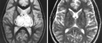
If the tumor has affected the brain, the patient suffers from headaches, nausea and vomiting. His gait is disturbed and convulsions are possible. If diagnosed during pregnancy, the risk of intracerebral hemorrhage is high.
What is a spinal hemangioma?
Reference. Spinal hemangioma is a benign formation that is located inside the vessels in the vertebral body. This is a plexus of capillaries and large blood vessels that forms blood cavities.
Hemangioma never progresses to the stage of cancer, despite the fact that it is a tumor. It only slowly increases in size over time.
Most often, hemangiomas occur in the thoracic region. They differ from other diseases in that they do not cause back pain or any other unpleasant symptoms.
Such vascular tumors can be congenital or acquired. Women are five times more likely to develop this disease than men. However, such a tumor can occur in anyone.

