Agiosarcomas (hemangiopericytomas and hemangioendotheliomas) are oncological diseases that affect blood vessels, in other words, it is vascular cancer. They are characterized by a high degree of malignancy, metastasis, and invasion into the depths of other tissues and organs.
The vascular tumor externally looks like a purple nodule with a soft-elastic consistency, without clear boundaries. The formation does not shift upon palpation and is painful. On a section, angiosarcoma consists of a large number of cavities filled with blood.
Causes and risk factors
The main cause of angiosarcoma is considered to be stagnation of lymph (lymphostasis). Violation of lymph outflow leads to the accumulation of carcinogenic substances in tissues. By affecting endothelial cells, they cause mutations, resulting in the appearance of malignant cells.
The risk of developing angiosarcoma increases when the body is exposed to thorium, radium, arsenic and its compounds, and polyvinyl chloride.
The high degree of malignancy of angiosarcoma and its ability to quickly metastasize to other organs make therapy ineffective.
Provoking factors also include:
- ionizing radiation;
- violations of local immunity;
- vascular factors.
The risk of liver angiosarcoma increases in the presence of the following diseases:
- Recklinghausen's disease;
- idiopathic chronic hemochromatosis.
The medical literature describes cases of the occurrence of cutaneous forms of angiosarcoma after mechanical injuries and viral skin lesions, against the background of benign angiogenic tumors.
A rare type is post-radiation angiosarcoma. They sometimes develop 15-25 years after radiation therapy for malignant tumors (cervical, breast, ovarian, endometrial, penile, testicular cancer).
Types and localization
The formations are formed from atypical endothelial cells. Another type of cell that has become undifferentiated creates muff-like growths around the vessels.
There is a generally accepted classification of pathology, according to which tumors are divided into four types.
- The idiopathic cutaneous type of angiosarcoma occurs on the head, selecting areas: the scalp,
- face.
Also, angiosarcoma tumor can be located in soft tissues: liver, heart, kidney, etc.
Symptoms
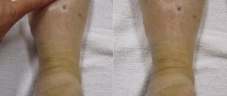
Malignant tumors caused by lymphostasis are usually localized on the lower extremities, the anterior chest wall, on the forearm, shoulder and in the area of the antecubital fossa. They can be single or multiple. Initially, the tumor appears as a blue-violet spot, similar to a regular hematoma. Then in its place a node forms, resembling a conglomerate of polyps, the surface of which is covered with erosions and ulcers and bleeds easily. The pathological process quickly spreads to surrounding tissues.
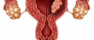
Angiosarcoma of the liver is usually observed in men over 60 years of age, and angiosarcoma of the mammary glands is observed exclusively in women 35-45 years of age.
Idiopathic cutaneous angiosarcoma is usually localized on the face, scalp, tonsils, pharynx or larynx, lower or upper jaw. At first, the swelling looks like an orange peel (looks like wrinkled, bumpy skin) or a hematoma. After this, painful nodes are formed, covered with erosions and prone to bleeding. This form of angiosarcoma quickly metastasizes to the lungs.
Angiosarcoma of the mammary gland has the appearance of a node with an uneven color from light blue to blood-red.
Post-radiation angiosarcoma is usually located in the pelvis, chest or abdomen. In terms of clinical manifestations, it does not differ from other types.
In addition to local ones, there are general symptoms that accompany the development of angiosarcoma:
- weakness, fatigue;
- signs of intoxication (pallor, nausea, decreased general health);
- increased body temperature;
- lack of appetite, weight loss;
- anemia.
Angiosarcoma of the liver is also characterized by:
- liver enlargement;
- abdominal pain;
- jaundice;
- ascites.
Angiosarcoma of the liver
In approximately every fourth case, patients with liver angiosarcoma experience massive bleeding into the abdominal cavity, quickly leading to death.
ANGIOSARCOMA
ANGIOSARCOMA
(
angiosarcoma
; Greek angeion - vessel + sarcoma; synonym
angioplastic sarcoma
) - a group of malignant tumors from elements of the wall of blood (hemangiosarcoma) or lymphatic (lymphangiosarcoma) vessels. These include, depending on the elements of the vascular wall: angioendotheliomas (see), angiopericytomas (see Hemangiopericytoma), angioleiomyomas (see Leiomyoma) of blood or lymphatic vessels and so on. Actually, angiosarcoma means a malignant vascular tumor, which, due to severe anaplasia (see), cannot be attributed to a specific element of the vascular wall.
Some pathologists are inclined to abandon the term “angiosarcoma” altogether and, if it is impossible to clarify the histogenesis of a malignant vascular tumor, they prefer to define it as an undifferentiated sarcoma (see), rich in blood vessels.
Histological picture
The tumor consists of spindle-shaped, round and polymorphic cells, forming cords and intimately associated with abundantly developed newly formed vessels and vascular cavities. Some cells may maintain a muff-like arrangement around the vessels.
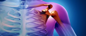
Angiosarcoma is a relatively rare tumor. Localization is varied: internal organs, retroperitoneal space, mediastinum, orbit, bones. Angiosarcomas most often occur in the soft tissues of the extremities, trunk, head and neck. Angiosarcoma of the soft tissues of the extremities most often occurs on the thigh in young people.
Rice. Angiosarcoma of the soft tissues of the leg with an ulceration that caused bleeding. The location of the lesion is indicated at the top right
Angiosarcomas grow quickly and tend to invade surrounding tissue and skin early, causing heavy bleeding (Fig.). Radiographs may reveal secondary destruction of the adjacent bone and an intense shadow of the tumor, sometimes containing calcareous inclusions. An important role in the diagnosis of angiosarcoma is played by cytological studies of punctate samples from the tumor.
Angiosarcomas are highly malignant tumors. Up to 60% of tumors metastasize hematogenously and up to 20% of tumors lymphogenously; after radical excision, relapses are observed in 50% of patients. Despite the use of combined treatment methods, the prognosis is unfavorable: 50% live for 3 years, 40% for 5 years, and 25% for radically operated patients live for 10 years.
See also Tumors.
Bibliography:
Wichert A. M., Galil-ogly G. A. and Poroshin K. K. Soft tissue tumors, microscopic diagnosis, p. 89, M., 1969; Guide to pathological diagnosis of human tumors, ed. N. A. Kraevsky and A. V. Smolyannikov, p. 72, M., 1971; Smolyannikov A.V. Morphological diagnosis of soft tissue tumors, p. 114, M., 1969.
A. A. Klimenkov.
Diagnostics
To diagnose angiosarcoma, a comprehensive laboratory and instrumental examination is carried out, including:
- general blood analysis;
- blood chemistry;
- blood test for tumor markers;
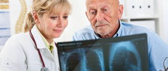
- radiography;
- Ultrasound;
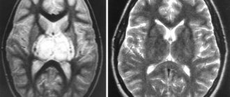
- computed tomography or magnetic resonance imaging.
Angiosarcoma can be localized in any part of the body and in any organ, but most often it affects soft tissue and skin (in 60% of cases).
The final diagnosis is made based on the results of histological analysis of the tumor area obtained during the biopsy.
Diagnostic plan for angiosarcoma at Top Clinic Ichilov
The cost of the examination is $3,045.
1st day
The examination begins with an appointment with a diagnostician. First, the doctor measures the patient's vital signs. After examining the patient and studying his medical documentation, the doctor enters the necessary data into the medical history in Hebrew. If there are slides and blocks with biopsy materials, they are transferred to the laboratory for revision.
In conclusion, the diagnostician gives the patient directions for further research.
2nd day
The patient undergoes the prescribed tests:
- blood tests (general and biochemical) - necessary to assess the patient’s general condition and plan treatment;
- general urinalysis - to identify possible pathological changes associated with the disease;
- PET-CT – to determine the location of possible tumor metastases.
3rd day
The patient is seen by one of the leading oncologists at Top Clinic Ichilov - Prof. O. Merimsky / Prof. Sh. Shneybaum , a specialist in the treatment of sarcomas. For an experienced specialist, examining a patient and talking with him are no less informative diagnostic methods than imaging studies.
At the end of the appointment, the doctor gives the patient a final 100% accurate diagnosis and determines treatment tactics.
The cost of diagnosis and prescribing a treatment protocol is $3,045.
Ofer Merimsky at Top Clinic Ichilov
Treatment
The high degree of malignancy of angiosarcoma and its ability to quickly metastasize to other organs make therapy ineffective.
When a malignant tumor is diagnosed early, it is removed, after which radiation therapy is prescribed. In some cases, radiation therapy may be used in the preoperative period. This combined approach reduces the growth of metastases, reduces the likelihood of relapses, and helps alleviate the patient’s general condition.
In the presence of metastases in distant organs, chemotherapy is indicated.
Free quota surgery: how to get it and what’s changing in 2020
Cancer treatment >> Types of cancer >> Angiosarcoma is a very malignant and often metastatic neoplasm of blood vessels.
Occurs from degenerated cells of the endothelial layer of the inner wall of blood or lymphoid vessels. The cells of formation are characterized by a high degree of genetic mutations.
Angiosarcoma is a fairly rare cancer pathology that occurs equally often in men and women, more often at the age of 40-50 years.
The most common location of angiosarcoma is the lower extremities.
Prevention
There are no specific preventive measures to prevent the onset of a malignant disease.
Every person should be attentive to their health, avoid factors that provoke disease, maintain the state of the immune system, and balance the diet.
Systematic preventive examinations play an important role, as well as visiting a doctor at the first symptoms of pathology.
Advantages of Israeli medical centers
- Compliance with strict international patient safety standards
- Application of modern antitumor therapy protocols
- Prompt and high-quality diagnostics
- Wide range of minimally invasive surgery options
- Providing a personal assistant
Contact our consultants to learn more about the possibility of treating angiosarcoma in the most prestigious medical centers in Israel.
- 5
- 4
- 3
- 2
- 1
(3 votes, average: 5 out of 5)
Attending doctors Top Clinic Ichilov
Professor Ofer Merimsky
– Head of the Department of Skeletal and Soft Tissue Tumors, the most famous Israeli specialist in the treatment of sarcomas. Thanks to his 30 years of experience in oncology, the professor will select the optimal treatment program for you, which will allow you to save the affected limb (for angiosarcomas of the bone) and several times increase your chances of a complete cure.
Professor Shlomo Schneibaum
– a leading Israeli orthopedic surgeon, has been performing operations for angiosarcomas of the skin and breast for 30 years. Performs organ-preserving operations to completely remove tumors and avoid breast amputation.
Professor Einat Even-Sapir
– Head of the Department of Nuclear Diagnostics, the best Israeli specialist in interpreting PET-CT results. For more than 25 years he has been diagnosing angiosarcomas of various locations.