Hemorrhagic shock is essentially pathological loss of blood. When blood volume decreases sharply and by a significant amount, the body falls into a stressful state. Usually the body saturates with about 5-6 liters of blood, even a slow loss of about 400 milliliters, which is usually taken from a donor, causes immediate weakness and dizziness. That is why, after donating blood, to stimulate the restoration of the full volume of fluid circulating through the vessels, doctors strongly recommend drinking sweet warm tea with hematogen.
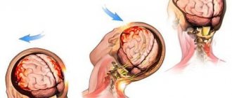
This reaction is provoked by slow blood loss, to say nothing of rapid blood loss. With a sudden loss of blood, the tone of the veins is increased, and the body is immediately thrown into shock from an instant decrease in blood volume. When the blood supply decreases, the body begins to function differently. More than 15% of the leak includes a kind of energy saving mode - the body switches forces to life-supporting organs: the heart, lungs, brain, and the remaining parts are considered secondary. There is hemorrhagic and hypovolemic shock. They are distinguished by and large only by the rate of decrease in blood volume. Hypovolemia does not provoke a catastrophic outcome, because the recovery algorithm is activated. This means that only shock during a rapid decrease in volume can be considered hemorrhagic.
Causes of hemorrhagic shock
Hemorrhagic shock is based on serious vascular damage. Acute leakage of fluid in the vessels implies the absence of half a liter to a liter of blood, combined with a rapid decrease in the amount of circulating fluid. This situation is usually provoked by serious injuries, which are accompanied by severe damage to blood vessels. Often, hemorrhagic shock is a consequence of gynecological pathologies: trauma during childbirth, postpartum hemorrhage, prematurely separated placenta, intrauterine fetal death, ectopic pregnancy. Of course, severe bleeding can occur after surgery, when the cancerous tumor disintegrates, a through hole appears and, as a result, a gastric ulcer.
Clinical manifestations
The manifestation of acute blood loss directly depends on the amount of fluid lost. Doctors distinguish three stages of hemorrhagic shock. The division occurs in direct proportion to the volume of blood lost:
- Stage I. The degree to which compensation for lost fluid is still possible. The victim is conscious, maintains sobriety, looks quite pale, the pulse is weak, low blood pressure and a decrease in the temperature of the extremities are observed. In this case, the lost volume does not exceed 15–25% of the total volume. The heart muscle tries to compensate for the missing fluid with the heart rate, so the heart rate increases to 90–110 per minute;
- Stage II. At this stage, normal organ functions are disrupted. The lack of a large volume of blood forces the body to distribute life support processes in accordance with the priority of specific organs. There is oxygen starvation of the brain, the heart pumps out blood noticeably weaker. Symptoms appear when there is a loss of 25 to 40% of circulating blood volume. The victim’s consciousness is disturbed - the person’s thinking is inhibited. The fluid in the vessels has a critically low level of oxygen, so the face, hands, and feet turn bluish, and sticky sweat appears throughout the body. A thread-like pulse appears, blood pressure decreases, and the heart rate reaches 140 beats. The kidneys stop filtering fluid normally, urination decreases;
- Stage III. This is an irreversible shock. The patient's condition is considered extremely critical. Consciousness is completely absent, the skin acquires a marble tint, the pressure in the arteries decreases to 60-80 millimeters of mercury or is not detected at all. Tachycardia occurs - the heart contracts up to 140-160 times per minute.
What influences the severity of this condition?
The effectiveness of the compensation mechanism depends on the following components:
- blood clotting;
- state of the immune system;
- environmental conditions (there is more oxygen in the forest than in the office);
- vascular tone;
- states of the nervous system;
- the ability of the heart to work in conditions of lack of oxygen.
A person with multiple chronic diseases is unlikely to be able to endure serious blood loss. The hardest situation is for those people who have lost blood in areas with thin air or in a room where carbon dioxide predominates. On average, a healthy person circulates 5 liters of blood. 75% of the fluid runs through the venous system, so the speed of adaptation of the veins to its loss will determine whether a person survives or not.
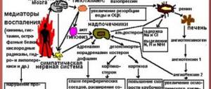
A loss of 20% of the total circulating blood volume does not allow the compensation mechanism to quickly activate. Venous pressure drops sharply. Blood circulation is centralized around the heart, brain and lungs. The body takes the muscles, intestines, and skin as “superfluous” and “disconnects” them from the general blood supply system. Systolic contractions are only sufficient to energize the coronary arteries. In response to this, the body turns on the endocrine system, due to which fluid is retained inside the organs. This process leads to an increase in the concentration of chlorides with sodium and a decrease in the amount of potassium.
How is the degree of blood loss determined?
Doctors determine the levels of shock stages using the Algover index. This number shows the proportional ratio of the number of contractions of the heart muscle to the upper blood pressure. The numerical value of the index directly depends on the severity of the victim’s condition. The normal value is within 1.0. Doctors further divide the severity of the indicator into:
- light, within 1.0 to 1.1;
- moderate severity, within 1.1 to 1.5;
- severe, within 1.5 to 2.0;
- critical severity, ranging from 2.0 to 2.5.
MedGlav.com
Bleeding can occur not only from injury, but also from disease and blunt trauma.
Nose bleed. Nosebleeds can sometimes be significant and require emergency treatment. The causes of nosebleeds are varied. Bleeding occurs as a result of local changes (trauma, scratching, ulcers of the nasal septum, with strong nose blowing, skull fractures), and as a result of various diseases: blood diseases, heart defects, infectious diseases (scarlet fever, influenza, etc.), hypertension . When a nosebleed occurs, blood flows not only out through the nasal openings, but also into the pharynx and oral cavity. This causes coughing and often vomiting. The patient becomes restless, which increases bleeding.
The person providing assistance must first eliminate all causes that increase bleeding. It is necessary to reassure the patient, convince him that sudden movements, coughing, talking, blowing his nose, and straining increase bleeding. The patient should be seated, given a position in which there is less opportunity for blood to enter the nasopharynx, an ice pack, a ball of snow wrapped in a scarf, a handkerchief moistened with cold water, a bandage, a ball of cotton wool, etc. should be placed on the area of the nose and bridge of the nose. it is necessary to ensure a sufficient supply of fresh air; if bleeding occurs from overheating, move the patient to the shade, apply cold compresses to the head and chest.
If the bleeding does not stop, you can try to stop it by firmly pressing both halves of the nose to the nasal septum. In this case, the patient’s head is tilted slightly forward and possibly higher, and the nose is squeezed with force. The patient must breathe through his mouth. You need to squeeze your nose for 3-5 minutes or more. The patient should spit out blood that gets into the mouth.
Instead of pressing, you can tamponade the nasal passages with a dry ball of cotton wool or a ball of cotton wool moistened with a solution of hydrogen peroxide. Cotton balls are inserted into the nasal passages, and the patient's head is tilted forward. On cotton wool, the blood clots quite quickly and the bleeding stops. Usually, these measures will stop the bleeding; otherwise, the patient must be immediately taken to the hospital.
Bleeding after tooth extraction. Significant bleeding may occur after tooth extraction. It is stopped by filling the defect in the gum with a ball of cotton wool and pressing it tightly with the teeth.
Bleeding due to damage to the ear canal and internal structures of the ear (impact, scratches, fracture of the skull). It is stopped by inserting gauze into the external auditory canal, folded into a funnel, which is held in place with a gauze bandage over the ear.
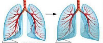
Pulmonary hemorrhage. If the lungs are damaged (a strong blow to the chest, broken ribs), or a number of diseases of the lungs and heart (tuberculosis, cancer, lung abscess, mitral heart disease, etc.), pulmonary hemorrhage may develop. A patient with sputum and coughing begins to secrete scarlet foamy blood - hemoptysis. Sometimes pulmonary bleeding is very severe.
If blood appears in the sputum of the patient, it is necessary to free him from clothes that impede breathing and immediately place him in a semi-sitting position. If possible, the patient should be reassured and convinced that he needs complete rest for treatment. There should be a lot of fresh air in the room where the patient is. The patient is prohibited from moving, talking, and is advised to breathe deeply and hold back his cough. It is advisable to place an ice pack on your chest. Anti-cough tablets are prescribed as medications.
Any pulmonary bleeding is a terrible symptom of any serious illness, so the task of first aid is to quickly deliver the patient to a medical facility. Patients with pulmonary hemorrhage are overly sensitive to transportation. Delivery of such patients from home to a medical facility should be carried out by special ambulance transport in a semi-sitting position, and special care must be taken to avoid shaking and sudden movements, which can increase coughing and bleeding.
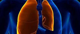
Bleeding into the chest cavity. With a blow to the chest, broken ribs, and some lung diseases, blood vessels can be damaged and one or both pleural cavities can fill with blood. The accumulating blood compresses the lung, causing breathing problems. Due to blood loss and exclusion of the lung from the act of breathing, the patient’s condition quickly deteriorates: breathing becomes more frequent and becomes difficult, the skin becomes pale, with a bluish tint.
The patient must be urgently transported to a medical facility. Help consists of giving the patient a semi-sitting position. An ice pack is applied to the chest.
Gastrointestinal bleeding. Bleeding into the cavity of the stomach and intestines is a complication of a number of diseases (peptic ulcer, stomach cancer, varicose veins of the esophagus, etc.) and injuries (foreign body, burn, etc.). It can be significant and lead to death. Symptoms of gastric bleeding, along with the general symptoms of acute anemia (pallor, weakness, sweating), are bloody vomiting or vomiting contents the color of coffee grounds, frequent loose stools and black stool (tarry stool).
To improve the patient's condition and reduce bleeding, it is necessary to create rest for the patient, give him a horizontal position, put an ice pack on his stomach, and completely prohibit the intake of food and liquid.
The main task of first aid is to organize the immediate delivery of the patient to a medical facility. Patients with gastrointestinal bleeding should be transported in a supine position with the leg end of the stretcher raised - this prevents bleeding of the brain.
Bleeding into the abdominal cavity. Occurs with blunt trauma to the abdomen, most often due to ruptures of the liver and spleen. The cause of intra-abdominal bleeding may be some diseases of the liver and spleen; In women, bleeding is possible as a result of a rupture of the fallopian tube during an ectopic pregnancy.
Bleeding into the abdominal cavity is manifested by severe abdominal pain. The skin is pale, the pulse is frequent. If there is significant bleeding, loss of consciousness may occur. The patient should be laid down, an ice pack should be placed on the stomach, and food and water intake is prohibited. Such patients should be immediately transported to the hospital in the supine position.
Acute anemia. Develops with significant blood loss. Patients tolerate blood loss in different ways. Children and the elderly are most sensitive to blood loss. People who have been ill for a long time, hungry, tired, or in a state of fear do not tolerate blood loss well.
An adult may hardly feel the loss of 300-400 ml of blood at all, but for a child this blood loss will be fatal. Instant blood loss (2-2.5 liters) is fatal.
The loss of 1-1.5 liters of blood is very dangerous and is manifested by the development of a severe picture of acute anemia, expressed by a sharp circulatory disorder and the development of oxygen starvation. A similar condition can develop with relatively small blood loss, but it occurs very quickly. The severity of the patient’s condition is judged not only by the amount of blood shed, but also by the level of blood pressure.
Symptoms of acute anemia are very characteristic and do not depend on whether the patient has external or internal bleeding. The patient complains of increasing weakness, dizziness, tinnitus, darkening and flickering of spots in the eyes, thirst, nausea, and vomiting. The skin and visible mucous membranes become pale, facial features become sharper. The patient is inhibited, sometimes, on the contrary, excited, breathing is rapid, the pulse is weak or not detected at all, blood pressure is low. Subsequently, as a result of blood loss, loss of consciousness may occur due to bleeding of the brain, the pulse disappears, blood pressure cannot be determined, convulsions appear, and involuntary separation of feces and urine. If appropriate measures are not taken urgently, death occurs.
With large blood loss and low blood pressure, bleeding may stop; however, when providing first aid, it is necessary to apply a pressure bandage to the wound, and then begin anti-shock measures. The victim should be laid on a flat surface to prevent anemia of the brain. In case of significant blood loss that causes fainting or shock, the patient (wounded) is placed in a position in which the head is lower than the body. In some cases, it is useful to perform a “self-blood transfusion”: all the limbs of a lying wounded person are raised, thereby achieving a temporary increase in the amount of circulating blood in the lungs, brain, kidneys and other vital organs. If consciousness is preserved and there is no damage to the abdominal organs, the patient can be given hot tea, mineral water or, if it is not available, plain water.
In terminal conditions and cardiac arrest, resuscitation is performed. The main method of treating acute anemia is an urgent transfusion of donor blood, so the victim must be taken to a medical facility as quickly as possible. When transported by a special ambulance, blood transfusion can be performed in the car, since such vehicles have a supply of donor blood.
Severity
Of course, only the index indicator cannot be considered as absolute. Doctors see it in combination with blood loss. The classification of shock severity types is named in the same way as the indices, but requires the presence of a certain volume of blood. So, a mild degree implies a shock index of 1.0-1.1 and a blood loss of 10 to 20% of the volume, but not more than 1 liter. Moderate severity - shock index up to 1.5, loss from 20 to 30% of volume, but not more than 1.5 liters. Severe degree - index up to 2.0, loss up to 40% or up to 2 liters. Extreme severity - index up to 2.5, loss more than 40% or more than 2 liters.
Treatment of hemorrhagic shock
Intensive therapy after stopping bleeding and catheterization of veins is aimed at:
- Elimination of hypovolemia and restoration of circulating blood volume.
- Detoxification.
- Ensuring adequate microcirculation and cardiac output.
- Restoring the initial parameters of osmolarity and oxygen transport capacity of the blood.
- Normalization and maintenance of normal diuresis.
- Prevention of DIC syndrome (erythrocyte aggregation).
To achieve these goals, priority in infusion therapy of HS was given to:
- HES solutions up to 1.5 liters per day and normalization of blood oncotic pressure;
- intravenous crystalloid solutions in a volume of up to 2 liters, until blood pressure normalizes;
- packed red blood cells and other blood substitutes under the control of central venous pressure to a hematocrit level of 32-30%;
- colloidal solutions (gelatins and dextrans) in a 1:1 ratio to the total volume of infusions;
- donor blood;
- glucocorticosteroids in maximum dosages (up to 1.5 mg).
An important role in the treatment of HS is assigned to vasodilator drugs necessary to eliminate vasospasm (papaverine, aminophylline); prevention of reperfusion syndrome, for which alkalizing solutions, antioxidants, GHB, trental and antihistamines and proteolysis inhibitors are used.
Criteria for treatment effectiveness
Intensive therapy for HS is carried out to the level of indicators indicating the elimination of a life-threatening condition:
- Blood pressure up to 100/60 mm Hg. Art. and higher;
- Heart rate up to 100 beats per minute;
- CVP 4 and above mm water column;
- minute diuresis is more than 1 ml, and hourly diuresis is more than 60 ml;
- hemoglobin level 60 g/l;
- blood oxygen concentration 94 -96%;
- protein content in blood plasma more than 50 g/l;
- venous blood hematocrit 20% or higher.
Possible complications
Against the background of decompensated HS, the following may develop:
- DIC – syndrome (erythrocyte clumping);
- reperfusion syndrome (oxygen paradox);
- myocardial ischemia;
- coma;
- ventricular fibrillation;
- asystole.
Consequences. Several years after massive blood loss accompanied by HS, endocrine pathology and chronic diseases of internal organs may develop, resulting in disability.
Diagnosis of the disease
Hemorrhagic shock (ICD 10 code - R 57.1) refers to conditions similar to dehydration, which are characterized by a sharp decrease in the amount of blood that is in the vessels of the body. The focus of diagnosing the symptoms of hemorrhagic shock is to determine the amount of blood lost, the source of the leak, and its intensity.
The first step is to inspect the source of fluid leakage from the vessels. The doctor assesses the extent of the damage. Blood may flow out in a pulsating stream or spurt out. It is important to understand that leakage occurs suddenly, in large volumes and over a short period.
How to provide first aid
It is very important to correctly assess the condition of the victim. It is necessary to find the cause of bleeding and eliminate it as quickly as possible. Correctly provided first aid helps the victim recover from shock more quickly, and sometimes can even save his life.
So, let's figure out what needs to be done in case of hemorrhagic shock. First of all, it is necessary to localize the source of the loss. The area above the source of blood leakage should be bandaged with a bandage or tourniquet. The tourniquet usually puts a lot of pressure on the vessels and can damage them, so emergency doctors recommend using a rag or gauze bandage. It needs to be tightly bandaged over the wound, with a tight bundle wrapped on top, which after 1 hour will need to be gradually untwisted to avoid tissue death below the bandaged area. It is not recommended to take any further measures without doctors. You need to wait for the ambulance to arrive and be sure to write on the victim the time a tight bandage was applied so that doctors understand how long the wound is localized from the blood supply.
Emergency care for hemorrhagic shock
Since the disease is serious and can cause irreversible complications, the patient must be given proper first aid. It is the timely provision of first aid that will influence the positive outcome of therapy. The basics of such treatment will focus on addressing the following issues:
- Emergency care for hemorrhagic shock is aimed, first of all, at stopping the bleeding, and for this it is necessary to establish its causes. Surgery may be required for this purpose. Or the doctor may temporarily stop the bleeding using a tourniquet, bandage, or endoscopic hemostasis.
- The next stage of emergency treatment is to restore the blood volume (BCV) that is necessary to save the patient’s life. Intravenous infusion of solutions should be at least 20% faster than the rate of ongoing bleeding. For this purpose, the patient’s blood pressure, central venous pressure and heart rate readings are used.
- Urgent measures for HS also include catheterization of large vessels, which is done to ensure reliable access to the bloodstream, including ensuring the required rate of infusions.
Possible complications
Hemorrhagic shock can cause quite serious complications. It all depends on the amount of fluid lost, its intensity, and the speed of localization of the source. Most complications occur due to oxygen deprivation. This is damage to the mucous membrane of the lungs, mild brain exhaustion, damage to the functions of the brain, kidneys, and liver. If shock occurs due to labor, irreversible damage to the reproductive organs is possible.
So, we found out how hemorrhagic shock manifests itself, what its degrees and stages are, and how to provide first aid to the victim. If you still have questions after reading the article, feel free to write them in the comments.
What complications are possible?
Patients who have survived hemorrhagic shock may experience the following complications:
- systematic clumping of red blood cells;
- ischemia;
- coma;
- gastric fibrillation;
- asystole.
For your information. After suffering hemorrhagic shock and heavy blood loss, patients in some cases develop pathologies of the endocrine system or internal organs. These factors can provoke subsequent disability of the patient.
Hemorrhagic shock is a sudden and extremely serious condition that can significantly worsen a person’s health and put his life at risk. Pregnant women should be especially attentive to their well-being, because not one, but two whole lives depend on the state of their health. The shock state of blood leaving the vessels develops quite rapidly, so the provision of timely first aid for hemorrhagic shock largely determines the future outcome of the situation.