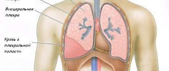
Pleurisy is a pathology of the lungs in which the inflammatory process localized between the pleural layers is activated. In this case, fibrous deposits or serous fluid form there, and exudate can accumulate in the pleural cavity.
The pleura is the lining of the lungs, which has 2 layers (outer and inner). There is an intermediate cavity between these leaves. A small amount of liquid and protein compounds can enter this cavity. A small amount of liquid can be absorbed, with the exception of proteins, they remain in the cavity as sediment. This process can lead to the pleural layers thickening and inflammation appearing, but there may also be a large amount of exudate, this provokes exudative pleurisy.
Quite often, pleurisy occurs as a complication from other pulmonary pathologies. Less commonly, it occurs as an independent pathology. Because of this, the symptoms and treatment of pulmonary pleurisy may differ in different situations.
There is also the concept of “hemorrhagic pleurisy”; this pathology is distinguished by the fact that the fluid that fills the pleural cavity contains red blood cells in high concentrations. And this pathology is characteristic of the tuberculous nature of pleurisy.
Quite often, segments of the lungs are involved in the general pathological process developing in the pleural cavity. This condition is called pleural pneumonia.
Classification
Modern medicine knows of pleurisy of various types and forms, and there are several classifications of this pathology. But in Russian practice, the classification scheme of Professor N.V. Putov is traditionally used. In accordance with it, the following types of pleural pathologies are distinguished.
By etiology:
- Infectious (staphylococcal, tuberculous pleurisy, etc.);
- Non-infectious (indicating the disease that became the cause);
- Unclear etiology (idiopathic).
According to the presence of effusion and its nature:
- Exudative pleurisy (with serous exudate, serous-fibrinous, cholesterol, putrefactive, etc., as well as purulent pleurisy);
- Dry pleurisy (including adhesive pleurisy, in which adhesions are fixed between the pleural layers).
According to the course of inflammation:
- Acute pleurisy;
- Subacute;
- Chronic.
According to the location of the effusion (degree of pleural damage):
- Diffuse (total inflammation);
- Enclosed pleurisy, or delimited (diaphragmatic, parietal, interlobar, etc.).
The types of disease are also distinguished according to the scale of distribution: unilateral (left- and right-sided) or bilateral inflammation of the pleural membrane.
Reasons for development
It must be said that the disease in its pure form is rare. For example, its development can be caused by trauma to the chest or hypothermia. In most cases, it accompanies any disease or occurs as a complication of it.
Pulmonary pleurisy is characterized by the formation of fibrinous deposits on the surface of the pleural layers and/or accumulation of exudate in the pleural cavity. Symptoms depend on the form of the disease.
Infectious pleurisy is the most common. Sensitization of the body also plays a major role in the mechanism of development of pathology. Microbes and their toxins lead to changes in the body's reactivity and allergization of the pleura. The immune system begins to “send” produced antibodies to the site of inflammation, which, when combined with antigens, affect the production of histamines.
About 70% of forms of pathology are caused by bacterial agents:
- Streptococci;
- Pneumococci;
- Mycobacterium tuberculosis;
- Anaerobes;
- Mushrooms;
- Legionella;
- Tuberculosis.
The causes of non-infectious pulmonary pleurisy are as follows:
- malignant tumors of the pleural layers,
- metastasis to the pleura (in breast cancer, lung cancer, etc.),
- connective tissue lesions of a diffuse nature (systemic vasculitis, scleroderma, systemic lupus erythematosus),
- pulmonary infarction.
Is pleurisy contagious? To answer this question unambiguously, you need to know the cause of pleurisy itself. If the suffering is associated with a chest injury, then, naturally, such pleurisy is not contagious. With a viral etiology, it can be quite contagious, although the degree of contagiousness is low.
Pleurisy causes
Pleurisy does not appear just like that, out of nowhere; it mainly acts as an accompaniment of inflammation in the human body. The causes of its appearance are divided into infectious and non-infectious.
Infectious causes:
- viral diseases (staphylococcal or pneumococcal infection),
- the presence of fungal microorganisms (candidiasis, blastomycosis),
- syphilis,
- tuberculosis,
- typhoid fever,
- surgery,
- injuries in the chest area.
Non-infectious causes:
- malignant formation of the pleura,
- metastases in the pleura (breast cancer, lung cancer, etc.),
- pulmonary infarction,
- TELA.
In addition, there are factors that contribute to the occurrence of the disease. These include stress, hypothermia, alcoholism, unbalanced nutrition, and an allergic reaction to medications.
Inflammation of the pleura occurs due to microorganisms that can enter it in various ways: through blood, lymph, or through various injuries, as well as during surgery.
Symptoms
In most cases, dry pleurisy develops acutely. Patients usually clearly indicate the time of onset of the disease. Characteristic complaints are chest pain, increased body temperature, and severe general weakness.
Chest pain is associated with irritation of the nerve endings of the pleura by fibrin. The pain is often unilateral on the affected side, quite intense, with a tendency to intensify with a deep breath, coughing, or sneezing. Body temperature rises to 38°C, rarely higher. With a gradual onset of the disease at first, the body temperature may be normal. Also concerned about general weakness, sweating, headache, intermittent pain in muscles and joints.
With exudative pleurisy, symptoms are caused by the accumulation of fluid in the pleural cavity. Complaints vary depending on the onset of the disease. If exudative pleurisy occurs after fibrinous pleurisy, then it is possible to trace a clear chronology of events. At the beginning of the disease, the patient is bothered by intense unilateral chest pain, which intensifies with deep inspiration. Then, when exudate forms, the pain disappears, and in its place comes a feeling of heaviness, pressure in the chest, and shortness of breath. A dry cough, increased body temperature, and general weakness may also occur. If exudative pleurisy occurs primarily, then in this case pain syndrome is not typical. In this case, patients complain of general weakness, sweating, fever, and headache. After a few days, shortness of breath appears, a feeling of heaviness in the chest with little physical activity, and with a large amount of exudate - at rest. At the same time, nonspecific symptoms of intoxication intensify.
If the above complaints occur, you should immediately consult a therapist. With progressive deterioration of the condition (increase in body temperature, difficulty breathing, increased shortness of breath), hospitalization in a hospital is indicated.
Dry pleurisy of the lungs
It is a consequence of an existing primary disease. But its symptoms come to the fore, masking the picture of the underlying pathology.
The disease is characterized by a sudden onset. The dominant symptom is pain in the chest on the side where the inflammation is localized. During breathing and coughing, there is an increase in pain. Patients complain of pain in the side, which is stabbing in nature. This is usually observed in anterolateral views of the chest. The pain is most severe at the very beginning of the disease. The patient tries to limit chest movements as much as possible. He has to lie on his healthy side, because the pressure intensifies the pain. If exudate appears, the pain subsides. On examination, there is a pronounced lag in the affected half of the chest. On palpation, the intercostal muscles are painful and tense.
Patients usually do not have any special problems with their general condition. There may be no temperature rise at all. Sometimes the temperature numbers are in the nature of a low-grade fever.
On auscultation, a pleural friction noise is clearly audible. But this is not crepitus or fine wheezing. A clear distinction must be made here. On the side of the inflammatory process, there is a weakening of breathing. The noise that occurs when the pleura rubs is rough in nature. If you put pressure on the chest, it may get worse. At the apex, where lung excursion is minimal, the noise may not be audible. The same situation can occur with diaphragmatic pleurisy. After coughing, the nature of the noise does not change. This is a differential sign of differentiation from wet rales, in which after coughing their character becomes different.
The blood picture shows all the signs of inflammation. An X-ray examination shows that the movement of the diaphragm is limited. Suffering ends with recovery or transforms into another form.
Stages of disease development
Pleurisy develops in 3 stages.
The first, mildest phase of the disease is characterized by dilation of blood vessels and the release of a large volume of pleural fluid. The functioning of the lymphatic system is not impaired, which facilitates the complete removal of excess fluid from the pleura.
When pleurisy moves to the second stage of development, adhesions begin to form, impeding the outflow of pleural fluid. In the absence or ineffectiveness of treatment, pus and fluid gradually accumulate in the pleural zone.
The third stage is the recovery period. At this time, the foci of the inflammatory process gradually resolve. Sometimes fibrous tissue can form around them, protecting the previously affected area from healthy areas of the pleura. A similar process can be the beginning of chronic pleurisy.
Causes of the disease
Pleurisy can be primary in nature - when the inflammation is isolated and includes only the pleura (without occupying the pulmonary parenchyma). However, most often it is a complication of diseases occurring in surrounding tissues, such as:
- pneumonia;
- tuberculosis;
- pulmonary embolism;
- lung cancer.
The disease can also be caused by heart, liver and kidney failure, hormonal imbalance, pancreatitis and gastrointestinal diseases. Its development can be facilitated by chest trauma with rib fractures.
Complications
Pleurisy is a serious disease that significantly impairs the function of the respiratory system. In most cases, this pathology indicates a complication of the underlying disease (pneumonia, tuberculosis, tumor process, allergies). Correct and timely elimination of the cause of pleurisy allows you to completely restore lung function without any consequences.
However, in many cases, pleurisy can cause partial or complete structural and functional restructuring of the pleural or lung tissue.
The consequences of pleurisy include:
- Adhesions between the layers of the pleura. Adhesions are connective tissue strands between the layers of the pleura. They are formed in the area of inflammatory foci that have undergone organization, that is, sclerosis. Adhesions, called moorings in the pleural cavity, significantly limit the mobility of the lungs and reduce the functional tidal volume.
- Overgrowth of the pleural cavity. In some cases, massive pleural empyema can cause complete “overgrowth” of the pleural cavity with connective tissue fibers. This almost completely immobilizes the lung and can cause serious respiratory failure.
Diagnostics
Pleurisy can be unilateral or bilateral, occur in acute or chronic form. Diagnosis of this disease is carried out using the following methods:
- X-ray examination;
- thoracoscopy;
- Ultrasound of the pleural area;
- computed tomography;
- laboratory testing of blood, composition of exudate.
Using radiography, fluid accumulation in the interpleural cavity is detected when its volume exceeds 300 ml. A more informative study that allows us to identify the nature of pleurisy is thoracoscopy and pleural therapeutic and diagnostic puncture.
A study such as thoracoscopy allows us to determine what caused pleurisy of the lungs or its complications, and how to treat this disease.
The thoracoscope is inserted through a puncture in the chest directly into the pleural cavity. Using this procedure, the attending physician is able to visually assess the condition of the patient’s pleural area.
The therapeutic and diagnostic procedure thoracoscopy allows:
- take a sample of exudate for histological analysis;
- destroy the adhesions between the pleural sheets;
- remove exudate.
Purulent pleurisy of the lungs
Such conditions are associated with the accumulation of pus in the pleural cavity. As a rule, such a pathology is secondary and is observed after pneumonia, being its complication.
Patients complain of pain and a feeling of heaviness in the side. There is a cough. Breathing is difficult, it is impossible to take a full breath. The temperature can rise to significant levels. Patients complain of weakness. At the beginning of the disease, the pain is severe, but as exudate accumulates, it subsides, but the feeling of heaviness in the side increases.
As the disease progresses, shortness of breath becomes more pronounced. The cough is often dry. But if pleurisy develops after pneumonia, it is accompanied by sputum production. Sputum is characterized by a purulent consistency. The cough gets worse and can occur even at night. Sometimes the cough can take on the character of attacks. If the patient does not receive help, then an increase in cough is observed. Body temperature is hectic in nature and can rise to maximum levels. The pulse quickens, tachycardia becomes pronounced. If a spontaneous opening of an abscess occurs, air enters the pleural cavity. This leads to the development of pneumothorax and pleural shock.
Treatment of pleurisy
The doctor chooses a treatment regimen depending on the type of pleurisy and the nature of its course. Thus, it is necessary to treat infectious pleurisy using antibiotics.
For pleurisy caused by a tumor or the penetration of its metastases, antitumor treatment is required. And inflammation of the pleura caused by scleroderma or systemic lupus erythematosus must be treated with glucocorticosteroids.
The following medications are used in the treatment regimen for pleurisy of various origins:
- Antibacterial drugs, even before obtaining bacteriological results - drugs and analogues of Bigaflon, Levofloxacin, Cefepime or Ceftriaxone, followed by their replacement with drugs for a specific pathogen.
- Painkillers and anti-inflammatory drugs used for diseases of an inflammatory and degenerative nature (Mefenamic acid, Indomethacin or Nurofen);
- Antifungal therapy for fungal causes of pathology.
- In case of pleuresia, as a consequence of tumor processes, preparations of natural hormones and antitumor medications are prescribed.
- In the treatment of exudative pleurisy, the use of diuretics is justified. And vascular drugs (as indicated).
- For the dry form of pleuresia, suppressive cough medications (Codeine or Dionine), thermal physiotherapy techniques, and tight bandaging of the sternum are prescribed.
- To prevent the development of pleural empyema, as a consequence of complications of exudative pleurisy, puncture removal of purulent exudate is carried out, followed by washing the cavity of the pleural leaves with antibiotic solutions.
After acute pain symptoms subside, physiotherapeutic procedures, physical therapy exercises, and breathing exercises are prescribed.
Treatment Basics
Timely prevention of pleurisy promotes complete recovery. The consequences of the disease are expressed by adhesions between the pericardium and the lung, diaphragm, etc. If a medical examination reveals inflammation of the pleura, immediate hospitalization occurs.
The nature of the complication of pleurisy determines the duration of treatment. Dry pleurisy lasts up to 5 days, elimination of purulent ones is accompanied by several months of therapy. If the pathology is mild, the patient is prescribed bed rest and mustard compresses are applied to the chest area.
To reduce pain, the thoracic region is tightened with bandages and painkillers are administered. The inflammatory process (if present) is localized and eliminated with broad-spectrum antibiotics and anti-inflammatory drugs.
The therapeutic course is accompanied by a decrease in fluid intake and abundant consumption of vitamin-containing foods.
In case of acute purulent pleurisy, an operation is immediately performed, where liquid pus is removed from the respiratory chamber and the cavities are treated with antiseptics.
Folk remedies
Treatment with home recipes must be consistent with the main course and approved for use by a doctor. It includes procedures for using decoctions, infusions or tinctures, relaxing and nourishing compresses made from beneficial substances.
- To relieve pain, you can use a sponge soaked in warm salt or sea water.
Compresses made from decoctions of lentils and cabbage, with the addition of vinegar in small quantities, on woolen cloth soaked in oil help.
- A mustard bandage will help localize pain - mustard sachets should be applied to the irritated areas.
- Cottage cheese compress is useful as a source of protein and calcium. For absorption through the skin, it can be placed on the back for 1.5-2 hours at least twice a day.
Therapeutic drink recipes:
- Dried coltsfoot leaves, licorice root, three-leaf leaves, swamp cudweed, and knotweed, 1 tbsp each, are mixed. l., 2 tbsp. l. elecampane root and St. John's wort herb are added. After mixing, the mixture is poured with 250 ml of boiling water and left for 6 hours. The decoction is strained and served on an empty stomach, one teaspoon at a time. three times a day. Not recommended for people with poor circulation and high blood pressure.
- Cushion herb, elecampane root and licorice root are mixed 1 tbsp each, 2 tbsp are added. l. calendula flowers, horsetail and birch buds. Preparation and use as in the previous recipe. Prohibited for kidney disease.
- The roots and rhizomes of Caucasian hellebore (half a teaspoon each) must be evaporated in 0.5 liters of water over low heat until the volume of liquid is equal to a glass. Take half a teaspoon in the morning, before lunch, before bedtime. The decoction strengthens the walls of blood vessels and helps stop suppuration. It is also used for tuberculosis and heart failure.
Squeezed red beet juice is mixed with honey at the rate of 1/2, stirring the mixture with hot water (1/1). Can be consumed on an empty stomach or during meals. The “cocktail” gives tone to the body, promotes the renewal of blood cells and enriches with lactose and sucrose. Restrictions on intake for personal lactose intolerance, diabetes mellitus and allergies to components.
- The resin resin is cleaned of additives, placed in a jar and filled with purified drinking alcohol until it completely covers it. The composition must settle until the resin is completely dissolved. To one part of the infusion, 2 parts of raw lard are added - all this is mixed and melted in a clean container on the stove.
After melting, linden honey is added to the hot mass (proportion 1/1). The product is taken one teaspoon 3 times a day for up to 6 months. Helps with purulent pleurisy, tuberculosis, chronic bronchitis. The product is contraindicated for diabetics, lactose and honey allergy sufferers!
- Infusion of plantain leaf or common plantain. For half a liter of boiling water add 2 tbsp. l. dried plant. The liquid is filtered and drunk warm, 100-120 ml 4 times a day. The drink is harmless, has a healing and antibacterial character.
You can help your body fight germs with onions. To do this, you need to chop the onion and put the resulting porridge on a plate. The patient closes his eyes, the plate is brought slightly below the chin so that the smell is strongly noticeable, but does not cut into the mucous membranes of the mouth and nose with pain. Vapors must be inhaled for at least 4 minutes.
Eating vegetables rich in active antiseptic components will help speed up your recovery. These include:
- onion,
- garlic,
- horseradish,
- leek,
- mustard,
- hot peppers.
Folk remedies
The use of alternative medicine recipes for pleurisy is possible only if they are combined with medications prescribed by the attending physician. They are not suitable as independent remedies.
The following remedies will be effective.
- Mix crushed marshmallow root with coltsfoot leaves (2 g each) in equal proportions. Add 1 g of dry oregano herb, take 1 tbsp. l. raw materials and pour ½ liter of boiling water. Leave until completely cooled, after which the finished medicine is well filtered. Drink half a glass warm 3-4 times a day. Take the infusion as an expectorant.
- Dry crushed leaves of large plantain in the amount of 1 tsp. pour 200-250 ml of boiling water. Leave until completely cooled, filter. Drink 15 ml 3-4 times a day, regardless of meals. The infusion works well to liquefy and remove phlegm.
- Take dry leaves of coltsfoot, lungwort and plantain in equal proportions. Mix with crushed licorice roots and fragrant violet herb (in the same quantities). 1 tbsp. l. Pour the mixture without a slide with cold water and leave for 2 hours. After the time has passed, boil the mixture, cool and filter. Drink the entire decoction during the day in several doses.
It is advisable to use these recipes for different types of pleurisy, but only as thinners and expectorants.
Treatment of pleurisy with folk remedies at home
Medicinal plants used in traditional medicine include:
- increasing the body's defenses: rose hips, sea buckthorn, licorice root, pine needle infusions, black and red currants , etc.,
- allergy relievers (nettle, chamomile , etc.),
- enhancing the secretion and dilution of sputum (coltsfoot, calamus root, sweet clover , etc.).
To relieve pain when breathing, you can use the following method.
Take the processed skin of any small animal (rabbit, dog, muskrat, cat) and put it in brine for three days (concentration so that the egg floats).
Then the skin must be dried in the shade for 3 days. After drying, the skin is ready for use for 2-3 years. Tie the skin to the place with the greatest pain and keep it as a compress for 7-8 hours. Usually, after such a procedure, the pain is either greatly reduced or removed completely. Repeat three to five times until your general condition improves. A skin compress can be used for all inflammatory diseases of internal organs (when heat is not contraindicated), for radiculitis, osteochondrosis and any neuralgic pain (myositis, myalgia, inflammation of the sciatic nerve, etc.).
Lesser periwinkle . Prepare a decoction: add 1 tablespoon of crushed leaves to a glass of water and cook in a glass of boiling water for 20 minutes. Take a third of a glass three times a day.
Forest mallow . Used for inflammatory diseases of the digestive system and respiratory diseases. Mallow thins mucus, acts as an expectorant and as an antispasmodic.
Blackberry, bramble, azhina . The main effect is astringent, anti-inflammatory and fixing. Blackberry leaves and fruits are used. Blackberry wine, as well as blackberries and blackberry tea are used in folk medicine to treat influenza, pharyngitis, sore throats, and bronchitis. Pour 2 tablespoons of leaves into 500 ml of boiling water and leave for an hour. Drink 100-150 ml 4-5 times a day hot.
Common fig tree ( fig ). Has a weak laxative and anti-inflammatory effect. Used as an antitussive. Figs boiled with milk are used in folk medicine as a cough suppressant and for gargling for laryngitis, pharyngitis, and sore throat.
Althaea officinalis . The main effect is emollient, expectorant and anti-inflammatory. Pour 2 tablespoons of crushed marshmallow roots into 500 ml of cold water (cold infusion) and leave for 1 day. Strain the resulting infusion, add a little sugar. Take a tablespoon every two hours.
Boil the onion in milk and drink half a glass of the broth 3-4 times a day (drink hot).
Eucalyptus blue . The main effect is anti-inflammatory, especially during inflammatory processes in the respiratory tract (bronchitis). Pour a tablespoon of eucalyptus leaves into half a liter of boiling water and drink the prepared hot infusion 100 ml before meals three to four times a day for chronic bronchitis and bronchial asthma.
A decoction of rye or wheat bran (cook with honey) helps with an exhausting, severe cough.
Common pikulnik . The main action is expectorant. In folk medicine, hot infusions from this plant are used as an expectorant for bronchitis. Pour 3 teaspoons of the plant with 2 cups of boiling water, leave for 1 hour, strain. Drink the infusion throughout the day.
Boil 60 g of pepper roots with 0.25 liters of white wine, strain. Drink hot 3-4 times a day.
Glaucium ( yellow poppy ). Glaucine serves as the starting material for the production of antitussive drugs (glauvent and glauterpine). In folk medicine, an infusion or decoction of the whole plant is used (a tablespoon of crushed raw materials per 300 ml of boiling water). Take one third of a glass three to four times a day.
Coltsfoot . The main action is expectorant, emollient, anti-inflammatory, antiseptic. Coltsfoot acts as an antitussive and promotes sputum production during acute inflammatory processes in the respiratory tract. Pour 400 ml of boiling water over a tablespoon of dry leaves and leave to steep for 1 hour. Strain and drink 100 ml. 4 times a day.
In Bulgarian folk medicine, the following composition is popular against cough:
Collection 1:
- centaury – 50 g,
- scolopendra foliage – 50 g,
- coltsfoot – 50 g,
- linden flowers – 50 g.
Pour 2 tablespoons of this mixture with 1 teaspoon of flaxseed into 500 ml of boiling water and cook for 10 minutes. Leave for 1 hour, strain. Drink half or one third of a glass 3-4 times a day.
Ancient healers on the treatment of pleurisy... Odo of Mena in the poem “On the Properties of Herbs” writes:
Wormwood: “It’s better if you add honey and pepper to the grated nettle, and, as stated above, take it together with wine. Its seed will heal the lungs and chests of those suffering from pleurisy if taken in combination with honey...”
Ruta: “...If you boil it in water in combination with hot vinegar, this decoction will successfully soothe colic in the stomach, it will heal the lungs and the chest, and it will heal the pain in the side that is called pleurisy...”
Black initial letter: “Black initial letters take the powder with boiled honey - it will heal empyema and cough, as well as shortness of breath... with honey, the herb also expels cough, softens the stomach; If a patient with a fever trembles every day, take an ounce of plantain and combine two ounces of black cap with it and drink it, crushed, with warmed water, before there is a chill - the always harbinger of fever ... "
When is puncture necessary for pleurisy?
Pleural puncture (thoracentesis) is a procedure in which a certain amount of fluid accumulated there is removed from the pleural cavity. This manipulation is carried out for both therapeutic and diagnostic purposes, therefore it is prescribed in all cases of effusion pleurisy.
Relative contraindications to pleural puncture are the following conditions:
- pathologies of the blood coagulation system;
- increased pressure in the pulmonary artery system;
- chronic obstructive pulmonary disease in a severe stage;
- having only one functional lung.
Thoracentesis is performed under local anesthesia by inserting a thick needle into the pleural cavity at the level of the eighth intercostal space on the side of the scapula. This procedure is carried out under ultrasound control (with a small volume of accumulated fluid), or after a preliminary x-ray examination. During the procedure, the patient sits (as this allows you to maintain the highest level of fluid).
With a significant volume of pleural effusion, puncture allows you to drain part of the pathological fluid, thereby reducing the degree of compression of the lung tissue and improving respiratory function. The therapeutic puncture is repeated as necessary, that is, as the effusion accumulates.
Prevention
Prevention of pleurisy is the prevention and timely diagnosis of diseases that can provoke the development of inflammation of the pleural layers.
To do this, you need to follow simple recommendations:
- Strengthen the immune system: exercise regularly, take multivitamins, eat right;
- Train the respiratory system: simple breathing exercises along with morning exercises will help avoid inflammation of the respiratory system;
- Avoid complications of seasonal ARVI;
- At the slightest suspicion of pneumonia, you need to take an x-ray and begin full-fledged complex therapy;
- Stop smoking: nicotine often causes tuberculosis and tuberculous lesions of the pleura.
Strengthening the immune system, paying attention to your health and timely consultation with a doctor will help not only protect yourself from inflammation of the pleura, but also prevent such dangerous consequences as pleural adhesions, empyema, pleurosclerosis and overgrowth of the pleural cavity.