Causes of defeat
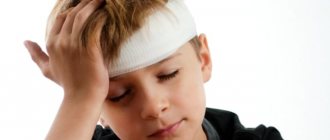
Head and neck injuries are included in the collective term “traumatic brain injury.” This includes damage to the structural parts of the brain and its membranes, blood vessels and nerves, as well as the bones of the skull. About 25% of victims do not survive a head injury. According to statistics, in the summer months, young people from 20 to 40 years of age who are injured in criminal disputes are more likely to suffer. In winter, head injuries are more often diagnosed in older people who are injured as a result of a fall.
Head injuries can result from a combination of unfavorable circumstances, in particular, these can be:
- injuries to professional athletes;
gunshot wound;- blow to the head with a blunt object;
- suicide;
- injuries from falling from a height;
- road accident;
- accidents at work and at home;
- cutting the head with a cold weapon.
Separately, birth trauma of newborns is distinguished. During pregnancy, sometimes obstacles to natural childbirth arise. Doctors do not advise a woman to give birth on her own if she has a narrow pelvis or breech presentation of the fetus, and suggest a caesarean section. However, the birth process does not always go according to plan; head injuries can be the result of an obstetrician’s error or occur accidentally. After a woman gives birth, doctors may notice compression or hemorrhage in the newborn because the soft bones of the skull cannot withstand the excessive pressure.
Psychosomatics gives its own version of the causes of traumatic brain injury. Well-known psychoanalysts collected information about the people with whom the accident occurred. The psychosomatics of trauma is that the victims unconsciously encouraged themselves to be injured. The reason for this behavior, scientists believe, is anger at oneself or a feeling of guilt, and the resulting injury is a well-deserved punishment. The psychosomatics of trauma combines two multidirectional desires - to destroy the prohibition and the fear of punishment.
According to some psychologists, head injuries are more often suffered by adults and children who hold back emotions: joy, anger, fear, irritation, resentment.
Regarding birth injuries, the science of psychosomatics believes that in this way a person chooses his own parents. It is difficult to prove such a fact practically, but such an opinion has a right to exist. Whether psychosomatics are right or not, the consequences of brain damage can lead to changes in the psyche, physical abnormalities, disability and death.
Treatment of lacerations
An injured person should be quickly taken to a hospital, where he will receive qualified care.
First aid for a laceration in an outpatient setting:
- Small injuries are treated on an outpatient basis;
- The surface is washed abundantly with antiseptics, non-viable edges swell, drain or suture;
- If the outcome is successful, the suture material is removed on the 10th day;
- If the wound is infected, it is washed, if necessary, opened and expanded, freed from purulent contents, non-viable tissue is removed, and drainage is applied without sutures.
Victims who have extensive lacerations are subject to hospitalization in traumatology. Most likely, such patients experience traumatic shock and require urgent anti-shock measures. The earlier the measures are taken, the more favorable the prognosis.
In the intensive care unit, the condition of the victim, the nature of the injury, and the severity of the injury are taken into account. The patient is given active pain relief, all measures are used to restore blood circulation, cardiac activity, and breathing.
Qualified doctors for serious laceration injuries:
- After recovery from shock, if possible, surgical treatment of the wound is performed under local or general anesthesia, the wound is washed, then bandages with furatsilin are applied;
- The scalped flaps are sutured to maintain the outflow of fluid; a perforation is applied to this area;
- If the flap is stretched too much, relaxing incisions are made along the edges, after which drainage is placed;
- In order for the wound to cleanse faster and begin to epithelialize, laser and ultrasound treatment, cryotherapy and other techniques are used;
- In the postoperative period, antibiotics are necessarily prescribed based on the sensitivity of the pathogen, as well as painkillers.
During the healing and epithelialization phase, patients are given restorative treatment, dressings are carefully made using antibacterial drugs, which further increase tissue regeneration.
If the injured area is very extensive and a large skin defect is observed, free skin grafting or grafting with a displaced flap is performed.
Clinical forms
Minor cuts and bruises are also considered head injuries. There is a classification of head injuries depending on the type of injury:
- closed - characterized by damage to the brain structure, but without compromising the integrity of the soft tissues of the head;
- open - contact of parts of the brain with the external environment occurs; if the membrane of the brain is injured, the injury is considered penetrating.
The classification of injuries according to the severity of the injuries inflicted includes mild, moderate and severe. Depending on the nature of the impact, injuries can be combined, isolated, or combined. Combined head trauma is more common than others: in addition to the head, a person is diagnosed with fractures of the ribs or limbs.
Head injuries can take the following forms:
- Shake. Occurs in 80% of cases of injuries in children and adults. After a blow to the occipital, temporal or frontal part of the head, the interaction of nerve cells and brain areas is temporarily lost. Classic symptoms are usually observed and treatment takes a short period of time. When a concussion occurs:
fainting for a maximum of 20 minutes;
- weakness;
- nausea, sometimes vomiting;
- In rare cases, temporary memory loss may occur. All signs of a concussion disappear on their own after 7-10 days. Children aged 5 to 7 years experience a concussion without loss of consciousness, recovery takes 3-4 days.
dizziness;
- loss of consciousness (in severe cases, a coma is likely);
skull deformation;
Compression. Occurs after hemorrhage, cerebral edema or pressure from fragments of the skull bones. Signs of a head injury of this clinical form do not appear immediately. As a result of progressive pressure on the brain, the patient may experience seizures of epilepsy, asymmetrical dilation of the pupils, and a decrease in pulse rate. The patient must remove the resulting hematomas, otherwise death is inevitable.
Preparation for the procedure
Before providing first aid, carefully wash your hands and treat them with medical alcohol or any other alcohol-containing liquid to prevent infection from entering the wound. You need to clean the head wound with a sterile gauze swab. You should not use cotton wool; its particles can remain in the wound, which will provoke additional complications. When the scalp is damaged, you need to cut the hair around at a distance of two centimeters, rinse the damaged area with chlorhexidine, three percent hydrogen peroxide or a weak solution of potassium permanganate.
Examination of the victim
For all head injuries (fell, hit, cut the head, got into an accident, was injured), the victims require hospitalization. Only an examination by a qualified doctor can establish the correct diagnosis and assess the consequences.
To understand what to do with an admitted patient, specialists conduct a special examination. Diagnosis begins with interviewing the patient (who is conscious); for unconscious victims, vital signs are measured: pulse, respiration, heart rate, blood pressure.
Evaluation of patients with severe injuries includes:
intracranial pressure analysis if bleeding or swelling is present;
- determination of clarity of consciousness according to the Glasgow scale;
- examination by a neurosurgeon and neurologist;
- X-ray of the skull if an injury to the occipital region is detected;
- examination of the fundus for hemorrhages, congestion of the optic nerve head;
- lumbar puncture (prescribed to all patients with signs of concomitant and combined injuries, with the exception of patients with cerebral compression);
- echoencephaloscopy;
- CT is a mandatory procedure for victims admitted to the hospital; tomography determines the lesions and the severity of damage to the brain and its elements.
With this approach, the diagnosis will be established quickly. Treatment and its results depend on the professionalism and speed of decision-making by the team of doctors. The consequences of damage can appear immediately or after a certain time. Sometimes complications are completely unpredictable and largely depend on the person’s age, concomitant diseases and the severity of the injuries.
Even a minor head injury can lead to changes in the psyche. In particular, there may be:
depression;
- epileptic seizures;
- memory loss;
- deterioration of mental abilities;
- change in personal qualities.
Physiological complications of injury include: meningitis, bleeding, purulent infections, hematomas. The leading cause of death after head injury is cerebral edema. The long-term prognosis for all injuries other than concussion and mild contusion is poor. The negative consequences of injury can be fully assessed only a year after the injury. As a rule, after this time the patient’s condition stabilizes.
Algorithm for providing first aid to a victim with a foreign object in a head wound
1.
Estimate the likely speed of arrival of the ambulance. If the ambulance can arrive within half an hour, you should call it immediately and then begin providing first aid to the victim. If the ambulance does not arrive within 20 - 30 minutes, then you should begin providing first aid, and then organize the delivery of the victim to the hospital on your own (in your own car, on passing transport, by calling friends, acquaintances, etc.);
2.
3.
If a person is unconscious, his head should be tilted back and turned to the side, since it is in this position that air can freely pass into the lungs, and vomit will be removed outward without threatening to clog the airways;
4.
If any foreign object is sticking out of your head (knife, rebar, chisel, nail, axe, sickle, shell fragment, mine, etc.), do not touch or move it.
Do not try to remove an object from the wound, as any movement can increase the amount of damaged tissue, worsen the person's condition and increase the risk of death; 5.
First, inspect the head for bleeding. If there is one, it should be stopped. To do this, you need to apply a pressure bandage as follows: put a piece of clean cloth or gauze folded in 8 to 10 layers on the bleeding site. Place a hard object on top of the gauze or fabric that will put pressure on the vessel, stopping the bleeding. You can use any small, dense object with a flat surface, for example, a box, a TV remote control, a bar of soap, a comb, etc. The object is tied to the head with a tight bandage made of any available material - bandage, gauze, piece of fabric, torn clothing, etc.;
6.
If it is impossible to apply a pressure bandage, then you should try to stop the bleeding by pressing the vessels with your fingers to the bones of the skull near the site of the injury.
In this case, the finger should be held on the vessel until blood stops oozing from the wound; 7.
An object protruding into the wound should simply be fixed so that it does not move or shift while transporting the victim.
To do this, make a long ribbon (at least 2 meters) from any dressing material at hand (gauze, bandages, fabric, pieces of clothing, etc.) by tying several short pieces into one. The tape is thrown over the object exactly in the middle to form two long ends. Then these ends are tightly wrapped around the protruding object and tied into a tight knot; 8.
After fixing the foreign object in the wound and stopping the bleeding, if any, cold should be applied as close to it as possible, for example, an ice pack or a heating pad with water;
9.
The victim is wrapped in blankets and transported in a horizontal position with the leg end raised.


