How gangrene begins on the toes, what to do and how to treat it
Gangrene is a serious disease that causes necrosis of the skin, subcutaneous layers and even bones in severe cases.
This pathology can develop for various reasons, often due to disorders in various systems of the human body, for example, circulatory, endocrine, and problems with nerve conduction. Gangrene requires mandatory surgical intervention, and the longer it is not treated, the worse the outcome for the patient will be; in severe cases, gangrene becomes a reason for amputation of the limb. To prevent this, you need to know the signs of the disease and go to the hospital in time and get treatment.
Gangrene of the legs in diabetes mellitus: photo of toe necrosis
Have you been struggling with DIABETES for many years without success?
Head of the Institute: “You will be amazed at how easy it is to cure diabetes by taking it every day...
Read more "
Everyone knows that gangrene of the leg in diabetes mellitus often causes amputation of the limb. As a result of damage to soft tissues, a strong inflammatory process can begin, which negatively affects the functioning of all organs.
For example, very often gangrene of the fingers causes amputation of this part of the body. But the worst thing in this situation is that the inflammatory process does not stop there. Usually, in the case of amputation of a finger, the patient faces other troubles that cause the loss of the entire leg.
There is a high probability that the inflammatory process that occurs in the soft tissues will cause blood poisoning.
The sooner doctors begin to treat this disease, the greater the likelihood that the patient will be able to overcome this trouble and save his lower limb.
There are many illustrations showing gangrene of the leg; photos of the initial stage of diabetes indicate that this is a very serious problem that requires immediate treatment.
Causes of gangrene development
Without a doubt, diabetes is a very serious disease that is accompanied by a number of negative consequences.
Negative phenomena that accompany the development of diabetes mellitus are various diseases of internal organs, diseases of the nervous and immune systems, as well as viral and bacterial infections.
For example, more than half of patients suffer from gangrene; it develops in cases where a number of negative processes are triggered in the human body, which entail certain changes.
Such changes in the body can be:
- Changes in joint and bone structure.
- Ulcerative formations.
- Disorders of the circulatory system, which most often appear as a result of the development of a disease such as atherosclerosis.
If any patient detects any of the above changes, he should immediately seek help from a doctor. It is important to remember that treatment for gangrene should begin immediately, otherwise there is a high chance of losing the leg.
There are several types of this soft tissue lesion, in this case we are talking about diabetic wet gangrene. It is characterized by the death of local tissue, resulting in infection of living tissue, as well as human blood. Corpse and toxic substances lead to the destruction of internal organs. Most often they suffer from this:
- kidneys;
- lungs;
- liver.
And, of course, the worst thing is that if timely treatment is not carried out, the patient’s death may occur.
Reasons for the development of infection
There are certain factors that influence the occurrence of this complication.
Developed atherosclerosis, as a result of the vessels being severely clogged, the patient may develop ischemic gangrene. As a result of this process, tissue cells do not receive the proper amount of oxygen and begin to die.
A diagnosis such as diabetic foot may also be a cause. It is characterized by the formation of ulcers directly on the foot, as well as in the lower leg area. The most dangerous thing in this situation is that these formations do not heal for a very long time, as a result of which gangrene begins to develop.
Another sign that indicates that the patient may have problems with the limbs is polyneuropathy. The reason is a malfunction in sugar metabolism.
This process is accompanied by damage to microvessels and their too rapid aging, a decrease in the level of permeability of capillary walls, osteoporosis, aseptic necrosis and abscess.
In general, the patient's lower limb suffers from any immune disorders. Therefore, any patient who has been diagnosed with the above should especially carefully monitor their health and regularly visit their doctor. After all, it is impossible to say what exactly is the cause of the development of gangrene.
Any of the changes could serve as an impetus for the development of this complex problem.
But, if you undergo a timely inspection and know about the danger in advance, you will still be able to defeat it and cope with the problem.
Diagnosing gangrene in the presence of diabetes
It is difficult to identify this problem at the earliest stage.
With the onset of infection, tissue sensitivity decreases significantly and it is almost impossible to understand that the disease has begun to actively develop.
But still, there are some signs that can tell the patient and his attending physician that treatment needs to be started urgently.
The main signs of the development of complications are:
- Feeling of constant fatigue and heaviness in the lower extremities.
- Chills appear and the leg may become numb.
- Severe muscle pain appears, and the limb may become numb.
- The skin color changes slightly, and the patient's body temperature also increases.
Visually, you can notice some redness or, conversely, severe pallor of the skin. Sometimes patients notice a cyanosis of their feet. By the way, calluses, severe redness and swelling almost always form on the limb.
Diabetes fears this remedy like fire!
Sugar goes down instantly! Diabetes is “afraid” of this. 5 minutes a day - and your sugar levels return to normal!
If any of the listed signs are present, or even all of them, then you need to understand that the prognosis in this case is not pleasant. Of course, it is quite logical that if you do not seek help from a doctor in a timely manner, it is unlikely that you will be able to do without leg amputation if you have diabetes.
If the situation drags on, patients will also notice other symptoms of the problem. Namely:
- problem areas become black;
- lesions do not receive the proper amount of blood;
- a strong inflammatory process begins, which is accompanied by infection;
- there is a feeling of nausea, severe chills, aches and pain in the body.
In this case, the forecast becomes even more dangerous. Therefore, you need to consult a doctor immediately.
How to treat the complication?
For some reason, many patients are sure that it is almost impossible to save their leg.
I would like to refute this opinion and clarify once again that if you start treatment immediately, you will still be able to solve this problem.
In this case, there are several treatment options.
You can try to cope with the disease using conservative methods of therapy, or you can resort to surgical intervention. The second method is the most effective. It is recommended to first try to overcome the disease with standard methods of therapy.
If we talk about standard methods, we should start with this:
- Return the course of diabetes to the compensation stage.
- Reduce the level of infection and try to relieve inflammation in general; special antibiotics are prescribed for this.
- Improve immunity with the help of special vitamin complexes, as well as other healthy foods. The vitamins Olijim and Doppelgerz are useful.
If all these methods do not provide adequate effectiveness, then you need to resort to surgery. In this case, it is initially important to conduct a complete examination of the patient’s body. Find out what secondary diseases he may have, whether he has any allergic reactions to anything, and much more.
It is most effective to be treated in a hospital, where sterility and the necessary care are provided at the proper level.
What should you remember when treating complications?
So, based on everything that has been said above, it becomes clear that even if gangrene is detected, it is quite possible to save the leg. But for this you need to follow all the recommendations of experts and carry out correct and timely treatment.
Any patient knows that if you have diabetes, it is important to regularly see your doctor and always monitor any changes in your health.
If you still have to face the problem described above, then it is very important to properly treat the disease. It is necessary to promptly start taking special antibiotics and other drugs that will help restore health and save the limb.
But if the condition of the skin has reached the point where necrosis has appeared on it, you need to immediately contact a surgeon. Of course, in this case it is unlikely that amputation of the limb will be avoided, but here it is much more important to stop further infection and prevent the death of the patient.
In addition to all this, you should follow the doctor’s other recommendations. For example, the patient should remain calm, eat right, not drink alcohol, and follow a number of other recommendations. The video in this article will tell you what complications diabetes can have.
Causes of gangrene and risk factors
All causes of gangrene can be divided into the following groups:
- physical and chemical factors (bedsores, extensive injuries, exposure to temperatures above 60 ˚С or below -15 ˚С, electric shock, burns with acid or alkali, etc.);
- infectious damage (infection with Escherichia coli, streptococci, clostridia, Proteus, etc., which can occur with knife or gunshot wounds, crushed tissue, as well as with minor injuries against the background of concomitant tissue nutritional deficiency);
- circulatory disorders (with cardiovascular diseases, prolonged spasm or blockage of blood vessels, vascular sclerosis, embolism, obliterating endarteritis of the lower extremities, too long a tourniquet, ergot poisoning, etc.).
Factors that may influence the rate of development of gangrene and the spread of the pathological process include the anatomical and physiological characteristics of the patient’s body, as well as environmental influences. At the same time, a more severe and rapid course of the disease is observed with exhaustion of the body, intoxication, anemia, vitamin deficiency, acute and chronic infectious diseases, hypothermia, and metabolic disorders. The development of gangrene is influenced by the condition of the walls of blood vessels (changes that occurred as a result of endarteritis or sclerosis), the anatomical features of the vascular system, and the presence or absence of infection in the affected area. The progression of necrosis can be facilitated by low or high ambient temperatures.
Gas gangrene develops when infected with bacteria of the genus Clostridium. These microorganisms live in street dust, soil, water, and wastewater. The risk of developing gas gangrene increases when wounds that have pockets and areas of necrotic tissue become infected, as well as tissues that are insufficiently supplied with blood. Endotoxins secreted by clostridia contribute to a more rapid spread of infection in tissues.
Bacteria of the genus Clostridium cause the development of gas gangrene
Risk factors for the development of gangrene include: old age, surgical interventions, childbirth, strangulation of the hernial sac, allergic processes, smoking, wearing narrow rings and tight shoes (especially against the background of diabetes), long-term chronic inflammatory processes with impaired tissue trophism.
Dry and wet gangrene (etiology, clinic, treatment)
Gangrene. Death of a body part. Any tissue and organs can be affected - skin, subcutaneous tissue, muscles, intestines, gall bladder, lungs, etc. There are dry and wet gangrene.
Dry gangrene often develops when the blood circulation of the limb is impaired in exhausted, dehydrated patients. With the slow development of necrosis, the tissues dry out, wrinkle, mummify, become dense and acquire a dark brown or black color with a bluish tint. Dry gangrene usually does not progress, being limited to part of the limb segment. The onset of clinical manifestations is characterized by the appearance of severe ischemic pain below the site of vessel blockage. The limb becomes pale, then the skin takes on a marbled appearance, becomes cold to the touch, and the pulse cannot be felt. Sensitivity is lost, and a feeling of numbness occurs in the leg. Painful sensations last a long time, which is explained by the preservation of nerve cells among dead tissue and reactive swelling of tissues above the focus of necrosis. Simultaneously with the obstruction of the patency of the main main vessel, a spasm of the collateral arterial branches is usually observed, which accelerates and expands the necrotic process. Starting from the peripheral parts of the extremities, gangrene spreads upward to the level of blockage of the vessel or slightly below. With a favorable course, a demarcation (protective) shaft gradually develops at the border of dead and healthy tissue. Complete tissue rejection and recovery are a long process. Microorganisms in dry tissues develop poorly, however, in the initial phases, putrefactive microflora that gets into them can cause the transition of dry gangrene to wet. In this regard, it is especially important to maintain asepsis before the tissues dry. With dry gangrene, there is almost no decay of dead tissue, and the absorption of toxic products is so insignificant that intoxication is not observed. The general condition of the patient suffers little. This makes it possible, without great risk, to postpone the operation of removing dead tissue (necrectomy) or amputation until the demarcation shaft is completely and clearly visible.
Wet gangrene, putrefactive, is caused by the same reasons as dry gangrene, but more often develops with a rapid circulatory disorder (embolism, vessel injury, etc.) in obese, pasty (edematous) patients. In these cases, the dead tissues do not have time to dry out and undergo putrefactive decay, which leads to abundant absorption of decay products into the body and severe intoxication of the patient. Dead tissue serves as a good breeding ground for microbes, which rapidly develop, leading to the rapid spread of gangrene. Wet gangrene develops with necrosis of internal organs (intestines, gall bladder, lungs), thrombosis of large veins (iliac, axillary, mesenteric, etc.) and insufficiency of venous collaterals while maintaining arterial blood flow. Its prolonged stagnation and tissue swelling when the veins are blocked by a blood clot cause spasm and then paralysis of the capillaries, which leads to tissue hypoxia and necrosis followed by melting. The clinical picture of wet gangrene begins with blanching of the skin of the affected limb and the appearance on it of a visible network of bluish veins, dark red spots, and blisters of exfoliated epidermis filled with sanguineous contents. The disintegrating tissue turns into a fetid, wet mass of gray-dirty green color. Common symptoms include severe general condition, pain in the affected limb, rapid small pulse, low blood pressure, dry tongue, high temperature, lethargy, lethargy, etc. Severe intoxication of the body with tissue breakdown products and bacterial toxins, emanating from the source of gangrene, leads to Because demarcation does not have time to develop, the process of necrosis progressively spreads, which creates a threat to the lives of patients who may die from sepsis. Wet gangrene is especially difficult in patients with diabetes mellitus due to sharply reduced body resistance and increased blood sugar.
Prevention of necrosis. Consists of activities:
for the prevention, early diagnosis and treatment of a number of diseases, a complication or consequence of which is gangrene (acute surgical, heart diseases, trauma, endarteritis, etc.);
to restore impaired blood circulation (development of collateral vessels, relief of spasm of the great vessels, embolectomy - removal of a blood clot, etc.).
Treatment. Aimed at combating intoxication, infection and improving the functions of the cardiovascular system. Large amounts of fluids (glucose, isotonic solution, blood substitutes) are administered in various ways (subcutaneously, intravenously, orally), antibiotics, cardiac drugs, blood transfusions, plasma, etc. Local treatment consists of removing dead tissue and organs. For dry gangrene of a limb segment, surgery may be delayed until dead tissue is completely contained. With wet gangrene, the increase in intoxication and high temperature force early amputation of the limbs within healthy tissue to save lives. For patients with gangrene of the abdominal organs, immediate transsection is indicated to remove the affected organ.
Ticket No. 31
1. Theories of anesthesia.
Theories of anesthesia. Currently, there is no theory of anesthesia that clearly defines the mechanism of the narcotic action of anesthetic substances. Among the existing theories, the following are of greatest importance.
Narcotics cause characteristic changes in all organs and systems. During the period of saturation of the body with a narcotic drug, a certain pattern (stages) is observed in changes in consciousness, breathing, and blood circulation. In this regard, certain stages are distinguished that characterize the depth of anesthesia. The stages appear especially clearly during ether anesthesia.
There are 4 stages: I - analgesia, II - excitement, III - surgical stage, divided into 4 levels, and IV - awakening.
Analgesia stage (I). Sick in . consciousness, but inhibited, dozing, answers questions in monosyllables. There is no superficial pain sensitivity, but tactile and thermal sensitivity is preserved. During this period, it is possible to perform short-term interventions (opening phlegmons, ulcers, diagnostic studies). The stage is short-term, lasting 3-4 minutes.
Excitation stage (II). At this stage, inhibition of the centers of the cerebral cortex occurs, while the subcortical centers are in a state of excitement: there is no consciousness, motor and speech excitation is expressed. The patients scream and try to get up from the operating table. The skin is hyperemic, the pulse is rapid, and blood pressure is elevated. The pupil is wide, but reacts to light, lacrimation is noted. Often there is a cough, increased bronchial secretion, and vomiting is possible. Surgical manipulations cannot be performed against a background of agitation. During this period, it is necessary to continue saturating the body with a narcotic drug to deepen anesthesia. The duration of the stage depends on the patient’s condition and the experience of the anesthesiologist. Excitation usually lasts 7-15 minutes.
Surgical stage (III). With the onset of this stage of anesthesia, the patient calms down, breathing becomes even, pulse rate and blood pressure approach the original level. During this period, surgical interventions are possible. Depending on the depth of anesthesia, there are 4 levels of stage III anesthesia.
First level (III, 1): the patient is calm, breathing is even, blood pressure and pulse reach their original values. The pupil begins to narrow, the reaction to light is preserved. There is a smooth movement of the eyeballs and their eccentric location. The corneal and pharyngolaryngeal reflexes are preserved. Muscle tone is preserved, so performing abdominal operations is difficult.
Second level (III,2): the movement of the eyeballs stops, they are located in a central position. The pupils begin to gradually dilate, the reaction of the pupil to light weakens. The corneal and pharyngolaryngeal reflexes weaken and disappear by the end of the second level. Breathing is calm and even. Blood pressure and pulse are normal. A decrease in muscle tone begins, which makes it possible to perform abdominal operations. Usually anesthesia is carried out at level III.1-III.2.
The third level (III.3) is the level of deep anesthesia. The pupils are dilated, react only to a strong light stimulus, and there is no corneal reflex. During this period, complete relaxation of skeletal muscles occurs, including intercostal muscles. Breathing becomes shallow, diaphragmatic. As a result of relaxation of the muscles of the lower jaw, the latter may sag; in such cases, the root of the tongue sinks and closes the entrance to the larynx, which leads to respiratory arrest. To prevent this complication, it is necessary to bring the lower jaw forward and maintain it in this position. The pulse at this level is rapid and of low filling. Blood pressure decreases. You need to know that performing anesthesia at this level is dangerous for the patient’s life.
Fourth level (III,4); maximum dilation of the pupil without its reaction to light, the cornea is dull and dry. Breathing is shallow, carried out due to movements of the diaphragm due to the onset of paralysis of the intercostal muscles. The pulse is threadlike, frequent, blood pressure is low or not detectable at all. Deepening anesthesia to the fourth level is dangerous for the patient’s life, as respiratory and circulatory arrest may occur.
Awakening stage (IV). As soon as the supply of narcotic substances is stopped, the concentration of the anesthetic in the blood decreases, the patient goes through all stages of anesthesia in reverse order, and awakening occurs.
2. Organization of donation. Requirements for donor blood.
Blood donation (from the Latin donare - “to give”) is the voluntary donation of one’s own blood or its components for subsequent transfusion to patients in need or to receive components of medications.
One and a half million Russians receive blood transfusions every year, according to statistics. Blood is needed by victims of burns and injuries, during complex operations, during difficult childbirth, and by patients with hemophilia and anemia - to maintain life. Blood is also vital for cancer patients during chemotherapy. Every third inhabitant of the Earth needs donor blood at least once in his life.
According to statistics for 2007-2008, there is a shortage of domestically produced blood components and products in Russia, which is why the federal Blood Service program has been opened to develop gratuitous blood donation.
Benefits of donation
Thanks to donation, the hematopoietic system—red bone marrow cells—is activated and the immune system is stimulated. There is some unloading of the organs that are involved in the disposal of dying red blood cells: the spleen, liver.
prevention of the body: resistance to blood loss in accidents, accidents, burns, severe operations;
prolongation of youth due to stimulation of hematopoiesis and self-renewal of the body;
prevention of diseases of the cardiovascular system;
prevention of diseases of the immune system, digestive disorders, atherosclerosis, liver and pancreas activity;
removal of excess ballast from the body: excess blood and its elements.
Types of donation
Autodonation
Certificate of blood donation in Moldova
Autodonation is the collection of the patient’s own blood before a subsequent planned operation. Transfusion of foreign blood is stressful for the body, and transfusion of your own allows you to minimize the negative effects.
Autoplasma
Own, pre-prepared blood plasma. Used during obstetrics and other operations.
Blood donation
Certificate of blood donation to the PMR
It involves collecting blood, which is then resuspended in a special preservative solution, divided into components, transfused or processed.
Requirements for donors in Russia:
In accordance with the Law of the Russian Federation “On the Donation of Blood and Its Components,” every capable citizen over the age of 18 who has undergone a medical examination can be a donor.
Carry a passport with registration
In the morning of the day of delivery, have a carbohydrate-free, non-fat breakfast. The more liquid, the better. It is not recommended to donate blood on an empty stomach.
Depending on the place of delivery, they require a certificate from a therapist and/or an infectious disease specialist about the absence of diseases and contacts with infectious patients.
Restrictions on donating blood
Direct blood donation
Apparatus for hermetically sealing tubes
Donors are not recommended
The evening before donating blood, eat fatty, fried, spicy, smoked, dairy products, butter, eggs.
Drink alcohol two days (48 hours) before the procedure.
Take aspirin, analgin, and other drugs containing analgesics three days (72 hours) before the procedure.
Smoke less than an hour before the procedure. Donating blood involves some stress on the body, when the loss of fluid and pressure must be replenished. In this regard, official measures to support donors are being introduced: paid days off are provided, etc., and some restrictions are also imposed:
According to the current rules in Russia, you can donate whole blood no more than once every 60 days.
Men can donate whole blood no more than 5 times a year, women no more than 4 times a year.
After donating whole blood, you can donate plasma after 30 days.
Repeated donation of plasma or blood components is allowed after two weeks.
You should not donate blood after a sleepless night.
Women should not donate whole blood during menstruation, 7 days before menstruation and for a week after. And also during pregnancy and lactation (during lactation, hormonal levels have not yet returned to normal, and the body needs time to recover after childbirth and breastfeeding, usually 1.5-2 years)
The donor's weight must be at least 50 kg, blood pressure must be at least 100 over 80.
General conditions for delivery
To become a donor, you must first undergo a medical examination, which includes blood tests.
After the procedure, it is recommended to refrain from intense physical activity, drink more and eat well. To restore blood pressure, the following products are recommended for the donor: chocolate, coffee, hematogen.
Donor plasmapheresis
Plasma extractor of the Republican Blood Center in Tiraspol
Plasma extractor at work
Centrifuge separators. Blood plasma collection procedure:
With manual plasmapheresis, blood is collected into a sterile bag (as in a regular blood donation procedure), centrifuged, separated into red blood cells and plasma using a plasma extractor, after which the red blood cells are returned to the donor. The volume of circulating blood is replenished by administering an adequate amount of saline.
With automatic plasmapheresis, the donor is connected to a separator through a special system, the entire blood is taken, then it is divided into plasma and formed elements, and then the formed elements are returned to the donor back into the blood. Depending on the device, the volume of single-phase blood taken can vary, but it is always much less than the volume that is taken using the centrifugal (discrete) method, usually from several tens of ml to 300 ml. The return time for a single-phase blood volume taken also varies depending on the device and can range from several seconds to several minutes. Blood purification occurs in a similar way, using the cascade filtration method of plasma.
Donor plasma is transfused in case of severe burns and long-term compression syndrome (for example, caught under the ruins of buildings during an earthquake).
Decompensated ischemia - intestinal infarction
Decompensated intestinal ischemia is a severe degree of vascular damage, which can lead to irreversible phenomena - the occurrence of intestinal gangrene. It is customary to distinguish between two phases of decompensated ischemia.
The first phase is reversible, lasting up to two hours, the next 4 hours are characterized by relative reversibility with a high probability of an unfavorable outcome of events. After this period, necrosis inevitably begins - gangrenous lesions of the intestine or a separate part of it. At this stage, even if the blood supply can be restored, it will no longer be able to restore the functions of the necrotic intestine.
Intestinal necrosis, or a narrower concept that characterizes this condition - gangrene, has a vascular factor as its primary cause: when arterial blood flow stops, a spasm of the intestine occurs, it turns pale, and a so-called “anemic infarction” of the intestine occurs. During this period, toxic substances - products of incomplete metabolic transformation - begin to gradually accumulate in the affected organ. Thrombosis intensifies as a result of hypoxia, the vascular wall ceases to be impermeable to blood components. The intestinal wall becomes saturated with them and changes color to dark red. A hemorrhagic infarction develops. A section of the wall begins to collapse, which causes the penetration of blood components into the abdominal cavity, intoxication intensively develops, and peritonitis occurs. After 5-6 hours, complete tissue necrosis occurs, which is called gangrene. Now, even if blood flow is restored through surgery, tissue necrosis can no longer be eliminated.
Intestinal gangrene: symptoms, diagnosis, treatment methods
Intestinal gangrene is the death of organ tissue that occurs due to impaired blood supply. Due to ischemia and lack of oxygen, cells undergo necrotic changes. This is an extremely dangerous condition that requires emergency surgery. It is no longer possible to restore intestinal function and the dead part of the organ must be removed. Without treatment, patients die within the first two days. But even with timely surgical intervention, the prognosis of the disease remains unfavorable.
Causes of pathology
The cause of intestinal gangrene is ischemic disease of this organ. Due to narrowing or blockage of blood vessels, blood stops flowing into the intestinal tissues. Hypoxia occurs, and then tissue necrosis.
Ischemia can be acute or chronic. In the first case, the blood supply stops instantly due to sudden blockage of blood vessels. This form of the disease is rare and very quickly leads to gangrene. Acute ischemia requires emergency medical attention.
More often, ischemia develops gradually and is chronic. This disruption of blood supply is observed in elderly patients and is associated with atherosclerosis. In this case, at the initial stage it is still possible to restore vascular patency using conservative methods. However, if tissue necrosis has already begun, then surgery is the only option.
Ischemia most often occurs as a result of cardiovascular disorders. After all, the blood supply to the intestines directly depends on the work of the heart and the condition of the blood vessels. This condition can also be caused by injuries and pathologies of the gastrointestinal tract.
Forms of ischemia
What causes disruption of the blood supply to the intestines? Doctors distinguish two forms of ischemia: occlusive and non-occlusive.
Occlusive ischemia occurs due to blockage of the mesenteric veins and arteries. The following pathologies can provoke a violation of the blood supply:
- atrial fibrillation;
- heart defects;
- intestinal tumors;
- diseases of the gastrointestinal tract;
- cirrhosis of the liver.
Also, the occlusive form of the disease is observed in some patients who have undergone heart valve replacement.
Non-occlusive ischemia is observed in approximately half of the cases. Signs of pathology develop slowly. At present, the exact reasons for this violation have not been established. It is assumed that the following diseases and conditions can provoke non-occlusive ischemia:
- heart failure;
- chronic arterial hypotension;
- dehydration;
- taking medications that thicken the blood.
It is important to remember that any form of ischemia can lead to intestinal gangrene. Even if the blood supply disorder develops gradually, without treatment, sooner or later necrotic changes in the tissues occur.
Stages of ischemia
Necrosis of intestinal tissue develops in several stages. Doctors distinguish several stages of ischemia:
- Impaired blood supply. Due to a lack of nutrients in the intestinal tissues, metabolism deteriorates. Dystrophic changes occur in the epithelium. Because of this, the production of enzymes decreases and food digestion is disrupted, and peristalsis also changes. At this stage, the oxygen deficiency is compensated by the flow of blood through bypass routes.
- Intestinal infarction. This stage of ischemia is considered decompensated. Blood stops flowing even through the bypass branches of the vessels. Tissue necrosis occurs. At this stage, intestinal gangrene occurs. Photos of necrotic areas of the epithelium can be seen below.
It should be noted that with decompensated ischemia, the color of the intestinal wall changes. First, due to lack of blood supply, anemia occurs and the epithelium of the organ turns pale. Then the blood begins to leak through the vessels. The intestinal wall becomes red. Blood appears in the stool. In this case, doctors talk about hemorrhagic gangrene of the intestine, since tissue necrosis is accompanied by bleeding. As necrosis develops, the affected area becomes black.
Without surgery, necrosis very quickly leads to peritonitis. Tissue death is aggravated by inflammation. The wall of the organ becomes thin and ruptures. The contents of the intestines come out, and inflammation of the peritoneum occurs. This often causes death.
Symptoms of ischemia
Symptoms of circulatory disorders depend on the type of pathology. If ischemia develops suddenly and occurs in an acute form, then it is characterized by the following manifestations:
- Severe pain appears in the abdominal cavity. It is localized in the navel or right upper abdomen.
- Nausea and vomiting occur due to impaired digestion of food.
- Intestinal motility sharply increases, frequent urge to defecate and diarrhea mixed with blood appear.
- Fever occurs.
Acute ischemia threatens the patient's life and requires immediate medical attention. Approximately 6 hours after the first manifestations, irreversible changes occur and intestinal gangrene begins.
With chronic ischemia, symptoms develop over time and are less pronounced:
- The patient is bothered by paroxysmal abdominal pain that does not have a clear localization. They occur after eating. At the beginning of the disease, pain is relieved by taking antispasmodics, but in advanced cases, medications no longer help.
- Patients have a swollen abdomen, and due to increased gas formation, rumbling can be heard in the abdominal cavity.
- The patient is often bothered by nausea and vomiting.
- The process of defecation is disrupted, diarrhea alternates with constipation.
- Due to attacks of pain, a person cannot eat properly. This leads to sudden weight loss, even to the point of exhaustion.
Such symptoms should be a reason to immediately consult a doctor. Even the slow course of the disease is extremely dangerous. Chronic disruption of blood supply can lead to an attack of acute ischemia and the rapid development of gangrene.
Signs of necrosis
Symptoms of intestinal gangrene develop approximately 6 hours after an attack of acute ischemia. The patient's condition is extremely serious. The following pathological manifestations are noted:
- sudden severe weakness;
- pale skin;
- unbearable abdominal pain;
- flatulence;
- vomit;
- diarrhea or delayed bowel movements;
- rapid heartbeat;
- weak pulse;
- a sharp drop in blood pressure;
- loss of consciousness.
Signs of pathology also depend on the location of the area of necrosis. Gangrene of the small intestine is characterized by vomiting of bile and blood. As necrosis develops, fecal admixture appears in the vomit. When the colon is affected, bloody diarrhea occurs.
12-14 hours after the cessation of blood supply, peritonitis begins. The patient's pain disappears, as the nerve endings become necrotic. Gases and feces do not pass away. The patient is inhibited and apathetic. In severe cases, seizures occur and the patient falls into a coma. This condition can be fatal within 48 hours.
Diagnostics
In case of gangrene, the patient requires immediate surgery and there is very little time left for diagnosis. When palpating the abdomen, a swollen area of the intestine with the mesentery is determined. This is a specific manifestation of necrosis.
Upon admission to the hospital, the patient is given an X-ray of the intestine. No contrast agent is administered. If the image shows signs of tissue destruction or peritonitis, then surgical intervention is started immediately.
Treatment
Surgery is the only treatment for intestinal tissue necrosis. Dead parts of the organ have to be removed. First, the surgeon restores the blood supply and then resects the affected area. After this, sanitation of the abdominal cavity is performed.
After the operation, the patient is prescribed a course of antibiotics and anticoagulants. Special solutions are administered to dissolve blood clots. Novocaine blockades are given to prevent intestinal spasms. It is also necessary to administer medications to maintain heart function.
Drug therapy is only an additional method of treatment and is used after surgery. It is impossible to get rid of such a disease using conservative methods alone.
Disability
The most pronounced consequences are observed in patients after surgery for gangrene of the small intestine. Is there a disability group or not? This question often interests patients.
During surgery, part of the intestine is removed. As a result, the length of the organ changes and its function changes. Patients experience periodic abdominal pain, flatulence, diarrhea, and weight loss. The condition after surgery is called short bowel syndrome (SBS). The purpose of the disability group depends on the degree of its severity:
- 3rd group. It is prescribed if the manifestations of SCM are moderate or moderate, and body weight is no more than 5-10 kg below normal.
- 2nd group. It is established if the patient has a severe form of SBS. At the same time, in addition to diarrhea, there are signs of vitamin deficiency and metabolic disorders, and the person loses more than 10 kg of weight.
- 1 group. It is prescribed to the most severely ill patients in whom SBS occurs with complications and has intestinal fistulas. This disability group is also assigned to those patients from whom 4/5 of the small intestine has been removed.
Forecast
The outcome of the disease largely depends on how timely medical care was provided for intestinal gangrene. The prognosis of this pathology is always very serious.
Even with timely surgery, death occurs in more than 50% of patients. Without surgical intervention, mortality is 100%. Patients die from sepsis or peritonitis.
The sooner a patient is hospitalized and operated on, the greater the chance of survival. If assistance was provided on the first day of illness, the prognosis is more favorable.
Prevention
How to prevent intestinal gangrene? To avoid such a dangerous disease, you need to be careful about your health. It is necessary to treat pathologies of the heart, blood vessels and gastrointestinal tract in a timely manner. If a person is often bothered by abdominal pain, nausea, diarrhea and causeless weight loss, then he should immediately visit a doctor. At the initial stage of ischemia, it is still possible to normalize blood circulation and avoid serious surgery.
Body
Forms of the disease
Depending on the consistency of the necrotic areas, gangrene can be dry or wet.
Gas gangrene, in turn, is divided into emphysematous, edematous-toxic and mixed forms.
Gangrene can be complicated by secondary bacterial infection, the development of hemolytic anemia, sepsis, renal failure, intestinal obstruction, peritonitis and other life-threatening conditions with subsequent death.
Depending on the cause, infectious, allergic, toxic, and ischemic gangrene are distinguished.
Depending on the location of the pathological process, gangrene occurs:
- lower extremities (nails, fingers, feet, legs);
- upper extremities (nails, fingers, hands, arms);
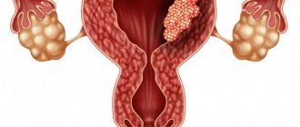
- internal organs (lung, intestines, gall bladder, appendix, etc.);
- genital organs (perineum, scrotum, penis, labia);
- areas of the face (noma);
- skin (bedsores);
- fetus
Causes of the disease
Based on established medical practice, the following list of causative factors is identified, the presence of which is a prerequisite for the onset of gangrene of the lower limb.
Atherosclerotic gangrene
Tissue necrosis, which occurs due to impaired conductivity of blood vessels, which makes it impossible to saturate cells with oxygen and nutrients. As a result of this process, they die and form necrotic lesions.
Infection
The entry into an open wound of purulent microbes, which were able to adapt to new environmental conditions, overcame the immune barrier and provoked acute inflammation with profuse suppuration, which turned into gangrene.
Diabetes
Patients suffering from elevated blood glucose levels are at high risk of developing dry gangrene. The fact is that sugar crystals, which are not extinguished by the hormone insulin, clog the smallest blood vessels and capillaries and impede blood circulation. As a result, a similar effect is formed as with atherosclerotic necrosis.
Thermal, chemical or radiation burn
Exposure of a limb to too high or low temperatures, aggressive chemicals or radiation from radioactive metals leads to a burn effect of varying degrees of severity and etiology, which subsequently ends in tissue death and the process of necrosis.
These are the main causes of gangrene, which are most often diagnosed in patients based on the results of a comprehensive examination of the body. Also, against the background of these pathological factors, in 70% of patients there is a decrease in the protective function of the immune system, which allowed the bacterial microflora to increase its population in the tissues of the leg to a critical level.
What is an intestinal hernia and how to treat it
VIDEO ON THE TOPIC: Intestinal necrosis - the parents of 12-year-old Denis from Kiev ask for help 03/20/19 Even ancient people faced such a disease as gangrene. Written sources describing this disease have survived to this day and they date back to the times of the ancient Greek physician Hippocrates. A manifestation of gangrene is the death of tissue in a living organism. Most often, doctors are faced with such types of diseases as gangrene of the extremities and intestinal gangrene, although this disease itself can occur in any human tissues and organs. Gangrene is very dangerous and quite often ends in death. The patient's death occurs rapidly due to intoxication by decomposition products and dehydration of the body.
Intestinal gangrene is a serious disease associated with impaired blood supply to the organ and subsequent tissue necrosis.
Lung gangrene
Lung gangrene
Abscess and gangrene of the lung are considered in pulmonology and thoracic surgery to be the most severe infectious destructive pulmonary processes. In the structure of nonspecific destructive lung diseases, gangrene accounts for 10-15%. It is known that lung gangrene develops much more often in middle-aged men. The danger of lung gangrene is associated with a high probability of multiple complications that can lead to the death of the patient: pleural empyema, phlegmon of the chest wall, pericarditis, pulmonary hemorrhage, sepsis, disseminated intravascular coagulation syndrome, respiratory distress syndrome, multiple organ failure.
Symptoms of gangrene
The manifestation of certain signs of gangrene depends on the form of the disease.
Dry gangrene
Dry gangrene usually occurs in patients with dehydration, as well as in malnourished patients. It develops slowly, sometimes over several years. Distally located areas (fingers, toes, feet) are primarily affected.
The first sign of developing gangrene is pain. At the initial stages, the pain is tolerable, but gradually the intensity of the pain increases and is not relieved by conventional analgesics. The pain worsens at night, and the patient takes a forced position in which the pain intensity is slightly less. Usually this is an elevated or, conversely, lowered position of the affected limb. With the development of the pathological process, due to loss of sensitivity in the area of necrosis, pain disappears, but some patients may experience phantom pain. The skin in the affected area turns pale, becomes cold to the touch, the affected limb becomes numb, and the pulse in the peripheral arteries is not detected. The necrotic area decreases in volume and darkens, acquiring a mummified appearance. Healthy tissues have a clear border with necrotic ones (demarcation shaft). An unpleasant odor is not typical for this type of disease. Dry gangrene is limited and does not spread to healthy areas with normal blood circulation. The patient's condition is usually stable, with the exception of cases when gangrene turns into a wet form.
The first sign of dry gangrene is pain
Wet gangrene
Wet gangrene develops quickly, due to an abrupt loss of blood supply to a certain area, often as a result of thrombosis or thromboembolism. Overweight patients are more susceptible to this form of the disease.
Gangrene: types, symptoms and diagnosis
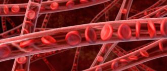
Gangrene is the death of part of your body tissue. This often occurs because the tissues are not receiving enough blood from your circulatory system.
Gangrene usually affects the extremities—the areas farthest from the heart, such as the fingers and toes. However, it can also affect other parts of your body. Gangrene can even affect your internal organs.
The condition usually starts in a specific part of the body, such as a leg, arm, or internal organ. Gangrene can spread throughout your body and cause shock if left untreated. Shock is a condition characterized by many symptoms, including low blood pressure. Shock can be life-threatening and is considered a medical emergency.
Gangrene is a medical emergency that can lead to amputation or death. Recognizing and treating this disease as quickly as possible will improve your outlook.
Dry gangrene
All of your organs (such as the liver, heart and muscles) need oxygen to function and survive properly. Oxygen is carried by the blood to different parts of your body. Dry gangrene occurs when one part of the body does not receive enough oxygen. Eventually, part of the body will begin to break down and die. In dry gangrene, the skin is closed and there are no signs of infection.
Wet gangrene
Wet gangrene occurs when the tissues of your body become infected with some type of bacteria. Fabrics react to the presence of bacteria by becoming damp and breaking down. This process causes the death of your tissue. It is more of an emergency than dry gangrene because of the possibility of the infection spreading to other parts of the body.
Gas gangrene
Clostridia bacteria cause gas gangrene. These bacteria cause an infection that causes gas bubbles and toxins to form inside the affected area. The resulting gases cause tissue death. This type of gangrene can be fatal, although it is rare in the United States.
You are more likely to develop gangrene if you have a history of certain medical conditions, including:
Certain other physical events may increase your risk of gangrene. You may be more likely to get this disease if you have:
- decreased immunity due to cancer disease or treatment
- recently had surgery
- suffered severe frostbite or head trauma, an animal bite, or a serious burn
- suffered injuries, including crushing of body tissue
- received an injection of promethazine hydrochloride, which resulted in tissue damage
Smoking, drinking alcohol and using intravenous drugs can also increase the risk of developing gangrene.
External gangrene
Sometimes the first sign of dry gangrene is the appearance of a reddish line around the affected tissue. This line may later turn black.
Other signs that may indicate gangrene include:
- red, painful, or swollen sore
- a wound that is filled with pus or has a foul odor
- feels cold in an isolated area of your body
- Lack of touch in isolated area
- Sores that keep coming back in the same place on your body
- Part of your skin has an unusual color (greenish-black, red, blue, or bronze)
Internal gangrene
Internal gangrene, which affects your internal tissues or organs, is also possible. In this case, you may not have any symptoms on your skin or limbs. However, you may have pain, an unexplained high temperature that lasts a long time, or low blood pressure. You may also experience confusion.
Your doctor may suspect gangrene based on your medical history and symptoms. They may also use a combination of additional diagnostic methods to determine your condition.
Laboratory analysis of tissue or fluid samples
Tissue scrapings from the affected part of the body can be examined under a microscope to look for dead cells.
Blood tests
An abnormally high white blood cell count may indicate a gangrenous infection.
Medical imaging
Some types of imaging help diagnose the spread of gangrene in internal tissues. These tests may include an X-ray, MRI, or CT scan.
An anarteriogram may be performed if doctors suspect your gangrene is due to a circulatory problem. This test uses X-rays to monitor the flow of a special dye through your arteries, showing whether any arteries are blocked.
Antibiotics
Your doctor may prescribe antibiotics if bacteria are present. They are usually given intravenously or through a needle directly into the bloodstream.
Vascular surgery
For people with poor circulation that leads to gangrene, vascular surgery (surgery on arteries or veins) may be recommended to improve the flow of blood through the veins to the body's tissues.
Hyperbaric oxygen chamber
Placing a person with gas gangrene in a special oxygen-rich environment can slow the growth of bacteria. This allows the skin to begin to heal. It also delivers oxygen to damaged tissue to speed up healing.
Fabric processing
In severe cases of gangrene, dead tissue or body part may need to be removed. This process is called wound debridement. Removal of the wound can be done with surgical instruments or chemicals. The purpose of this type of surgery is to remove the affected areas to prevent the spread of infection and rid the body of dead tissue.
One alternative form of wound debridement, known as maggot debridement, involves maggots eating away bacteria and dead tissue. Although this practice is rare, it is still used by doctors in the United States and abroad.
Sometimes doctors are able to restore oxygen flow to the affected area. Skin grafts can repair any damaged tissue. This procedure uses a piece of healthy skin from another area of the body to cover the damaged area.
Amputation
In severe cases, amputation of a limb, finger, or foot may be necessary to save a life. People who need to have part of an arm or leg amputated due to gangrene may be given artificial limbs or artificial limbs to replace the missing body part.
Sometimes gangrene can be treated without serious complications, especially if it is caught early. However, in some serious cases it can lead to amputation, especially if not treated quickly.
Gangrene can even be fatal for some people. This is rare, but can happen if:
- you have other serious health problems that complicate your treatment
- gangrenous area covers most of your body
- treatment is not carried out quickly enough
To save more tissue from dying, gangrene must be treated as early as possible to minimize damage. People with diabetes or blood vessel disease should regularly check their hands and feet for symptoms of gangrene. Pay attention to:
- any swelling, discharge or redness that may indicate infection
- a wound that doesn't seem to heal
- change in the color of your skin
Taking antibiotics before or after surgery, under your doctor's care, can help you prevent the development of gangrenous infections.
.
Characteristics of the disease
Diabetic gangrene
Gangrene of a finger or toe is necrosis, or death of living tissue, which appears as a result of a pathological process in the body. Under the influence of various factors, oxygen stops flowing to the leg, general blood circulation in the body worsens, which leads to tissue damage in large areas of the skin. If the symptoms are ignored, the affected areas begin to grow and affect healthy parts of the skin.
As gangrene develops, cells begin to die, which contributes to the further progression of the disease. The exact time of development of gangrene has not yet been established: the disease can develop slowly, gradually, or quickly go through all stages. During slow progression, cells gradually die off on the foot or toes, causing intoxication of the entire body and rapidly developing further.
Signs and treatment of gangrene of the leg in diabetes mellitus at the initial stage and subsequent ones
- The first symptoms of gangrene of the lower extremities
- Causes of gangrenous lesions
- Forms of the disease
- Diagnostic measures
- Treatment of gangrene in diabetes mellitus
- Treatment without amputation
- Surgical methods
- ethnoscience
Gangrene is a serious lesion of one or both lower extremities. The presented pathology quite often occurs in diabetics, and therefore it is important to know everything about treatment methods and other features. The initial stage of gangrene of the leg in diabetes mellitus and photos of all stages of the lesion deserve special attention.
The first symptoms of gangrene of the lower extremities
Gangrene in diabetes mellitus, even at the initial stage, is always associated with certain symptoms. First of all, we are talking about the rapid onset of leg fatigue even with short walking. Symptoms may also include sudden, spasmodic contractions. In addition, signs of gangrene in diabetes mellitus are freezing of the extremities, regardless of the time of year.
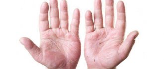
Another sign of pathology should be considered numbness of the toes and a general systematic loss of the optimal degree of sensitivity. Gangrene of the leg in diabetes mellitus may well be associated with the appearance of a waxy skin effect, in which the cover appears smooth and mirror-like.
In addition, ulcers will form on the skin that do not heal over a long period of time. Gradually they develop into an active gangrenous process. Taking this into account, it is recommended to take timely and full attention to the feet, their treatment and prevention.
Causes of gangrenous lesions
The first group of factors that provoke gangrene of the lower extremities in diabetes mellitus is associated with an unhealthy lifestyle. Pay attention to:
- absence or inadequate treatment of the underlying disease and related ailments;
- nicotine and alcohol addiction;
- wearing uncomfortable or tight shoes;
- neglect in the process of treating wounds, calluses, corns and other injuries.
In addition, gangrene sometimes begins due to overweight, obesity or frostbite. The second category of factors includes a decrease in the lumen in the area of blood vessels due to atherosclerosis or anemia. Gangrene in diabetes can develop under the influence of impaired regeneration processes and worsening immunity. Polyneuropathy, as well as problematic bone tissue formation, can also influence the presented process. This subsequently leads to osteoporosis and even necrosis.
Forms of the disease
There are several options for classifying pathology: depending on the nature of the dying tissue, according to the mechanism of development of the pathology, and the cause of the condition. In the first case, we are talking about the dry or wet variety, in the second - gas, hospital and fulminant (the most dangerous form, there is a high probability of losing a limb). In addition, diabetic gangrene of the lower extremities, as already noted, may be toxic, infectious or allergic.
According to experts, the development of dry gangrene is most often identified. Regardless of the specific form, it is important to start therapy as early as possible and ensure effective and correct treatment.
Diagnostic measures
In the vast majority of cases, a visual examination and examination of a diabetic’s complaints is more than enough to determine the diagnosis. In order to clarify the stage of pathology and identify the degree of susceptibility of pathogenic bacteria to antibiotic components, certain laboratory tests are prescribed:
- complete blood test - a decrease in the leukocyte ratio should be considered a symptom of worsening immunity;
- biochemical blood test - allows you to diagnose the initial stage of renal failure;
- complete urine analysis - with the development of anaerobic gangrene, either protein or glucose is noted in the urine;
- Bacterial seeding from the problem area is carried out within one week.
To confirm the diagnosis of gangrene of the foot, a Bethe test can be performed. It is useful for identifying the type of gangrene. For this purpose, a piece of tissue is removed from the affected area and placed in a 4–6% sodium chloride solution. If the sample floats, this should be considered a sign of anaerobic gangrene.
In a similar way, specialists interpret the information obtained from the results of an x-ray of the problem limb. If gas bubbles are present in the image, it means that we are talking about anaerobic gangrene. Even if the pathology is at the initial stage of its development in diabetes mellitus, it is recommended to start treatment as early as possible.
Treatment of gangrene in diabetes mellitus
Various techniques can be used to treat gangrene. Therapy without amputation is practiced; specialists resort to surgical intervention. In rare cases, we can talk about folk recipes.
In order for a particular method to be as effective as possible, it is very important to coordinate therapy with a specialist and not self-medicate.
Treatment without amputation
Therapy without amputation is a drug-assisted approach. Intravenous administration of a glucose solution of physiological composition is acceptable; plasma or even blood substitutes can be used. For restorative purposes, antibiotic injections may be prescribed. Gangrene can be treated through the use of various heart medications.
The use of novocaine blockades deserves special attention, which eliminates vascular spasm. Speaking about treatment without surgery, pay attention to:
- use of vitamins, anticoagulants and diuretics;
- use of drugs that have thrombolytic effects;
- carrying out a procedure such as blood transfusion according to indications.
Diagnostics
The diagnosis is usually not difficult due to the characteristic visual signs of the disease. To confirm it, the following methods are used:
- general blood test (there is an increase in the number of leukocytes, a decrease in erythrocytes and hemoglobin, and the absence of eosinophils);
- blood chemistry;
- microscopic examination of discharge from the wound;
- cultural examination of pathological discharge from the affected area;
- ultrasound duplex scanning of blood vessels;
- X-ray examination (with gas gangrene, intermuscular gas accumulations in the image look like “herringbones”; this phenomenon is called Krause’s symptom).
Differential diagnosis is carried out with putrid infection and fascial gas-forming phlegmon.
Diagnosis of lung gangrene
Diagnostic tactics for lung gangrene involves comparing clinical and anamnestic data, results of laboratory and instrumental studies.
When examining a patient with gangrene of the lung, attention is drawn to the general serious condition, adynamia, pale earthy skin tone, cyanosis of the lips and fingers, weight loss, and sweating. The lag of the affected half of the chest from the healthy half in the act of breathing, shortening of the percussion sound over the pathologically changed area of the lung, and increased vocal tremors are determined. On auscultation, with gangrene of the lung, various types of dry and moist rales, crepitus, and amphoric breathing are heard.
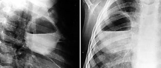
X-ray of the lungs in 2 projections reveals extensive darkening (a decay cavity of heterogeneous density) within a lobe with a tendency to spread to adjacent lobes or the entire lung. Using a CT scan of the lungs, tissue sequestration of various sizes is determined in large cavities. With gangrene of the lung, pleural effusion quickly forms, which is also clearly visible with fluoroscopy of the lungs and ultrasound of the pleural cavity.
Microscopic examination of sputum in lung gangrene reveals a large number of leukocytes, red blood cells, Dietrich plugs, necrotic elements of lung tissue, and the absence of elastic fibers. Subsequent bacteriological culture of sputum and bronchoalveolar lavage fluid makes it possible to identify pathogens and determine their sensitivity to antimicrobial drugs.
Bronchoscopy reveals signs of diffuse purulent endobronchitis; sometimes - obstruction of the bronchus by a foreign body or tumor. Shifts in the peripheral blood indicate a pronounced inflammatory process (increased ESR, neutrophilic leukocytosis, anemia). Changes in the biochemical profile of the blood are characterized by severe hypoproteinemia; Significant changes in lung gangrene are observed in the gas composition of the blood (hypercapnia, hypoxemia).
Prevention
In order to prevent gangrene, it is recommended to avoid factors that provoke its development. Men should avoid injuring the scrotum and observe personal hygiene rules to prevent infection from entering the body. It is also recommended to promptly treat diseases of the genitourinary system and monitor blood sugar levels.
Insufficient knowledge of the disease and the development of many complications make Fournier’s gangrene a rather dangerous disease that requires complex therapy. The study of predisposing factors for the development of pathology is the subject of further research aimed at developing effective methods of treatment and prevention of this serious dangerous disease.
Forecast and prevention of lung gangrene
Despite the successes of thoracic surgery, mortality in lung gangrene remains high - at the level of 25-40%. Most often, the death of patients occurs due to pneumogenic sepsis, multiple organ failure, and pulmonary hemorrhage. Only timely initiation of complex intensive therapy, supplemented, if necessary, with radical surgery, can count on a favorable outcome.
Prevention of lung gangrene is a complex medical and social task, which includes measures for health education, improving the standard of living of the population, combating bad habits, and organizing timely medical care for various infectious and purulent-septic diseases.
LiveJournal
Treatment of gangrene
Treatment of gangrene is carried out in a hospital setting and includes both general and local measures. Since gangrene is the death of tissue, the main goal of treatment is to preserve it and prevent further development of necrosis.
Bed rest is recommended for patients with gangrene. Conservative treatment is aimed at stimulating blood circulation, improving tissue trophism, and eliminating symptoms. Due to severe pain, taking analgesic drugs (non-narcotic or narcotic) is indicated for any form of the disease. If thrombosis is diagnosed, thrombolytics are prescribed. It may be necessary to carry out novocaine blockades, which eliminates spasm of collateral vessels; in some cases, a blood transfusion is required. If necessary, bypass surgery and stenting of blocked blood vessels, as well as vascular prosthetics, are performed.
Active measures to normalize blood circulation in the affected area make it possible to preserve it in the ischemic form of gangrene.
Novocaine blockades help eliminate pain during gangrene
With dry gangrene, self-amputation of the affected area may occur; in other cases, amputation is performed surgically after the formation of a demarcation shaft. The level of amputation is selected in such a way as to provide optimal conditions for healing of the stump while maintaining the maximum possible function of the affected limb. Wound healing occurs by primary intention. After complete formation of the stump, limb prosthetics is possible.
The prognosis for dry gangrene is favorable regarding the patient’s life, but unfavorable regarding the preservation of the affected area. Wet and gas forms of gangrene often have a lightning-fast course, which requires urgent surgical treatment.
In case of wet gangrene, excision of necrotic tissue (necrectomy) or amputation of the affected limb is indicated, which is carried out on an emergency basis. After cleansing the wound, a stump is formed. The main treatment can be supplemented with a course of antibiotic therapy to eliminate the infectious agent.
Gangrene of internal organs is an indication for emergency surgery with removal of the necrotic area or organ.
For gas gangrene, the affected limb is placed in a pressure chamber with high oxygen pressure (hyperbaric oxygenation method), which has a detrimental effect on anaerobic pathogens of the disease.
For gangrene of the lung, antibiotics and antiseptics are usually injected into the bronchi using a bronchoscope. Also used are drugs that dilate the bronchi (inhalation or parenteral), immunomodulators, and restoratives. Resection of part of the lung or amputation is indicated if there is no positive effect from drug therapy.
Classification of lung gangrene
According to the mechanism of development, the following forms of lung gangrene are distinguished: bronchogenic (post-pneumonic, aspiration, obstructive); thromboembolic; post-traumatic; hematogenous and lymphogenous.
Based on the degree of involvement of the lung tissue, lobar, subtotal, total and bilateral gangrene of the lung is distinguished. A number of authors consider segmental lesions of the lung as a gangrenous abscess. In clinical practice, there is a combination of gangrene and abscess of different lobes of one lung, gangrene of one lung and abscess of the other.
Taking into account the stage of the destructive process during lung gangrene, atelectasis-pneumonia, necrosis of the pulmonary parenchyma, sequestration of necrotic areas, purulent melting of necrotic areas with a tendency to further spread (lung gangrene itself) are distinguished.