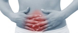
Symptoms of appendiceal infiltrate
The infiltrate develops 2-3 days after the onset of appendicitis, while the clinical picture of acute appendicitis itself is erased - there is no severe pain, general health does not suffer.
Upon examination, a dense, voluminous, painful formation is determined in the right iliac region. Body temperature rises to 38.5 degrees.
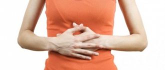
Appendiceal infiltrate has two development paths - abscess formation (suppuration of the infiltrate) and reverse development with “resorption” of the inflammatory tumor.
Abscess formation of the appendicular infiltrate is manifested by an increase in the inflammatory reaction: body temperature rises to 39-40 degrees, symptoms of general intoxication appear, pain in the right iliac region intensifies.
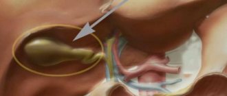
Gallbladder infiltration - what is it?
In the structure of acute diseases of the abdominal organs, it ranks second after acute appendicitis. In 95% of cases, it develops due to stone obstruction of the neck of the gallbladder or cystic duct. In other cases, the genesis is vascular (primary gangrenous cholecystitis), enzymatic or allergic. The pathogenesis is caused by overstretching of the gallbladder, impaired blood circulation in the vessels of the wall. Later, the microflora is activated, penetrating through the enterogenous, lymphogenous and hematogenous routes. In case of destructive cholecystitis, aerobic as well as anaerobic non-clostridial flora are detected in the gallbladder bile.
Local changes occur in the following sequence:
- Obstruction of the cystic duct;
- Increased pressure in the gallbladder;
- Stasis in the vessels of the gallbladder;
- Bacteriocholia;
- Destruction of the gallbladder wall (with or without perforation);
- Perivesical infiltrate;
- Local peritonitis (perivesical abscess);
- Diffuse peritonitis.
According to morphology, catarrhal (simple), phlegmonous, and gangrenous cholecystitis are distinguished. In elderly patients, against the background of widespread atherosclerosis and/or hypercoagulation, primary gangrenous cholecystitis occurs (due to thrombosis or embolism of the cystic artery).
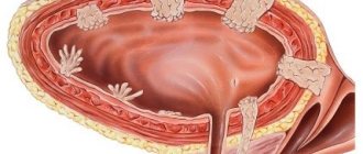
The clinical picture of acute cholecystitis is presented by the following symptoms: pain, fever, bitterness in the mouth, nausea, vomiting. With simple acute cholecystitis, the abdomen is soft; destructive cholecystitis is characterized by muscle deflation in the right hypochondrium. Symptoms of Murphy, Kehr, Ortner, Mussi are frequent, and later peritoneal symptoms appear (Shchetkin - Blumberg, Voskresensky). A fairly common symptom of acute cholecystitis is a palpable, painful fundus of the gallbladder or a palpable perivesical infiltrate. Laboratory findings show leukocytosis. The main method of instrumental diagnosis is ultrasound. The leading ultrasound signs of acute cholecystitis are: an increase in the size of the gallbladder, thickening of the wall, double-circuiting of the wall, the presence of infiltrate and fluid in the subhepatic space and abdominal cavity.
Surgical tactics for acute cholecystitis.
- All patients with acute cholecystitis must be hospitalized in a surgical hospital and treated as patients with acute surgical pathology of the abdominal organs.
- In case of acute cholecystitis complicated by peritonitis, emergency surgery is indicated in the first 2 hours.
- In the absence of peritonitis, conservative treatment (analgesics, antispasmodics, infusions, antibiotics) is indicated for 24-72 hours.
- If there is an effect and the acute process is stopped, a planned operation is indicated after 7-12 or more days. If the attack was stopped in a short time, the planned operation can be performed earlier, after 2-3 days.
- If there is no effect from conservative treatment, urgent surgery is indicated.
- If the risk of surgery is high, especially in elderly patients with destructive cholecystitis, palliative surgery – cholecystostomy – is possible.
The pathogenetic rationale for cholecystostomy is decompression of the gallbladder, which eliminates the disruption of intrawall blood flow, preventing further progression of destructive changes. Cholecystostomy can be performed by puncture percutaneous transhepatic drainage of the gallbladder under ultrasound control, laparoscopy and laparotomy. Local anesthesia is used, but the intervention is carried out under the supervision of an anesthesiologist.
Mortality in acute cholecystitis in case of emergency surgery is 2-3%, in case of emergency surgery it can exceed 4-6%.
For standards of diagnosis and treatment of acute cholecystitis, see the Appendix.
OTHER COMPLICATIONS OF GSD
Perivesical infiltrate. Forms on the 3-4th day of the disease. It is characterized by the presence in the right hypochondrium of a dense formation with clear boundaries. Infiltration is an indication for conservative treatment. Complete resorption of the infiltrate with drug therapy occurs after 1.5 - 2 weeks. With a virulent infection, the formation of a perivesical abscess is possible. With further progression, a subhepatic abscess is formed. If there is an abscess, the cavity is sanitized and cholecystectomy is performed. The operation ends with packing of the abscess cavity.
Hydrocele of the gallbladder is the outcome of obstruction with low-virulent flora. At the same time, the absorption of the constituent parts of bile occurs in the gallbladder, the bacteria die, and the contents become colorless and mucous in nature. It can exist for a long time without clinical symptoms or with minor manifestations. In some cases, an enlarged gallbladder is palpable. Treatment: cholecystectomy as planned.
Empyema of the gallbladder. It is an infected dropsy. In some cases, with conservative treatment of acute phlegmonous cholecystitis, chronic empyema is formed. Clinic: nagging pain in the right hypochondrium, occasional increases in body temperature. In some cases, palpation can detect an enlarged, moderately painful formation in the right hypochondrium. Treatment: cholecystectomy.
Bile duct strictures. The main cause is choledocholithiasis. There are high and low, limited and widespread. Often develop after intraoperative damage to the common bile duct. Clinical picture: transient obstructive jaundice. Cholangiography reveals concentric narrowing of the duct with prestenotic expansion. Treatment: choledochojejunostomy on a Roux-enhanced loop. The results are often unsatisfactory - recurrence of stricture formation.
Stricture (stenosis) of the large duodenal nipple. It occurs mainly when small stones pass through the major duodenal papilla (MDP), which leads to fibrous and scarring changes.
There are 3 degrees of stenosis of the abdominal joint:
1 - without functional impairment (compensated);
2 — narrowing to 3 mm with moderate expansion of the ducts (subcompensated);
3 - severe stenosis with cholestasis - complete block (decompensated).
In addition to choledocholithiasis, the causes of stenosis can be lesions of the pancreas, cholangitis, damage to the abdominal joint during surgery, duodenal ulcer, parafateral diverticula, etc.
Treatment is surgical. Various options for internal drainage are used: papillosphincterotomy (endoscopic, transduodenal, endocholedocheal, combined), choledochoduodenostomy, choledochojejunostomy.
Choledocholithiasis.
Choledocholithiasis occurs when gallstones pass from the bladder into the common bile duct or, much less frequently, when they form directly in the common bile duct. During cholecystectomy it is detected in 4-12% of cases. Clinic: severe attacks of pain in the epigastric region and right hypochondrium; after an attack, jaundice usually appears. At the same time, even with severe obstructive jaundice against the background of choledocholithiasis, in some cases there may be no pain syndrome. Jaundice can be remitting in the presence of a valve stone. In some cases, choledocholithiasis can be asymptomatic for a long time, and in 40% of cases it occurs without jaundice. In 4-5% of cases of choledocholithiasis, there are no gallstones. Main diagnostic methods: ultrasound, endoscopic retrograde cholangiopancreaticography (ERCP), percutaneous transhepatic cholangiography (PTCH), intravenous cholecystocholangiography (in the absence of obstructive jaundice). Ultrasound reveals stones in the bile ducts in an average of 75–80% of cases. The most reliable method is ERCP, however, its use limits the risk of developing acute pancreatitis.
Treatment of appendiceal infiltrate
Abscess formation of the infiltrate is an indication for emergency surgery. In the absence of abscess formation, conservative therapy is carried out in a hospital setting, followed (after 2-3 months) by a planned appendectomy.
Essential drugs
There are contraindications. Specialist consultation is required.
- Ceftriaxone (an antibiotic of the third generation cephalosporin group). Dosage regimen: IM, IV, adults and children over 12 years of age, the average daily dose is 1-2 g of ceftriaxone once a day or 0.5-1 g every 12 hours. In severe cases or in cases of infections caused by moderately sensitive pathogens, the daily dose can be increased to 4 g.
- Cefepime (IV generation cephalosporin antibiotic). Dosage regimen: adults and children weighing more than 40 kg with normal renal function 0.5-1 g intravenously (for severe infections up to 2 g) or deep intramuscularly at intervals of 12 hours (for severe infections - after 8 hours ).
- Metronidazole (antiprotozoal, antibacterial agent). Dosage regimen: IV for adults and children over 12 years of age, a single dose is 0.5 g. The rate of IV jet or drip administration is 5 ml/min. The interval between injections is 8 hours.
- Amoxiclav (broad-spectrum bactericidal antibacterial agent). Dosage regimen: intravenously, adults and children over 12 years of age or weighing more than 40 kg - 1.2 g of the drug (1000 + 200 mg) with an interval of 8 hours, in case of severe infection - with an interval of 6 hours.
- Tienam (antimicrobial, bactericidal, antibacterial agent). Dosage regimen: IV, by infusion: ≤ 500 mg - over 20-30 minutes, > 500 mg over 40-60 minutes. The average daily dose is 2000 mg (4 injections). The maximum daily dose is 4000 mg (50 mg/kg). The dose is adjusted taking into account the severity of the condition, body weight and renal function of the patient.
- Vancomycin (antibacterial, bactericidal agent). Dosage regimen: adults, 0.5 g intravenously every 6 hours or 1.0 g every 12 hours. Infusion duration is at least 60 minutes, rate is 10 mg/min.
Resection of infiltrate
3-4 hours
. Discharge one day after the operation. The price of the operation is from
100,000
rubles.
Description of the operation: Resection of infiltrate
Resection of infiltrate
Laparoscopy. Removal (excision) of endometrioid infiltrate (EI).
According to leading experts involved in endometriosis, the infiltrate that manifests itself must be removed. Namely, the indication for surgery is pain syndrome that is not amenable to drug therapy and infertility. The second indication is questioned by some experts. Asymptomatic infiltrates do not require surgical treatment.
This approach is due to the risk of severe intra and postoperative complications:
- inability to empty the bladder on your own
- ureteral injury
- rectal trauma with the development of peritonitis
- bleeding
- in rare cases, complete or partial paralysis of the lower extremities.
The operation begins with the classic entrance to the abdominal cavity:
- laparoscope (video camera) in the navel (10 mm trocar)
- three trocars in the lower abdomen 5 mm.
The next stage is creating conditions for comfortable work:
- separation of adhesions (they often accompany endometriosis)
- suspension of the appendages (ovaries and fallopian tubes are pulled to the anterior abdominal wall using a special T-Lift system. This is necessary for good access and visualization of the operation area.
After creating the conditions, the actual removal of the endometrioid infiltrate is carried out. Endometrioid infiltrate is usually located between the vagina and rectum and ureters. Favorite localization is the uterosacral ligaments. They stretch fan-shaped from the sacrum to the cervix, on the sides of the rectum.
Excision of endometrioid infiltrate can be divided into two main stages:
- Mobilization or so-called centralization of infiltrate
This is a technically very difficult stage. Its purpose is to separate EI from neighboring organs. In order to safely remove EI, it must be separated from the ureters and rectum. There is a standardized surgical technique that makes it possible to localize EI with minimal risk for its subsequent cutting off.The ureters are found in a place that is not involved in the pathological process (above the infiltrate) and are sequentially separated from the infiltrate from top to bottom. This approach allows you to keep the ureters under visual control, which significantly reduces the risk of injury. If it is impossible to separate the ureter from the infiltrate (the ureter is affected by endometriosis), part of the ureter is removed within its healthy tissue and an anastomosis is formed between the ureter and the bladder.
The rectum is separated from the posterior wall of the uterus and the uterosacral ligaments.
- Excision of EI itself
At this stage, the endometrioid infiltrate is cut off from the vaginal wall and rectum. A particularly difficult and dangerous step is separating the EI from the rectum. This stage is called “shaving” from the English word shaving (shaving).To exclude rectal injury, a so-called “bubble” test is sometimes performed at the end of the operation. The pelvis is filled with saline solution, and air is introduced into the rectum. If there is a penetrating injury to the rectum, bubbles appear in the water filling the pelvis.
In rare cases (if there is deep involvement of the ureter of the rectum or vaginal wall), it is necessary to excise part of the ureter of the vaginal wall or rectum (rectal resection).
EI is removed from the abdominal cavity.
Preparing for surgery
Before hospitalization, the patient must undergo a standard examination. If there are concomitant diseases that may affect the operation and postoperative period (diabetes mellitus, coronary heart disease, varicose veins of the lower extremities, etc.), it is necessary to undergo additional examinations and/or consultation with other specialists. A list of standard examinations is listed here.
What you need to take with you to the hospital: passport, compulsory medical insurance policy, tests, robe (or cotton suit), slippers, nightgown, pads (2-3 drops), hygiene items, stockings for the prevention of varicose veins of the lower extremities (1 degree of compression).
The patient is hospitalized on the day of the planned operation.
The day before going home, you need to shave your perineum and pubis. Preparation of the colon (cleansing enema or special laxatives) is necessary only before surgery for infiltrative endometriosis with suspected rectal involvement.
On the day of surgery, do not drink or eat anything in the morning. Before the operation, the patient is examined by an anesthesiologist.
List of examinations required for hospitalization:
You can download the exact list of tests and examinations in PDF
.
Rehabilitation
Depending on the severity of the operation, the patient remains in a horizontal position for several hours to a day. Most often in the evening you can get up. You cannot eat on the day of surgery, you can drink 2 hours after surgery. You can turn from side to side. Urine is drained through a urinary catheter. The next day, the urinary catheter is removed and the patient is allowed to walk and eat. The next day or one day after the operation, the patient is discharged home. There is no special diet. For two weeks, refrain from aggressive foods (fried, smoked, spicy, salty, fast food.)
Physical rest for 2 weeks after surgery.
Limiting physical activity means:
- do not exercise
- Do not lift weights exceeding 5 kg.
- Don't push on the toilet. Normal (ordinary) straining is acceptable. To maintain normal stool, follow a diet and, if necessary, use laxatives.
This operation corresponds to:
- Diseases Endometriosis
- Small pelvis
Incidence (per 100,000 people)
| Men | Women | |||||||||||||
| Age, years | 0-1 | 1-3 | 3-14 | 14-25 | 25-40 | 40-60 | 60 + | 0-1 | 1-3 | 3-14 | 14-25 | 25-40 | 40-60 | 60 + |
| Number of sick people | 0.01 | 2 | 2 | 2 | 2 | 2 | 2 | 0.01 | 2 | 2 | 2 | 2 | 2 | 2 |
Types of pathology
Depending on the depth of inflammation, keratitis is divided into superficial, which covers about 1/3 of the thickness of the cornea, and deep - inflammation reaches the stroma (the base of the cornea).
Depending on the location of the infiltrate, keratitis is:
- central – infiltrated tissue is located in the pupil area;
- paracentral - the infiltrate is located in the zone of the iris;
- peripheral - inflammation is localized in the limbus (edge of the cornea).
Classification of keratitis depending on the cause of occurrence:
- Internal keratitis is provoked by pathogens, allergic reactions, autoimmune processes, and vitamin deficiency. This group includes keratitis, the origin of which is unknown (filamentous keratitis or rosacea).
- External keratitis is provoked by injuries, viruses, keratomycosis, anomalies that appear due to the proliferation of bacteria due to conjunctivitis or meibomitis.
Abscess and infiltrate - what is the difference?
When the skin is injured due to surgical operations or inflammatory pathologies, complications arise. An infection enters the body and an abscess and infiltrate are formed. The latter is the accumulation of cellular elements in the tissue mixed with blood and lymph. Despite the common etiology and pathological anatomy, these are two different pathological processes. A soft tissue abscess differs from an infiltrate in the following: The presence of fluid in a closed cavity. In an abscess, the fluid is purulent exudate; in an infiltrate, there is no cavity at all, the tissue is saturated with decay products of the inflammatory process. Infiltrate can arise from tumor cells, and an abscess is caused only by pathogenic microorganisms. Infiltration can lead to the formation of an abscess, but the opposite does not happen.