Features of development (pathogenesis)
The development of suppuration in the lungs is caused by a mixed flora, usually coccal. These are pneumococci, staphylococci, streptococci. Sometimes the disease is caused by autoinfection of microbes, which are saprophytes of the upper respiratory tract.
Such microbes, penetrating the lungs, become pathogenic. They cause suppurations of all kinds. The manifestation of suppuration depends on the response of the patient’s body and the reactivity of the lung tissue.
Microorganisms enter the body in the following ways:
- Hematogenous. This is sepsis, that is, the pathogen spreads through the bloodstream. When foci of pus develop in various organs, the movement of bacteria through the blood vessels is called septicopyemia.
- Lymphogenic. This route is possible if the infectious focus is in the lungs and nearby tissues. In this case, microbes are transported by lymphatic flow.
- Bronchogenic. Microorganisms pass through the bronchi or are found in the mouth, from where they enter the organs.
Causes and risk factors
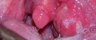
Typically, a lung abscess is caused by bacteria that normally live in the mouth or throat and are inhaled (aspirated) into the lungs, causing an infection. Often the source of bacteria that causes a lung abscess is gum disease (periodontal disease).
The human body has many ways of protecting itself (for example, coughing) from bacteria entering the lungs. Infection mainly occurs when a person is unconscious or very drowsy due to the use of sedatives, anesthesia, alcohol or drug abuse, or nervous system disease.
In people with weakened immune systems, a lung abscess can be caused by microorganisms not normally found in the mouth or throat, such as fungi or Mycobacterium tuberculosis (the microorganism that causes tuberculosis). Other bacteria that can cause lung abscesses are streptococci and staphylococci, including methicillin-resistant Staphylococcus aureus (MRSA), which causes a serious infection.
Blockage (obstruction) of the airways can also lead to the formation of an abscess. If the branches of the windpipe (bronchi) are blocked by a tumor or foreign body, secretions (mucus) can accumulate behind the obstruction, causing an abscess to form. Sometimes bacteria get into such secretions. The obstruction prevents secretions and bacteria from being coughed up through the airways.
Less commonly, abscesses occur when bacteria or infected blood clots are carried in the bloodstream to the lungs from another infected area of the body (septic pulmonary emboli).
Causes of the disease
Fungi and pyogenic bacteria act as etiological factors of the disease.
Lung abscess begins due to:
- tuberculosis;
- bronchiectasis;
- bacterial pneumonia;
- sepsis;
- gangrene or pulmonary infarction;
- purulent diseases of the dentofacial apparatus;
- aspiration of gastric mass during vomiting.
Inflammation acts as a secondary ailment. It signals that an endogenous type of infection is present in the body. Primary abscess appears rarely. In this situation, microorganisms penetrate from the environment.
What types of disease
There is a classification of lung abscess according to the route of infection. This is a traumatic, hematogenous and bronchogenic species.
There is also a division according to the form of flow:
- Spicy. It shows up brightly. The formation of an abscess occurs after the growth of the affected tissue. There are many microorganisms in it and pus. The disease can go away on its own, but becomes chronic.
- Chronic. It has a sluggish and unexpressed manifestation.
Classification
Lung abscesses are classified according to:
- cause of occurrence (type of microorganism that caused purulent inflammation);
- pathogenetic picture;
- the presence of one or more ulcers;
- by location: central or peripheral.
According to the flow, abscesses are divided into two types:
- Acute - arose less than two months ago, has a thin pyogenic membrane, has opened or not opened into the bronchus;
- Chronic - existing for more than two months, having a thick capsule with proliferation of connective tissue along the periphery, usually periodically drained by the bronchus.
For reference. An acute abscess does not necessarily become chronic. If the cavity is completely emptied, the process can be resolved.
The pyogenic membrane gradually resolves, and a patch of connective tissue forms at the site of the abscess. If the cavity was small in diameter, after it has healed, no residual effects may be visible. If the cavity was large, an area of fibrosis remains.
Chronic lung abscess is more dangerous; it has a protracted course and can exist for decades.
For reference. Chronic lung abscess is characterized by a recurrent course. The cavity is either filled with pus or emptied. Exacerbations may occur several times a year.
Each exacerbation is accompanied by the same symptoms that are characteristic of an acute abscess. At the same time, connective tissue gradually grows around the pathological focus, forming coarse fibrous cords.
Attention. The danger of the chronic course of the disease lies in the possibility of developing respiratory failure.
A long-term pathological focus in the lung leads to disruption of the functioning of the bronchopulmonary system. Symptoms such as constant shortness of breath, exercise intolerance, and cyanosis of the skin develop. Over time, the cardiovascular system also begins to suffer, which can lead to heart failure.
The presence of a cavity in the lung for a long time creates a risk of damage to the pleura. Any trauma to the chest can lead to rupture of the abscess wall and the formation of pneumothorax, which will have to be treated surgically.
For reference. Another danger is the development of complications associated with the long-term existence of a source of chronic infection in the body. These complications include amyloidosis of parenchymal organs, decreased immunity, and the gradual development of multiple organ failure.
A generally accepted classification of the disease has not yet been developed. The main ways of dividing it according to various criteria are presented in the table below.
| Classification | Description | Possible types |
| Etiological | Based on the isolation of the infectious agent | Pneumococcal |
| Staphylococcal | ||
| Streptococcal, etc. | ||
| Pathogenetic | Based on how the lung tissue became infected | Bronchogenic |
| Hematogenous | ||
| Post-traumatic | ||
| Topographical | Based on assessment of the location and number of purulent cavities | Central |
| Peripheral | ||
| Unit | ||
| Multiple | ||
| Unilateral | ||
| Double sided |
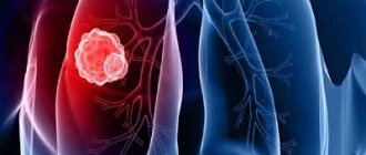
The photo shows a macropreparation with a fresh decay cavity
Risk group susceptible to the disease
There is a certain risk group of people who may be affected by this disease. These include those who suffer from alcohol and drug addiction.
The disease can also affect patients suffering from:
- diabetes mellitus;
- epilepsy;
- sinusitis;
- lung cancer;
- immunodeficiency;
- gastroesophageal reflux.
There is a possibility of getting sick after operations on the esophagus and stomach, if there are foreign objects in the respiratory tract.
Symptoms of a lung abscess
Lung abscess is a disease that is accompanied by respiratory and infectious manifestations. When the disease begins to develop, respiratory symptoms can be observed. These are weakness, fatigue, muscle aches, and fever. They are complemented by severe poisoning of the body with elevated temperature.
Respiratory manifestations look like a cough with sputum. Its quantity is affected by the size of the purulent focus and bronchus. The more extensive they are, the more phlegm comes out, sometimes up to a liter of foul-smelling greenish-yellow phlegm a day.
Sometimes other symptoms are observed, for example, severe pain in the sternum. It begins when the abscess is near the pleura. Also, before the pus breaks into the bronchi, a dry cough may begin.
The general infectious syndrome is expressed at the beginning of the disease, when the abscess is closed. Then the outflow of pus begins, and intoxication is greatly reduced, and the temperature gradually approaches normal levels.
Symptoms and signs
Symptoms usually develop slowly. However, depending on the cause of the abscess, symptoms may appear unexpectedly. Early symptoms include:
- fatigue;
- loss of appetite;
- sweating at night;
- fever;
- cough with sputum production.
The sputum may have an unpleasant odor (this is typical of bacteria from the mouth and throat) and may contain streaks of blood. In addition, the patient may experience chest pain when breathing, especially if the outer lining of the lungs and the inner lining of the chest wall (pleura) are inflamed. Many patients seek medical help only several weeks or even months after the first appearance of such symptoms. Such people develop chronic abscesses and, in addition to other symptoms, experience significant weight loss, daytime fever, and night sweats. Lung abscesses caused by Staphylococcus aureus, or MRSA, can be fatal within days, sometimes even hours.
Complications of the disease
An abscess in the lung can lead to complications. They occur in those who do not adhere to the regimen, are treated at home, and also with poorly selected medications. Often complications manifest themselves in the form of the disease becoming chronic, a purulent process on the other side, hemoptysis, accumulation of gases and pus in the pleural cavity.
There are also more rare complications. These include sepsis, thrombosis of the arteries and veins of the lungs, and the development of abscesses in other places. Any of the listed diseases greatly worsen the patient’s condition, cause great discomfort and interfere with the treatment process.
If treatment is not started in a timely manner, the following manifestations can be observed:
- oxygen deficiency in the respiratory system;
- bleeding in the lung;
- penetration of pus into the pleural cavity;
- emphysema;
- neoplasms;
- infection of other systems and organs;
- deformation of the bronchi.
Sometimes an abscess is the outcome of pneumonitis or pneumonia. Then it is not a complication, but the result of another disease.
Symptoms
The clinical picture of a lung abscess depends on the form and stage of the disease. In the acute phase, symptoms differ before and after opening the abscess. The abscessation process lasts on average a week and a half and is accompanied by severe manifestations.
Patients suffer from fever, chills, sweating and shortness of breath. The cough is dry and insignificant at first, then becomes annoying and is accompanied by chest pain. When you press on the chest in the area of inflammation, severe discomfort is felt. If the purulent cavity is shallow, hard breathing and moist rales can be heard. When infected with aerobic bacteria, the symptoms are more pronounced.
A couple of days before opening the cavity, a small amount of mucus and pus is released, and hemoptysis is possible. There is an unpleasant odor from the mouth. A breakthrough of the abscess is accompanied by copious discharge of yellow-green sputum. You may expectorate brown mucus. It comes out full of mouth, 250-400 ml of pus and mucus are released per day. In some cases, the volume of discharge reaches a liter. The patient's general condition improves, the fever subsides.
Sputum from a lung abscess is stratified. If you hold it in a container, at the bottom there will be a thick and dense layer of gray color with particles of lung tissue. Next comes liquid sputum mixed with saliva. The surface of the mass is foamy and consists of serous fluid.
Symptoms of chronic lung abscess depend on the number of cavities, the size of the capsules, and the degree of pneumosclerosis. The clinical picture may be blurry and consist of a cough with sputum and a slight increase in temperature. In other cases, the patient's condition is more severe. They experience hemoptysis and secrete a large amount of foul-smelling purulent mucus. At the same time, signs of fever, chills, shortness of breath, and sweating remain. Patients lose weight. Increased fatigue is noted.
External signs include changes in the hands. The phalanges thicken, the fingers become like drumsticks. Nails take on the appearance of watch glass. Patients also develop an unhealthy blush.
The main tool for diagnosing lung abscess is radiography.
How is an abscess diagnosed?
If an abscess is suspected, diagnostics are carried out in full. The patient donates urine and blood. If there is a disease in the blood, there will be a lot of leukocytes, ESR, and toxic granularity of neutrophils. When the next stage of the disease develops, the indicators improve.
In the chronic form, hemoglobin decreases significantly, biochemistry changes - albumin decreases, and fibrins, haptoglobins, and seromucoids increase. Urine examination shows changes in the level of microhematuria and albuminuria. If the diagnosis is confirmed, then a sputum examination is performed.
Be sure to check availability:
- tuberculosis bacteria;
- atypical cells;
- fatty acids;
- elastic fibers.
Which pathogen provoked the disease is determined by sputum bacterioscopy. Determine reaction and susceptibility to antibiotics. A quick and effective diagnostic method is fluoroscopy. When there are difficulties in performing the procedure, magnetic resonance and computed tomography are performed.
Diagnostics
Auscultation of the lungs
- lag of the diseased half of the chest during the act of breathing;
- pain when palpating the intercostal spaces, which indicates a reaction to the pathological process of the intercostal pleura - the membrane that lines the chest cavity;
- vocal tremors intensify;
- auscultation shows a weakening of normal (vesicular) breathing and the appearance of a bronchial tint on the affected side;
- tapping shows dullness of percussion sounds; normally, the sound above the surface of the lungs is clear, pulmonary.
The patient is prescribed additional tests, which include:
- general and biochemical blood tests;
- Analysis of urine;
- sputum analysis;
- chest x-ray;
A general blood test shows an increased content of leukocytes (up to 20x109/l), an increase in ESR (erythrocyte sedimentation rate), which indicates inflammation.
X-rays of light
The X-ray picture at the beginning of the disease is darkening of the lung tissue without reducing its volume.
The second period of the disease on the radiograph - against the background of decreasing infiltration of the lung tissue, a rounded clearing is noticeable, indicating a formed cavity.
Differential diagnosis is made using tomographic examination. The leading method remains a chest x-ray. As soon as the abscess can be accurately diagnosed in the emergency department, hospitalization is carried out in surgery to prescribe special treatment.
The examination is carried out by a pulmonologist. On visual inspection, the part of the chest with the affected lung lags behind during breathing, or, if the lung abscess is bilateral, the movement of the chest is asymmetrical. To clarify the diagnosis, the following procedures are prescribed:
- X-ray of the lungs. It is the most reliable test for making a diagnosis, as well as for differentiating an abscess from other bronchopulmonary diseases.
- Other instrumental techniques. In difficult diagnostic cases, CT or MRI of the lungs is performed. ECG, spirography and bronchoscopy are prescribed to confirm or exclude complications of a lung abscess. If the development of pleurisy is suspected, a pleural puncture is performed.
- Sputum tests. A general analysis of sputum is carried out for the presence of elastic fibers, atypical cells, Mycobacterium tuberculosis, hematoidin and fatty acids. Bacterioscopy followed by bacterial culture of sputum is performed to identify the pathogen and determine its sensitivity to antibacterial drugs.
- General blood test. In the blood there is pronounced leukocytosis, a band shift in the leukocyte formula, toxic granularity of neutrophils, and an increased level of ESR. In the second phase of a lung abscess, tests gradually improve. If the process becomes chronic, then the ESR level increases, but remains relatively stable, and there are signs of anemia.
- Blood chemistry. Biochemical blood parameters change - the amount of sialic acids, fibrin, seromucoid, haptoglobins and α2- and γ-globulins increases; the chronicity of the process is indicated by a decrease in albumin in the blood.
- Urine examination. In a general urine test - cylindruria, microhematuria and albuminuria, the severity of the changes depends on the severity of the lung abscess.
When the first signs of the disease appear, you should consult a pulmonologist. He will prescribe all the necessary tests and studies, with the help of which you can diagnose the degree of tissue damage, the body’s general response to the disease and choose an appropriate treatment regimen. You need to pay very close attention to the symptoms if you have a history of chronic respiratory diseases or other predisposing factors. If purulent inflammation of other organs is detected, the likelihood of damage to the respiratory system increases.
To obtain a clear clinical picture, it is necessary to conduct a number of tests and studies:
- general blood test, paying special attention to the number of leukocytes;
- blood chemistry;
- sputum analysis, identifying pathogens and determining their sensitivity to the effects of drugs (antibiotics);
- X-ray examination of the chest (localization of the lesion);
- computed tomography (more detailed diagnosis of the abscess);
- fibrobronchoscopy (to determine the condition of the respiratory tract tissues).
A preliminary diagnosis is made based on the clinical presentation (acute course, fever, cough with pus, chest pain) and physical examination data. During a physical examination, the doctor reveals the following signs of the disease:
- dullness of sound over the lesion is determined by percussion;
- during auscultation, weakened breathing is heard over the abscess cavity;
- if there is communication between the abscess and the bronchus - bronchial breathing.
To confirm the diagnosis and carry out differential diagnosis, additional tests are prescribed - laboratory and instrumental.
Treatment methods
Treatment begins with bed rest in a hospital setting. There are several basic principles of therapy that help speed up recovery and minimize the risk of complications.
Firstly, be sure to lie in bed, especially during the acute phase. Secondly, the patient needs to drink 1.5 liters of water, tea, herbal infusions and juices. Drinking this way reduces intoxication and thins sputum.
Thirdly, it is necessary to improve the drainage capacity of the bronchi. To do this, the patient takes a special position. He raises his legs 15-20 cm. He does this a couple of times a day for half an hour.
Fourthly, a high-calorie diet is indicated. Products should contain many vitamins, minerals and proteins. It is advisable to eat a lot of fish and meat products, fruits and vegetables, cottage cheese and dairy products.
Activities aimed at strengthening the body are also carried out:
- taking calcium containing products;
- intravenous glucose infusion;
- blood transfusion in small portions.
Treatment with sulfonamides and antibiotics is started immediately. The first drugs are prescribed to be taken over a long period of time, while the second are indicated intramuscularly, orally and in the form of an aerosol.
Do not forget about symptomatic therapy. The doctor prescribes medications that promote expectoration, thin mucus, and bronchodilators. Drainage is important in case of disease; it helps empty the cavity. The patient finds a position where the sputum is released, and the doctor recommends physical therapy.
It all depends on the characteristics of the disease and the patient’s condition; only a doctor can provide treatment.
If there is no improvement with conservative therapy, then surgical intervention is considered after 1-2 months. If the disease progresses rapidly, surgery is performed after half a month.
There are surgical methods:
- transthoracic puncture (removal of pus through a skin puncture);
- injection of antibacterial drugs into the problem area;
- bronchoalveolar lavage (removal of abscess and administration of antiseptics).
When the listed methods do not give a positive result or a life-threatening situation arises for the patient, the surgeon removes part of the lung or the entire organ under anesthesia.
Prevention
There is no prevention of the disease. Doctors strongly advise promptly treating diseases of the respiratory system and pneumonia, boils, purulent diseases on the body and mouth, and sanitizing chronic infectious foci. It is necessary to prevent the penetration of foreign bodies into the bronchi and lungs. It is important to combat bad habits such as smoking and alcohol abuse.
Sometimes the disease is completely cured. Occasionally, the acute form becomes chronic. According to statistics, death occurs in 5-10%. The likelihood of complications arises when treatment begins late, illnesses are present, doctors’ recommendations are not followed, or medications are not taken.
Author: Evgeniya Leshenkova Therapist, pulmonologist