Cystoscopy is an endoscopic examination of the walls of the bladder using an optical system. Cystoscopy is usually combined with urethroscopy (urethrocystoscopy), since on the way to the bladder the device allows you to examine the walls of the urethra along its entire length. In this way, the most complete picture of the condition of the urinary tract is compiled. Previously, these types of diagnostics were considered separately, since different devices were used for them; today their functionality is combined. There is no equivalent replacement for cystoscopy. Neither X-ray nor ultrasound will give such an accurate picture of the condition of the internal lining of the urethra and the walls of the bladder.
Cystoscope
Modern cystoscopes perform all the functions of urethroscopes. The main element of a viewing cystourethroscope is a long thin tube in which the visual (optical system), lighting, and irrigation (irrigation) channels are combined. The tube can be rigid or flexible.
Rigid cystourethroscope
The tube diameter ranges from 1.9 to 4 mm. A rigid cystoscope provides a greater overview.
Flexible cystourethroscope (fiber ureteroscope)
Video ureteroscope
Purpose of the procedure
Urethroscopy helps to identify the following pathologies:
- Chronic cystitis;
- Neoplasms and stones in the bladder, wall defects (diverticula - protrusions);
- Prostate enlargement (this changes the shape of the bladder);
- Ulcers and damage to mucous membranes;
- Strictures (narrowings, fusions) of the lumen of the urethra, as well as papillomas and condylomas.
The absolute indications for urethrocystoscopy are blood in the urine, renal colic, and urinary disorders that cannot be diagnosed by other methods.
There is a special toolkit for cystourethroscopes, so with the help of these devices the doctor can perform not only diagnostic, but also therapeutic manipulations:
- Remove small stones.
- Take material for a biopsy if suspicious areas are detected.
- Rinse your bladder.
- Dissect urethral strictures.
- Remove condylomas and other benign tumors.
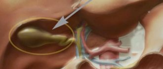
Cystoscopy is necessary for catheterization of the ureters in cases where the outflow of urine from the kidneys is difficult, as well as for relieving renal colic.
Catheterization of the left ureter
During cystoscopy, the doctor can also assess the condition of the seminal tubercle, the prostatic part of the urethra (how free its lumen is).
When is cystoscopy prescribed?
There can be many conditions for undergoing cystoscopy of the bladder in women and men:
- purulent or bloody discharge in the urine (hematuria or pyuria);
- frequent urge to empty the bladder;
- urinary tract infections;
- regular pain in the pelvic area;
- urolithiasis disease;
- chronic cystitis;
- abnormal cells in urine;
- polyps, tumors in the urethra or bladder detected by ultrasound, magnetic resonance or computed tomography;
- urinary retention due to inflammatory processes in the prostate gland or due to narrowing of the urinary canal.
Cystoscopy can also be used for therapeutic purposes:
- removal of stones;
- elimination of tumors and polyps;
- stopping bleeding;
- removal of obstructions.
Preparation and progress of the procedure
1-2 hours before cystoscopy, it is advisable to do a cleansing enema (the doctor may consider it necessary to conduct a rectal examination of the prostate for hyperplasia). On the day of the procedure, it is advisable not to consume food or irritating drinks. To exclude contraindications, before cystoscopy you need to undergo a general and biochemical blood test, a coagulogram, and it is advisable to perform an x-ray of the urethra (the study will show its shape and width of the lumen).
In case of acute inflammation of the genitourinary system, the procedure is not performed, and to prevent exacerbation of chronic pathologies, the patient is pre-administered a broad-spectrum antibiotic. As an alternative, you can take Monural at 10 pm on the eve of cystoscopy.
Immediately before cystoscopy, it is necessary to empty the bladder . Then the patient is placed on a urological chair (legs are spread apart and fixed on holders). Externally, the genitals are treated with an antiseptic solution.
For each patient, the diameter of the cystoscope tube is selected individually. Before insertion, it is lubricated with lubricant to facilitate insertion. This is usually medical glycerin, which does not impair the quality of the review. An anesthetic (2% warm solution of novocaine, gel with lidocaine or xylocaine) is poured into the urethra using a syringe with a rubber tip and manually massaged to the posterior urethra (inner part). If the patient has an unstable nervous system or there are painful changes in the mucous membrane of the urethra and bladder, then cystoscopy is performed under general or epidural anesthesia.
Stages of introducing a cystoscope into the bladder
In order not to injure the urethral mucosa during insertion, the tip of the cystoscope is protected by a special cover - an obturator, which is then removed, and its place is taken by the optical system. During the advancement of the device, the urethra may spasm (the sphincter in the bulbous part of the urethra will close), then the patient is asked to breathe deeply (with general anesthesia this is impossible, so doctors try not to use it). After passing through the urethra, the optics at the end of the device are replaced with cystoscopic ones.
With benign hyperplasia, the process of inserting a cystoscope is complicated because the prostatic part of the urethra becomes longer and its course changes.
To examine the walls of the bladder, you need to smooth them out as much as possible. Two methods are used for this:
- Dry, in which the bladder fills with air.
- Irrigation, when a special liquid (approximately 200 ml) acts as an expander.
Animation of bladder cystoscopy in men
Urine is not suitable as a filler, as it will significantly degrade image quality and can damage optical parts. After inserting the device, its remains are removed (suctioned off), and the bladder is washed with a warm solution of furatsilin. To take material for a biopsy, a special instrument is inserted that pinches off a piece of tissue (the process is painless). Cystoscopy lasts from 15 to 40 minutes. The conclusion is prepared within 15-20 minutes.
Side effects
The above consequences of the procedure are observed to one degree or another in almost all subjects.
But there are a number of unwanted complications, some of which may require surgery:
- Damage to the mucous layer of the urethra. Caused by improper insertion of the endoscope or restless behavior of the patient during manipulation. Manifested by bleeding from the urethra. It usually goes away on its own, but in some cases it requires repeated endoscopic intervention for electrocoagulation of blood vessels.
- False move. Inaccurate insertion of the cystoscope leads to injury to the walls of the urethra - the tube makes a hole and enters the tissue surrounding the urethra. If the damage involves the prostate, severe bleeding may occur.
- Perforation of the bladder walls. The main reasons are the low qualifications of the urologist, the presence of adhesions, scars in the walls, and abnormal structure of the organ.
POPULAR WITH READERS: Modern methods of treating prostatitis
Cystitis can also be a complication of cystoscopy. Inflammation most often occurs as a consequence of insufficient compliance with the rules of asepsis and antisepsis.
With a number of diseases, such as diabetes, the risk of cystitis increases. To reduce the likelihood of inflammation, such patients need to take antibacterial agents several days before the examination and for 5-7 days after.
Serious complications are indicated by prolonged retention of urination with a normal volume of fluid consumed, fever, pain in the lumbar region, in the lower abdomen, the appearance of bloody discharge or blood clots.
results
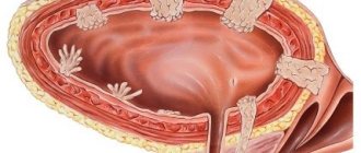
The healthy mucous membrane of the urethra is gray-red in color, blood vessels are visible through it. The color is uniform along the entire length. The shell is folded. There are no protrusions, ulcers, hemorrhages, ulcers, or protrusions on it.
In acute urethritis, the epithelium becomes bright red and swollen. Purulent effusion and loose growths may appear. Atrophic urethritis (changes in the structure of the walls due to hormonal imbalance) is characterized by the appearance of bluish tubercles - caruncles. If they do not cause concern, then treatment is not required.
In cases of chronic urethral infection, a diverticulum (pouch-like thickening) may be detected during cystoscopy. When squeezed or expanded, pus oozes out. Such formations are often found in patients with complaints of painful sexual intercourse.
In case of bladder injuries, cystoscopy will identify sites of vascular ruptures and hemorrhages. This diagnostic method also makes it possible to detect asymptomatic cystic cystitis (cysts in this case are small formations with fluid that arise at the sites of chronic inflammatory foci).
Picture of cystic cystitis Bladder tumor in the field of view of the cystoscope
Complications
The doctor will never forcefully advance the cystoscope ; he always strives to open the urinary canal, but in the first two days after cystoscopy, most patients still report discomfort when urinating and even bleeding. This is due to the fact that the mucous membrane of the posterior urethra is loose and easily damaged. Symptoms should subside with each passing hour. To relieve discomfort, you can use the urethral gel "Kategel". If the burning sensation does not go away, a dull pain appears in the lower abdomen, and the urine becomes cloudy, this means the development of cystitis. That is, during cystoscopy, an infection entered the bladder or an old, dormant infection became active. In rare cases, acute prostatitis, epididymitis, and urethritis may develop.
Urethral gel "Katejel" is a drug with local anesthetic and antiseptic effects for local use. Price in pharmacies from 197 rubles.
The most severe consequences of cystoscopy are a puncture of the bladder or ureter. This happens extremely rarely, usually during a biopsy. The reason is usually the doctor's inexperience. In such cases, acute pain immediately occurs, which does not go away after the procedure, and then the temperature rises. Surgery is necessary to correct the problem.
During the procedure, there is a low probability of developing so-called resorptive fever. This condition occurs immediately, usually during irrigation cystourethroscopy against the background of increased intraurethral pressure. The condition requires urgent intervention.
Contraindications
Since endoscopic examination using a cystoscope is a rather traumatic procedure, it has its contraindications. First of all, the patient is examined for the presence of inflammatory diseases of the bladder. To do this, it is enough to do a urine test and a blood test, interview the patient, and examine him. In the presence of cystitis and other similar ailments, cystoscopy is not performed.
If cystitis is a relative contraindication, then there are absolute contraindications. These include:
- Severe decompensated conditions - cardiac, renal, liver failure.
- Pregnancy.
Clinics and prices
Examples of clinics where cystoscopy can be performed:
- "Euromed" (St. Petersburg): 8800 rubles;
- “SM-clinic” (Moscow): dry urethroscopy – 4500 rubles, irrigation – 6000 rubles, cystoscopy – from 8500 rubles;
- “Unified Center for Spermograms and Problem Reproduction” (St. Petersburg): cystourethroscopy – 4400 rubles, cystoscopy with video recording – 3500 rubles.
The pain of the procedure itself and its consequences depend on the type of device, training and professionalism of the doctor.
Answers to frequently asked questions
- MRI or cystoscopy? Cystoscopy is a primary diagnostic examination of the mucous membrane with the possibility of simultaneous removal of a number of neoplasms, making it possible to accurately assess the condition of the organ. MRI of the bladder is mainly prescribed when cancer is diagnosed to determine its stage and determine the extent of spread.
- Is there an alternative to cystoscopy? If there are contraindications to cystoscopy, the doctor will prescribe an MRI or CT scan of the pelvis with contrast.
- Is it possible to have sex after cystoscopy? Protected sex and masturbation can be practiced after 7 days. Unprotected - no earlier than after 2 weeks (all microtraumas of the urethra must heal).
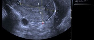
Ultrasound diagnostics
Ultrasound examination is the most common method in medical practice. It is completely harmless, publicly available, non-invasive.
Ultrasound diagnostics
This study does not require the use of special drugs, and its result does not depend on the functional state of the urinary system.
Special scanners are used for ultrasound examinations. Their work is based on the echolocation effect.
The reflected ultrasonic energy is captured by a special sensor and converted into electrical energy.
As a result, a black and white two- or three-dimensional image is formed on the monitor screen.
Medical research
Different echogenicity of tissues during examination may indicate either a normal state of health, or the presence of neoplasms, inflammation, or stones.
To get a complete picture of the condition of the bladder, an ultrasound examination is performed transvaginally (through the vagina), transrectally (through the rectum) and transabdominally (through the surface of the abdomen).
With this combination of methods, the doctor can draw a conclusion about the condition of all parts of the organ.
Also, for a more detailed examination of the bladder, special ultrasound sensors are used, which are attached to the urethroscope. This procedure is called endoluminal sonography.
To determine the degree of blood supply, a Doppler ultrasound examination of the vessels supplying the organ is performed. Deviations from the normal picture may indicate the development of a malignant tumor.
Reviews from men
Pavel, 38 years old: “I’ve been “ripe” for a cystoscopy for a long time (I suffered from cystitis of unknown origin). In the end, I decided to have the procedure and realized that I was afraid in vain: the room was quiet, warm, the staff was calm, the doctor told me not to think about anything and to breathe evenly. The sensations are unpleasant, but with painkillers they are quite tolerable. There was no blood in the toilet, discomfort when urinating bothered me until the evening.”
Yaroslav, 29 years old: “Cystoscopy for me personally was torture, not so much physical as moral. The doctor said that without it nothing is clear, and an ultrasound showed a suspicion of a tumor. Out of fear he agreed. Cystoscopy put everything in its place - the formation is benign.”