The phenomenon of stagnation of bile in the gallbladder is called cholestasis. This pathology indicates serious dysfunction of the biliary system, which, in addition to the gallbladder, also includes the liver. However, such a deviation from the norm can also be caused by other diseases of the internal organs. In simple cases, taking choleretic drugs helps, but advanced cases require more complex treatment.
In case of bile stagnation, a prerequisite for effective therapy is adherence to a special diet, which not only limits the consumption of certain types of foods, but also imposes certain requirements on the diet. Conservative treatment of cholestasis also involves drug therapy and certain medical procedures (for example, gallbladder tubing).
Drive bile with choleretic herbs or restore the liver?
Classic treatment of the gallbladder usually comes down to drug therapy using choleretic herbs and diet, and if stones still form, then to removal of the gallbladder. But there is a question: can the use of choleretic drugs and tubes on a schedule be considered reasonable if we know that normally the liver produces up to 2 liters of its digestive secretions every day. Even if we perform a series of tubes several times a year or take choleretic drugs, this will not affect everyday digestion. It will only give temporary relief. This is the same as an enema for the intestines!
The question is not that bile needs to be expelled. It is necessary to create conditions when it will be produced in a normal way and will freely stand out on its own! And for this, the liver cells simply need help to restore self-regulation and fluidity of bile, its ability to be released on time - in response to food intake will also be restored.
When we use the Sokolinsky System, the logic is to simply give the liver time to recover, reboot the microflora (so that it stops affecting the thickening of bile) and get used to a healthier diet. At the end of the course, there should be no tendency to stagnation.
How is it diagnosed?
Without special diagnostic measures, it is impossible to determine whether the outflow of secretions is increased or decreased. After the initial examination, the doctor needs the results of laboratory and instrumental examinations to make an accurate diagnosis. Doctors prescribe the following methods:
- General and biochemical blood test.
- Urine tests for bile pigment.
- Checking blood for cholesterol.
- Ultrasound. Determines deviations from the norm in the shape and size of the bubble, the presence of stones and kinks.
- Ultrasound with load. The first examination is carried out on an empty stomach. After breakfast, 30 minutes later, a second study is done to determine the degree of organ contraction.
- Duodenal sounding. Bile is collected using a probe that is inserted through the mouth or nose.
- X-ray and MRI. Used in cases of advanced forms of the disease.
How is bile formed?
Bile is produced by hepatocytes - liver cells. It collects in the bile ducts of the liver, then enters the gallbladder and finally into the duodenum, where it helps digest food. Congestion can occur both in the gallbladder and in the liver ducts. Of course, the risk is higher when the bile is thick.
Bile acids, of which almost 70% bile consists, break down fats, activate the movement of food through the small intestine, stimulate the production of mucus and some hormones: cholecystokinin and secretin, and prevent bacteria from attaching to the intestinal mucosa.
Without normally secreted bile, proper digestion and overall health are impossible.
Factors influencing the development of this pathology
There are many such factors, for example:
- physical inactivity (if a person leads a sedentary lifestyle);
- constant nervous stress leading to numerous stresses;
- diseases of the pelvic region (pathologies of the rectum);
- endocrine diseases and other pathologies of internal organs (gastritis, peptic ulcer, intestinal infections, pancreatitis, etc.);
- improper diet and regimen;
- excessive addiction to alcohol;
- changes in general hormonal levels during pregnancy;
- metabolic disorders in the body (for example, diabetes);
- stomach pathologies;
- obesity;
- atherosclerosis.
Read also: How is surgery to remove the gallbladder through the mouth performed?
In addition to these factors, the risk of cholestasis increases with:
- the presence of congenital pathologies of the gallbladder;
- exposure to stress hormones (adrenaline, cortisol, etc.), which suppress the contractility of the walls of the gallbladder, which causes stagnation in it.
With cholestasis, bile thickens in the gallbladder and its density changes. What makes it thick is excess cholesterol, which is formed with constant abuse of fatty, fried, spicy foods and alcohol. The size of the gallbladder in this case is increased.
Causes of bile stagnation
Causes of bile stagnation:
1. Increased lithogenicity (imbalance between cholesterol, phospholipids and fatty acids).
This is most positively influenced by improving the functioning of the liver itself, supporting the correct intestinal microflora, and correcting nutrition.
Disorders of the endocrine system, such as diabetes mellitus, dysfunction of the thyroid gland, hormonal imbalance caused by taking contraceptives, have a negative impact; smoking, drinking alcohol, an abundance of fatty and spicy foods, inflammation in the small intestine, intestinal dysbiosis;
This is primarily a love of rich fatty foods or foods rich in preservatives, dyes, flavor enhancers, irregular nutrition, and lack of minimal physical activity. This negatively affects both the formation and flow of bile.
2. Biliary tract tone (dyskenesia).
Increasing the amount of plant fiber in food has a positive effect. Approximately, it should be assumed that for 1 g of meat food there should be at least 4 g of vegetables or cereals. If it is difficult for you to adhere to such a regimen, then read about very successful helpers - NutriDetox and Redi Fiber.
Negative effects include neurosis and increased levels of stress hormones. Accordingly, it is necessary to maintain the correct emotional state, strengthen the nutrition of nervous tissue, and reduce the level of stress hormones. Read more about how you can do this naturally.
3. Anatomical anomalies in the structure of the bladder (bending, bending, pear-shaped, adhesions and constrictions).
Of all the causes of stagnation, this is the most problematic for correction, since it cannot be completely eliminated, and the risk of stone formation, fatty hepatosis and pancreatitis is high.
The only way to compensate for the negative is the same diet with a sufficient amount of plant fiber and regular inclusion of phospholipids (for example, LecithinUM) and omega-3 acids (for example, Megapolynol) in the diet according to the scheme of a month - one remedy, a month - a break, a month - another, month - break and then in a circle. But in some cases, when we use them to support nervous tissue or obtain an anti-inflammatory effect, a longer course is possible. The substances are 100% food grade and it is difficult to overdose on them.
Cholestasis - causes
Various factors can provoke the development of this disease. Intrahepatic stagnation of bile has the following causes:
- hypothyroidism;
- alcoholic liver damage;
- sepsis;
- viral hepatitis;
- liver damage caused by drugs or toxic agents;
- genetic diseases - Edwards syndrome or Patau syndrome;
- heart failure.
Extrahepatic cholestasis occurs due to:
- Caroli's disease;
- atresia;
- duct obstruction;
- transplant rejection;
- tuberculosis;
- pancreatitis;
- ductal dyskinesia.
Symptoms of bile stagnation
Normally, the gallbladder contracts after each meal, releasing bile and participating in the digestive process. At the slightest malfunction, symptoms of bile stagnation : dull, aching pain in the right hypochondrium, bitterness in the mouth, nausea, belching, itchy skin, alternating constipation and diarrhea.
In the acute stage, yellowing of the sclera and enlargement of the liver are observed. But this is a condition that requires close attention from a doctor.
The consequence of stagnation is the resulting vitamin deficiency and chronic fatigue syndrome. Stagnation of bile is not only momentary symptoms. It affects the quality of life and over time leads to the development of other diseases: cholelithiasis, atherosclerosis, pancreatitis, allergies, psoriasis, chronic constipation.
Thanks to the Sokolinsky System, you can break the vicious circle by setting the body to independently eliminate the causes of bile stagnation.
Cholestasis - what is it?
The term used to identify it will help you understand what this pathological condition is. In Greek, “cholē” literally means “bile,” and “stasis” means “standing.” All this sheds light on the characteristics of the disease. As a result, it becomes obvious that cholestasis is the slow production and stagnation of bile. This secretion can accumulate in the intra- and extrahepatic ducts.
Pathology provokes potentially reversible changes in the bile ducts:
- expansion of capillaries;
- disruption of hepatocyte membranes;
- formation of bile clots.
In the absence of timely treatment, the breakdown of fats supplied with food and their absorption are disrupted. These substances enter directly into the blood and prevent the transformation of glucose into glucogen. This situation can ultimately trigger the development of diabetes. Since bile contains cholesterol, due to a violation of its outflow, excess of this substance accumulates in the body. This leads to the development of atherosclerosis in the future. Stagnation of bile is fraught with the occurrence of an inflammatory process in the liver and in the ducts located outside it.
In the future, this situation may lead to the development of such complications:
- cholangitis;
- liver cirrhosis;
- osteoporosis;
- cholelithiasis;
- vitamin deficiency;
- liver failure;
- lethal outcome.
Intrahepatic cholestasis
This condition is characterized by impaired bile circulation. Moreover, this does not happen due to mechanical damage to the biliary tract. The pathological process can form either at the level of the bile ducts located inside the liver, or in hepatocytes.
All this causes the following problems:
- excessive release of bile into the blood;
- deficiency of this secretion in the intestines;
- pathological effect of bile on liver cells and the tubules located here.
As the disease progresses, such cholestasis may be:
- sharp;
- chronic.
Intrahepatic pathology can occur in the following forms:
- icteric;
- anicteric.
Hepatic cholestasis may be as follows:
- partial (with it the production of bile decreases);
- dissociated - a pathology characterized by a failure in the formation of individual components of the secretion;
- total (transport of bile to the duodenum is disrupted).
Extrahepatic cholestasis
This pathological condition is characterized by obstruction of the bile ducts. Cholestasis syndrome is often diagnosed in pregnant women. The reason for everything is considered to be the hormone estrogen, which takes an active part in many processes occurring in the female body. In addition, a pregnant woman increases the production of the hormone secretin, which stimulates the production of bile. At the same time, the level of somatropin, which blocks the contraction of the gallbladder, increases in the body. All this leads to the same bile being produced more intensively, but does not have time to be excreted through the ducts.
Cholestasis in pregnancy often resolves after childbirth. However, this does not mean that the disease does not require medical intervention.
If you leave things to chance, serious consequences can arise:
- bradycardia;
- premature birth;
- fetal distress.
Gallbladder treatment
The prescription of choleretic drugs, antispasmodics, and sometimes antibiotics is the basis of traditional treatment of the gallbladder. The existence of traditional medicine, oriental practice and European nutritional science allows us to influence the root cause of the disease, therefore, minimizing the chemical effects of classical drugs. You can learn in detail about how the Sokolinsky System works from our books and videos. We do not call this a “gallbladder cure” because the law does not allow this term to be applied to natural remedies.
For each case, the approach to getting rid of the causes of gallbladder dysfunction is different: for one it is more important to improve the nutrition of liver cells and cleanse the body, for another the primary measure is to eliminate chronic constipation, and for a third it is to calm the nervous system.
The Sokolinsky System uses natural plant complexes. We have experience in using Liver 48, herbal collection Fitocalcule and Phytobilin, and the export form of Ziflan since 2002.
The properties of milk thistle, nettle, immortelle, alpha-lipoic acid and other components have long been studied and were widely used in ancient times to treat the gallbladder in folk practice.
You can read in the book “Understandable methods of promoting health: for the busy and smart” or watch the video exactly how we use herbs to improve the functioning of the gallbladder and restore its self-regulation during bile stagnation.
If you want to get rid of the symptoms of stagnation, then it may be easier to use medications, and if you want to restore normal functioning for a long time, then get acquainted with natural nutritional remedies that affect the causes.
Treatment of cholestasis
Treatment of bile stagnation can be done using different methods, it depends on the cause. For particularly severe patients, surgery or endoscopy using a flexible fiber-optic surgical instrument is offered, but other options are also possible. If the drug is the cause of the disease, the doctor stops its use. If acute hepatitis is the cause, cholestasis and jaundice usually resolve after treatment of the hepatitis.
People with cholestasis are advised to avoid or stop using any substances that are toxic to the liver, such as alcohol and certain medications. Those patients who do not stop drinking alcohol only worsen their condition. Secondary complications, infections and other illnesses can also occur with improper treatment.
Drug therapy of any kind is most often ineffective, especially for pediatric patients. However, in some cases, ursodeoxycholic acid can be an effective drug. The drug consists of tertiary bile acid and has the property of displacing and eliminating toxic bile acid.
To eliminate itchy skin, drugs such as colelstipol and phenobarbital are commonly used.
Preventing cholestasis is often impossible, but there are precautions. It is worth getting vaccinated against hepatitis A and B in advance, limiting your intake of junk food, alcohol and nicotine.
The mechanism of stone formation in the gallbladder and its prevention
Larisa Viktorovna Tolokonnikova, Doctor of Intern, UMTS of the Administration of the President of the Russian Federation, Moscow [email protected] “The mechanism of stone formation in the gallbladder and its prevention” Abstract: In this article I approach the issue of drug therapy for gallstone disease by analyzing all stages of stone formation and the influence on the physiology of other internal organs, and also cite a clinical case from my practice with a favorable outcome. enterohepatic circulation, biliary pancreatitis, ursosan, duspatalin, sphincter of Oddi, motility, microflora, own practice, biochemical blood parameters, ultrasound. Gallstone disease (GSD) is one of the most common diseases humanity. Among diseases of the digestive system, it occupies a leading place, and its treatment involves not only gastroenterologists and therapists, but also doctors of other specialties, including surgeons. Epidemiological studies of the incidence of cholelithiasis indicate that the number of patients in the world increases at least every decade doubled. In general, in Europe and other regions of the world, cholelithiasis is detected in 10–40% of the population of various ages. In our country, the frequency of this disease ranges from 5% to 20%. In northwestern Russia, gallstones are detected on average in every fifth woman and every tenth man [2, 3, 6, 11]. The significant prevalence of this pathology is associated with the presence of a large number of risk factors that have become relevant recently. The most important of them include hereditary predisposition, developmental abnormalities of the biliary tract, inadequate nutrition, use of medications (oral contraceptives, drugs to normalize lipid metabolism, ceftriaxone, sandostatin derivatives, nicotinic acid), manifestations of metabolic syndrome (obesity, diabetes mellitus, dyslipoproteinemia) , pregnancy, inflammatory bowel diseases, chronic constipation, physical inactivity and others. It should be noted that the pathogenesis of stone formation is still being studied, but it is known that disruption of the mechanisms of enterohepatic circulation (EGC) of cholesterol and bile acids is of key importance. The reasons for the violation of the EGC are: violation of the rheology of bile (supersaturation with cholesterol with increased nucleation and formation of crystals);
violation of the outflow of bile associated with changes in the motility and patency of the gallbladder, small intestine, sphincter of Oddi, sphincters of the common pancreatic and bile ducts, combined with changes in peristalsis of the intestinal wall;
disruption of intestinal microbiocenosis, since when the composition and decrease in the amount of bile in the intestinal lumen changes, the bactericidal capacity of the duodenal contents changes with excessive proliferation of bacteria in the ileum, followed by early deconjugation of bile acids and the formation of duodenal hypertension;
indigestion and absorption, since against the background of duodenal hypertension and increased intraluminal pressure in the ducts, damage to the pancreas occurs, with a decrease in the outflow of pancreatic lipase, which disrupts the mechanisms of fat emulsification and activation of the chain of pancreatic enzymes, creating the preconditions for biliary pancreatitis [3, 8, 11 ]. An important unfavorable prognostic factor for cholelithiasis is the development of serious complications that affect the course of the disease. These include acute cholecystitis, choledocholithiasis, obstructive jaundice, cholangitis and chronic pancreatitis (CP). In addition, inadequately chosen treatment tactics for a patient with cholelithiasis often leads to the development of postoperative complications, the so-called postcholecystectomy syndrome, which significantly worsens the quality of life of these patients. The main reason for these circumstances is the lack of compliance between therapists and surgeons, while the former do not have clear tactics for managing patients with cholelithiasis, and the latter are interested in broad surgical treatment of all patients of this profile. Despite the long history of this disease, the only generally accepted classification tool remains a three-stage division GSD into 1) physicochemical stage, 2) asymptomatic stone-carrying and 3) stage of clinical symptoms and complications. This classification, developed with the direct participation of surgeons, however, does not answer the whole list of practical questions that arise for the therapist when treating patients of this profile, For example:
Is it necessary to carry out medicinal treatment of cholelithiasis; if such a need exists, then with what drugs and in the conditions of the department of what profile;
what are the criteria for the effectiveness and ineffectiveness of drug therapy;
what are the indications for surgical treatment in a particular patient;
Should the patient be observed after surgery, by which specialist, for how long and with what medications should postoperative treatment be carried out. That is, to date, generally accepted tactics for monitoring patients with cholelithiasis have not been developed. As evidenced by the analysis of the literature, the only algorithm for the management of patients with this pathology is the international recommendations of Euricterus on the selection of patients with cholelithiasis for surgical treatment, adopted at the Congress of Surgeons in 1997. The most important factors are those used in the Euricterus system to make decisions about surgical treatment. These include:
the presence of clinical symptoms (right hypochondrium syndrome or biliary pain, biliary colic);
presence of concomitant CP;
reduced contractile function of the gallbladder;
the presence of complications. Assessing the characteristics of clinical symptoms in patients with cholelithiasis requires differential diagnosis between right hypochondrium syndrome due to functional biliary disorder (FBD)
and biliary (hepatic) colic, which often causes difficulties even for qualified specialists. At the same time, a correct assessment of the clinical picture and, in particular, taking into account the number of colics in the anamnesis largely determine the tactics of managing a patient with cholelithiasis with the subsequent choice of direction for conservative therapy, sphincteropapillotomy or cholecystectomy. It should be noted that these clinical phenomena have fundamentally different mechanisms, so with FB pain is a consequence of a violation of the contractile function (spasm or stretching) of the sphincter of Oddi or the gallbladder muscles, which prevents the normal outflow of bile and pancreatic secretions into the duodenum. Whereas with biliary colic, it occurs due to mechanical irritation of the gallbladder wall by a stone, obstruction of the gallbladder, wedging into the neck of the gallbladder, into the common bile, hepatic or cystic duct. It should be emphasized, however, that part of the pain associated with colic is due to FBR. Having thus assessed the clinical picture of patients with cholelithiasis, it is possible to subdivide them into groups: the 1st group of patients with cholelithiasis should include patients without active complaints and obvious clinical symptoms. The diagnostic criteria will be the absence of biliary pain and the presence of biliary sludge (clots) detected by ultrasound. The 2nd group includes patients with biliary pain in the epigastric region and/or in the right hypochondrium, characteristic of a functional biliary disorder, and dyspeptic symptoms. Diagnostic criteria in this case are the presence of biliary/pancreatic pain, the absence of biliary colic, the presence of biliary sludge or stones on ultrasound. Occasionally, a transient increase in the activity of transaminases and amylase associated with an attack is also possible. Patients with cholelithiasis and symptoms of chronic pancreatitis deserve special attention, which, due to clinical prognostic and, most importantly, therapeutic features, constitute the 3rd group. Diagnostic criteria for this category of patients include: the presence of pancreatic pain, the absence of biliary colic, the presence of signs of pancreatitis, stones and/or biliary sludge with radiation methods, possible increased activity of lipase, amylase, decreased elastase1 and the presence of steatorrhea. Patients with cholelithiasis with symptoms of one and more attacks of biliary colic, belonging to group 4, are already patients with surgical pathology. Diagnostic criteria in this case are: the presence of one or more biliary colic, stones in the gallbladder, possible transient jaundice, increased activity of ALT, AST, GGTP, bilirubin level associated with hepatic colic. It should be emphasized the need for a detailed identification of biliary colic in the anamnesis, after the manifestation of which months and even years can pass. After determining the clinical groups, the directions of treatment for patients with cholelithiasis are both general and individual, group-specific. General directions include approaches that help improve EGC processes and suppress the mechanism of stone formation in the gallbladder. These approaches include:
impact on risk factors and factors for relapse of the disease;
improvement of the rheological properties of bile;
normalization of motility of the gallbladder, small intestine and restoration of the patency of the sphincter of Oddi, as well as the sphincters of the common pancreatic and bile ducts;
restoration of the normal composition of intestinal microflora;
normalization of digestion and absorption processes with restoration of the functioning of the pancreas. Impact on risk factors and factors for recurrence of the disease: A set of measures aimed at eliminating factors contributing to stone formation includes the abolition or dose adjustment of lithogenic drugs (estrogens, third-generation cephalosporins, drugs affecting the lipid spectrum , somatostatin, etc.), prevention of congestive gastrointestinal tract, including in pregnant women, treatment of biliary sludge, correction of hormonal levels [1, 11, 13]. The diet of patients with cholelithiasis should be balanced in protein content (meat, fish, cottage cheese) and fats, mainly vegetable. Thus, rational intake of protein and fat increases the cholate-cholesterol ratio and reduces the lithogenicity of bile. The polyunsaturated fatty acids contained in vegetable oils help normalize cholesterol metabolism, restore cell membranes, participate in the synthesis of prostaglandins and normalize the contractile function of the gallbladder. Prevention of excessive pH shifts to the acidic side by limiting flour and cereal products and prescribing dairy products (if tolerated) also reduces the risk of stone formation. High-calorie and cholesterol-rich foods are excluded. Following a diet helps reduce the likelihood of spastic contraction of the gallbladder muscles and the sphincter of Oddi, which can cause migration of stones, including small ones (sand). If there is a severe exacerbation of CP, in the first three days the patient is prescribed a complete fast with drinking water. Subsequently, meals should be frequent, fractional, with the exception of fatty, fried, sour, spicy foods and help normalize the patient’s body weight [3, 4, 7, 9]. Improving the rheological properties of bile: To date, the only pharmacological agent with a proven effect on the rheology of bile, is ursodeoxycholic acid. Our own experience in treating patients with cholelithiasis is associated with the drug Ursosan. With regard to determining the indications for the use of ursodeoxycholic acid drugs in cholelithiasis, it is important to take into account the achievement of remission of pancreatitis and the absence of extrahepatic cholestasis[10]. Therapy with this drug is carried out until the physicochemical and rheological properties of bile are normalized, the amount of microlites in the bile is reduced, further stone formation is prevented and the possible dissolution of stones is achieved. Its additional immunomodulatory and hepatoprotective effects are also taken into account. Ursosan is prescribed in a dose of up to 15 mg/kg body weight, the entire dose is taken once in the evening, an hour after dinner or at night. The duration of use depends on the clinical situation, amounting to approximately 6–12 months [12,13]. In the presence of abdominal pain and dyspeptic syndromes, the dose should be titrated, starting from a minimum of 250 mg, one hour after dinner, for approximately 7-14 days, with a further increase by 250 mg at similar time intervals to the maximum effectiveness. In this case, cover therapy is advisable, including the parallel use of a selective antispasmodic - Duspatalin (mebeverine). Normalization of the motility of the gallbladder, small intestine and restoration of the patency of the sphincter of Oddi, as well as the sphincters of the common pancreatic and bile ducts: The treatment manual includes measures to correct the outflow from the ductal system of the pancreas and biliary tract using endoscopy (in the presence of organic changes - cicatricial stenosis of the sphincter of Oddi, calcifications and stones in the ducts) and/or using medications. The means of conservative therapy are drugs that have antispasmodic and eukinetic effects. Frequently used non-selective antispasmodics (Noshpa, Papaverine) are drugs that do not have a dose-dependent effect, with low affinity for the biliary system and pancreatic ducts. The mechanism of action of these drugs generally comes down to inhibition of phosphodiesterase or activation of adenylate cyclase, blockade of adenosine receptors. Their disadvantages are significant differences in individual effectiveness; in addition, there is no selective effect on the sphincter of Oddi; there are undesirable effects caused by the effect on the smooth muscles of blood vessels, the urinary system, and the gastrointestinal tract [1, 3, 12]. Anticholinergics also have an antispasmodic effect ( Buscopan, Platyfillin, Metacin). Anticholinergic drugs that block muscarinic receptors on the postsynaptic membranes of target organs exert their effect by blocking calcium channels, stopping the penetration of calcium ions into the cytoplasm of smooth muscle cells and, as a result, relieving muscle spasm. However, their effectiveness is relatively low, and a wide range of side effects (dry mouth, urinary retention, tachycardia, impaired accommodation, etc.) limit their use in this category of patients [1, 5, 9]. Separately in this series is an antispasmodic with a normalizing effect on the tone of the sphincter of Oddi - Duspatalin (mebeverine). The drug has a dual, eukinetic mechanism of action: reducing the permeability of smooth muscle cells to Na+, causing an antispastic effect, and preventing the development of hypotension by reducing the outflow of K+ from the cell. At the same time, Duspatalin has affinity for the smooth muscles of the pancreatic and intestinal ducts. It eliminates functional duodenostasis and hyperperistalsis without causing hypotension and without affecting the cholinergic system [1, 4]. The drug is usually prescribed 2 times a day 20 minutes before meals, at a dose of 400 mg/day, for a course of up to 8 weeks. Restoring the normal composition of intestinal microflora: Antibacterial therapy is an important part in the treatment of cholelithiasis. A completely adequate requirement is the prescription of antibiotics in cases of exacerbation of cholecystitis, as well as in concomitant disorders of the intestinal microbiocenosis. Empirically used are 8-hydroxyquinoline derivatives (ciprofloxacin), which create a secondary concentration in the biliary tract, imipenem, cefuroxime, cefotaxime, Ampiox, Sumamed, fluoroquinolones in combination with metronidazole. A limitation for the use of ceftriaxone is the formation of biliary sludge when taking it. At the same time, a number of antibacterial drugs (tetracycline, rifampicin, isoniazid, amphotericin B) have a toxic effect on pancreatic acinar cells. As a rule, all patients with cholelithiasis combined with CP have varying degrees of severity of disturbances in the intestinal microbiocenosis, which significantly affect the course of the disease , the rate of regression of abdominal pain and dyspeptic syndromes. To correct it, the antibiotic rifaximin (Alfanormix), which is not absorbed in the intestine, is used, which is prescribed 3 times a day, at a dose of 1200 mg/day, for a course of 7 days. It is mandatory to combine the stage of intestinal sanitation with the use of probiotics (live cultures of symbiont microorganisms) and prebiotics ( preparations that do not contain living microorganisms and stimulate the growth and activity of symbiont intestinal flora). Lactulose (Duphalac) has a proven prebiotic effect. Duphalac is a drug with the highest lactulose content and the least amount of impurities. It belongs to synthetic disaccharides, the main mechanism of action of which is associated with their metabolism by colon bacteria to short-chain fatty acids that perform important physiological functions - both local, in the colon, and systemic, at the level of the whole organism. Clinical studies have proven that Duphalac has pronounced prebiotic properties, realized due to bacterial fermentation of disaccharides and increased growth of bifidobacteria and lactobacilli, as well as a physiological laxative effect. Normalization of digestion and absorption processes: Buffer antacids and multienzyme preparations are used for this purpose. The indication for prescribing buffer antacids (Maalox, Phospholugel) in patients with cholelithiasis is their ability to:
bind organic acids;
increase intraduodenal pH levels;
bind deconjugated bile acids, which reduces secretory diarrhea and their damaging effect on the mucosa;
reduce the absorption of antibacterial drugs, which increases their concentration in the intestinal lumen, enhances the antibacterial effect and reduces side effects. Indications for multienzyme drugs are:
damage to the pancreas due to duodenal hypertension, increased intraluminal pressure in the ducts;
violation of fat emulsification;
impaired activation of the chain of pancreatic proteolytic enzymes;
violation of the time of contact of food with the intestinal wall against the background of changes in peristalsis. To correct these changes, it is advisable to use enzyme preparations with a high content of lipase, resistant to the action of hydrochloric acid, pepsin, with an optimum action at pH 5–7, in the form of minimicrospheres with a maximum surface of contact with chyme of the Creon type 10,000–25,000 units. Taking into account the stated approaches to the treatment of cholelithiasis in practice, their individualization is expected in specific groups. These schemes are presented in the form of stepwise therapy, which can be carried out either simultaneously or sequentially, depending on the clinical situation. Group 1 - patients with cholelithiasis without clinical symptoms, stage 1. Normalization of bile rheology and prevention of stone formation: ursodeoxycholic acid (Ursosan) 8–15 mg/kg once in the evening until sludge resolution (3–6 months). 2nd stage. Correction of intestinal dysbiosis: Duphalac 2.5–5 ml per day, 200–500 ml per course, for prebiotic purposes. Prevention. 1-2 times a year for 1-3 months, maintenance therapy with Ursosan at a dose of 4-6 mg/mg body weight per day in combination with Duspatalin 400 mg/day orally in 2 doses 20 minutes before breakfast and dinner - 4 weeks. Group 2 - patients with cholelithiasis with symptoms of functional biliary/pancreatic disorder or gallbladder disorder, stage 1. Correction of motor-evacuation function and intraduodenal pH: Duspatalin 400 mg/day in 2 doses 20 minutes before meals - 4 weeks. Creon 10,000–25,000 U, 1 capsule 3 times a day at the beginning of meals - 4 weeks. Antacid drug, after 40 minutes after meals and at night, up to 4 weeks. 2nd step. Correction of intestinal dysbiosis: Alfanormix 400 mg 3 times a day for 7 days. Duphalac 2.5–5 ml per day 200–500 ml per course with a probiotic. 3rd stage. Normalization of bile rheology and prevention of stone formation: Ursosan - intake from 250 mg/day (4-6 mg/kg), then weekly increase in dose by 250 mg, up to 15 mg/kg. The drug is taken once in the evening until the sludge resolves (3-6 months). 3rd group - patients with cholelithiasis with symptoms of chronic disease. Diet 1-3 days of fasting, then table No. 5P. 1st step. Correction of pancreatic function: Omeprazole (Rabeprazole) 20–40 mg/day in the morning on an empty stomach and at 20:00, 4–8 weeks. Duspatalin 400 mg/day in 2 doses 20 minutes before meals - 8 weeks. Creon 25,000–40,000 ED 1 capsule 3 times a day at the beginning of meals - 8 weeks. 2nd stage. Correction of intestinal dysbiosis: Alfanormix 400 mg 3 times a day, 7 days. Duphalac 2.5–5 ml per day, 200–500 ml per course, with a probiotic. 3rd stage. Normalization of bile rheology and prevention of stone formation: Ursosan - from 250 mg/day (4–6 mg/kg), followed by a 7–14-day dose increase to 10–15 mg/kg body weight, lasting up to 6–12 months. Subsequently, 2 times a year for 3 months or continuous maintenance therapy at a dose of 4–6 mg/kg/day in combination with Duspatalin 400 mg/day orally in 2 divided doses 20 minutes before breakfast and dinner for the first 4 weeks. Group 4—patients GSD with symptoms of one or more attacks of biliary colic. Diet - hunger, then individually. Hospitalization in a surgical hospital, where conservative treatment is carried out jointly with a gastroenterologist. When relieving colic, patients are treated as group 3. If ineffective, laparoscopic cholecystectomy is performed. The choice of an adequate type of treatment for cholelithiasis is largely determined by the mutually agreed upon tactics of the therapist (gastroenterologist), surgeon and patient.
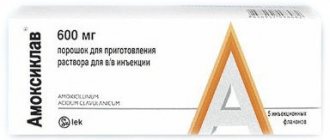
From my own practice, my relative suffered from chronic calculous cholecystitis, had diffuse changes in the tissue of the liver and pancreas. She was periodically bothered by severe pain in the right hypochondrium and dyspeptic symptoms. Bilirubin stones with a diameter of 8 mm (on ultrasound), I decided to carry out therapy for 6 months. She withstood the diet only for 2 weeks (table 5 a). Treatment included: 1. Polyphepan. A herbal preparation obtained from hydrolytic lignin. Binds various microorganisms, their metabolic products, toxins of exogenous and endogenous nature, allergens, xenobiotics, heavy metals, radioactive isotopes, ammonia, divalent cations and promotes their excretion through the gastrointestinal tract. It has an enterosorbing, detoxifying, antidiarrheal, antioxidant, hypolipidemic and complexing effect. Compensates for the lack of natural dietary fiber in human food, positively influencing the microflora of the large intestine and nonspecific immunity. 2. Hydrocholeretics. This group includes mineral waters - “Essentuki” No. 17 (highly mineralized) and No. 4 (weakly mineralized), “Jermuk”, “Izhevskaya”, “Naftusya”, “Smirnovskaya”, “Slavyanovskaya”, etc. Mineral waters increase the amount of secreted bile and make it less viscous. The mechanism of action of choleretic drugs of this group is due to the fact that, being absorbed into the gastrointestinal tract, they are secreted by hepatocytes into primary bile, creating increased osmotic pressure in the bile capillaries and contributing to an increase in the aqueous phase. In addition, the reabsorption of water and electrolytes in the gallbladder and bile ducts decreases, which significantly reduces the viscosity of bile. The effect of mineral waters depends on the content of sulfate anions (SO42), associated with magnesium (Mg2+) and sodium (Na+) cations, which have a choleretic effect. Mineral salts also help to increase the colloidal stability of bile and its fluidity. For example, Ca2+ ions, forming a complex with bile acids, reduce the likelihood of a sparingly soluble precipitate. Mineral waters are usually consumed warm 20–30 minutes before meals. 3. Amoxicillin. An antibacterial drug for the treatment of exacerbation of calculous cholecystitis must meet the following requirements: 1) penetrate well into the bile; 2) have a wide spectrum of action; 3) have the least hepatotoxicity. 4. Ursosan. Possessing high polar properties, UDCA is integrated into the membrane of hepatocytes, cholangiocytes and epithelial cells of the gastrointestinal tract, stabilizes its structure and protects the cell from the damaging effects of toxic bile acid salts, reducing thus. their cytotoxic effect. Forms non-toxic mixed micelles with apolar (toxic) bile acids, which reduces the ability of gastric refluxate to damage cell membranes in biliary reflux gastritis and reflux esophagitis. By reducing concentrations and stimulating bicarbonate-rich choleresis, UDCA effectively promotes the resolution of intrahepatic cholestasis. In case of cholestasis, it activates Ca2+-dependent alpha protease and stimulates exocytosis, reduces the concentration of toxic bile acids (chenodeoxycholic, lithocholic, deoxycholic, etc.), the concentrations of which are increased in patients with chronic liver diseases. Competitively reduces the absorption of lipophilic bile acids in the intestine, increases their fractional turnover during enterohepatic circulation, induces choleresis, stimulates the passage of bile and the excretion of toxic bile acids through the intestine. Reduces the saturation of bile with cholesterol by inhibiting its absorption in the intestine, suppressing synthesis in the liver and reducing secretion into bile; increases the solubility of cholesterol in bile, forming liquid crystals with it; reduces the lithogenic index of bile, increases the concentration of bile acids in it, causes increased gastric and pancreatic secretion, enhances lipase activity, and has a hypoglycemic effect. Reducing the saturation of bile with cholesterol promotes its mobilization from gallstones, resulting in the dissolution (partial or complete) of cholesterol gallstones and preventing the formation of new stones. The immunomodulatory effect is due to inhibition of the expression of histocompatibility antigens (HLA1 on the membranes of hepatocytes and HLA2 on cholangiocytes), normalization of the natural killer activity of lymphocytes, the formation of IL2, a decrease in the number of eosinophils, suppression of immunocompetent Ig, primarily IgM. Delays the progression of fibrosis. Regulates the processes of apoptosis of hepatocytes, cholangiocytes and epithelial cells of the gastrointestinal tract. 5. Hilak forte. Regulates the balance of intestinal microflora and normalizes its composition. Due to the content of metabolic products of normal microflora in Hilaka forte, the drug helps restore normal intestinal microflora biologically and allows you to preserve the physiological and biological functions of the intestinal mucosa. The biosynthetic lactic acid and its buffer salts included in Hilaka forte restore the normal acidity value in the gastrointestinal tract, regardless of whether the patient suffers from high or low acidity. Against the background of accelerating the development of normal intestinal symbionts under the influence of Hilak Forte, the natural synthesis of vitamins B and K is normalized. The short-chain volatile fatty acids contained in Hilak Forte ensure the restoration of damaged intestinal microflora in infectious diseases of the gastrointestinal tract, stimulate the regeneration of epithelial cells of the intestinal wall, restore the disturbed water-electrolyte balance in the intestinal lumen. There is evidence that Hilak forte enhances the body's protective functions by stimulating the immune response. 6. Pancreatin. Digestive enzyme agent, replenishes the deficiency of pancreatic enzymes, has proteolytic, amylolytic and lipolytic effects. The pancreatic enzymes included in the composition (lipase, alpha-amylase, trypsin, chymotrypsin) promote the breakdown of proteins into amino acids, fats into glycerol and fatty acids, starch into dextrins and monosaccharides, improves the functional state of the gastrointestinal tract, and normalizes digestive processes. Trypsin suppresses stimulated pancreatic secretion, producing an analgesic effect. Pancreatic enzymes are released from the dosage form in the alkaline environment of the small intestine, because protected from the action of gastric juice by the membrane. The maximum enzymatic activity of the drug is observed 30-45 minutes after oral administration. 7. Denol. Antiulcer agent with bactericidal activity against Helicobacter pylori. It also has anti-inflammatory and astringent effects. In the acidic environment of the stomach, insoluble bismuth oxychloride and citrate are precipitated, and chelate compounds are formed with the protein substrate in the form of a protective film on the surface of ulcers and erosions. By increasing the synthesis of PGE, mucus formation and bicarbonate secretion, it stimulates the activity of cytoprotective mechanisms, increases the resistance of the gastrointestinal mucosa to the effects of pepsin, hydrochloric acid, enzymes and bile salts. Leads to the accumulation of epidermal growth factor in the defect area. Reduces the activity of pepsin and pepsinogen. 8. Dibazol. Vasodilating agent; has a hypotensive, vasodilating effect, stimulates the function of the spinal cord, and has moderate immunostimulating activity. It has a direct antispasmodic effect on the smooth muscles of blood vessels and internal organs. Facilitates synaptic transmission in the spinal cord. Immunostimulating activity is associated with the regulation of the ratio of cGMP and cAMP concentrations in immune cells (increases the cGMP content), which leads to the proliferation of mature sensitized T and B lymphocytes, their secretion of mutual regulatory factors, cooperative reaction and activation of the final effector function of cells. I took it at night, and in combination with Ursosanam the effect is enhanced. 9. Vitamin therapy.10. Duspatalin is a dual-action myotropic antispasmodic that relieves spasms and does not cause atony. It has a selective effect on the sphincter of Oddi. Duspatalin is 2040 times more effective than papaverine in its ability to relax the sphincter of Oddi. It is important that duspatalin does not act on the cholinergic system and therefore does not cause side effects such as dry mouth, visual disturbances, tachycardia, urinary retention, constipation (which worsens the course of the disease) and weakness. The autonomic nervous system is not involved in the mechanism of action of mebeverine, therefore duspatalin in therapeutic doses does not have typical anticholinergic side effects. This allows the use of duspatalin in patients with concomitant diseases such as prostatic hypertrophy and glaucoma. To correct motor disorders of the gastrointestinal tract, drugs that reduce the content of cytosolic Ca2+ in myocytes are widely used in course therapy. Such a remedy is duspatalin, when taken, preservation of normal tone and
peristalsis after relief of muscle spasm. Duspatalin has two effects. The first of them comes down to the blockade of fast sodium channels of the myocyte cell membrane, which disrupts the processes of sodium entry into the cell, slows down the depolarization processes and stops the entry of calcium into the cell through slow channels. As a result, the processes of myosin phosphorylation are reduced and muscle fiber spasm is eliminated. The second effect is due to a decrease in the replenishment of intracellular calcium stores, which leads only to a short-term release of potassium ions from the cell and its hyperpolarization. The latter prevents the development of hypotension of the muscle wall. This effect of duspatalin distinguishes it favorably from the action of other myotropic antispasmodics that cause prolonged hypotension, and allows the drug to be used in clinical practice. As a result of treatment and observation after 7 months:
no attacks of pain in the right hypochondrium
stool returned to normal
dyspepsia were rare
Compared to previous tests (before therapy), the blood parameters showed total and indirect bilirubin reduced to within normal limits, C-reactive protein 0 (previously 24!), erythrocyte sedimentation rate 5 (previously 43!)
Ultrasound examination: echo signs of diffuse changes in liver tissue. No stones were found. Nothing else has been revealed.
Links to sources: 2. Burkov S.G. On the consequences of cholecystectomy or postcholecystectomy syndrome // Consilium medicum, gastroenterology. 2004. T. 6, No. 2, p. 24–27.3. Burkov S.G., Grebenev A.L. Gallstone disease (epidemiology, pathogenesis, clinic) // Guide to gastroenterology. In three volumes. Under the general editorship of F.I. Komarov and A.L. Grebenev. T. 2. Diseases of the liver and biliary system. M.: Medicine, 1995, p. 417–441.6. Lazebnik L.B., Kopaneva M.I., Ezhova T.B. The need for medical care after surgical interventions on the stomach and gall bladder (literature review and own data) // Ter. archive. 2004, No. 2, p. 83–87.11. Sherlock S., Dooley J. Diseases of the liver and biliary tract: Practical. hand: Per. from English edited by Z.G.Aprosina, N.A.Mukhina. M.: Geotar Medicine, 1999.864 p.8. McNally P.R. Secrets of gastroenterology: Trans. from English M.–SPb.: ZAO “Publishing House BINOM”, “Nevsky Dialect”, 1998. 1023 pp. 1. Diseases of the liver and biliary tract: A guide for doctors / Ed. V.T.Ivashkina. M.: LLC Publishing House MVesti, 2002. 416 p. 13. Yakovenko E.P. Intrahepatic cholestasis - from pathogenesis to treatment // Practitioner. 1998. No. 2 (13), p. 20–24.4. Grigoriev P.Ya., Yakovenko A.V. Clinical gastroenterology. M.: Medical Information Agency, 2001. 693 pp. 7. Leishner U. Practical guide to diseases of the biliary tract. M.: GEOTARMED, 2001. 234 p. 9. Petukhov V.A. Gallstone disease and indigestion syndrome. M.: Vedi, 2003. 128 p.
10. Sokolov V.I., Tsybyrne K.A. Cholepancreatitis. Chisinau: Shtiintsa, 1978. 234 p. 12. Yakovenko E.P., Grigoriev P.Ya. Chronic diseases of the extrahepatic biliary tract. Diagnosis and treatment. Methodological manual for doctors. M.: MedpraktikaM, 2001. 31 p. 5. Grigoriev P.Ya., Soluyanova I.P., Yakovenko A.V. Gallstone disease and consequences of cholecystectomy: diagnosis, treatment and prevention // Attending Physician. 2002, No. 6, p. 26–32.10. Sokolov V.I., Tsybyrne K.A. Cholepancreatitis. Chisinau: Shtiintsa, 1978. 234 pp. Tolokonnikova LarisaDoctorintern UNMTS President Administration [email protected] "The mechanism of stone formation in the gall bladder and its prevention" Abstract: In this article, I approach the issue of medical treatment of gallstones by reviewing all stages of stone formation and the impact on the physiology of the rest of the internal organs, as well as bring a clinical case from his own practice with a good outcome.enterohepatic circulation, biliary pancreatitis, Ursosan, Duspatalin, sphincter of Oddi, motility, microflora, private practice, biochemical blood tests, ultrasound.
Causes
Stagnation of bile, the symptoms of which will be described below, usually occurs with diseases of the stomach, liver, nervous disorders, and diseases of the rectum. The reasons for stagnation may lie in a person’s sedentary lifestyle, if he is not involved in the port. Stagnation of bile in the gallbladder occurs with constant neuroses, which lead to a weakening of the muscular system in the gallbladder, and endocrine disruptions occur in the body. Stagnation can occur if gastritis is observed in the stomach, there is an intestinal infection, or an ulcer.
Exercise - daily walks, jogging, swimming, dance classes and other types of physical education prevent bile stagnation; be sure to have breakfast, lunch and dinner, and not go to the hepatologist later; eat less fatty, sweet, salty and spicy foods, avoid alcohol; try not to eat too much, even healthy food should be in moderation; Temperament and irritability can cause spasms in the gallbladder; it is not in vain to keep them in your hands. Cholestasis is not just a disease, but a symptom of many diseases.
Causes of bile stagnation
Defined as bile stasis in the gallbladder or intrahepatic bile duct obstruction and possibly external ligation. Cholestasis can be divided into several subtypes. Clinical - creates a delay of various components in the blood, which change in bile. functional - its main characteristic is a decrease in the bile ducts and, as a consequence, a decrease in bilirubin.
- Morphological - in the form of accumulation of bile components in the bile ducts.
- Extrahepatic - stagnation of bile in the extrahepatic canals.
This disease occurs because at some point the digestive fluid is produced by the liver, preventing it from interacting with liver cells to produce bile from the duodenum.
Diet for bile stagnation is very important; first of all, it is recommended to give up fatty, spicy, fried, and alcoholic drinks. Add dietary fiber to your diet. If bile stagnates in the body, then it is recommended to consume food in small quantities, but as often as possible. With intense contraction of the gallbladder, its tone increases, while not all sphincters work fully, which leads to poor bile production.
As a result, it actively stops the production of bile pigment bilirubin and enters the human blood, and then accumulates in it. Typically, bilirubin passes through the liver and bile tract, then it is excreted into the gastrointestinal tract naturally.
The causes are divided into two groups: those that originate in the liver and those that occur abroad. Acute hepatitis and various types; Liver diseases associated with alcoholism; primary biliary cirrhosis with inflammation or scarring; Cirrhosis due to hepatitis B or C, in the stage of inflammation or scarring of the bile ducts; Inflammation of the bile ducts; Use of various hormone-based medications; hormonal effects on the liver during pregnancy; Cancer spreads to the liver; bacterial abscess in the liver; primary sclerosing cholangitis; severe infections that entered the liver through the bloodstream; tuberculosis; Sarcoidosis and amyloidosis. Possible presence of stone in the canal; reduce the channel; Inflammation or cancer of the pancreas; Pressure flows through other places that are much higher than normal. Another known cause of the disease may be excessive or prolonged consumption of certain medications such as ampicillin, anabolic steroids, pills, chlorpromazine, cimetidine, estradiol, imipramine, prochlorperazine, terbinafine and sugar-lowering drugs.
Diet for stagnation of bile is extremely important, because when eating food, the gallbladder actively contracts, and after eating, bile is released from the liver. Stagnation of bile, the treatment of which is given below, is dangerous because it does not absorb fat-soluble vitamins A and D. As a result, a child or adult may develop osteoporosis.
Symptoms of biliary stasis are not immediate; it all depends on the cause of the disease. The most common symptoms of the disease that we can immediately recognize are. Yellowing of the skin occurs from the accumulation of excess bilirubin, darkening of urine due to excessive output and bilirubin from the body. then the patient may have light stools and typical itching. The stool will be a bright shade, due to the lack of bilirubin in the stool, this is due to the lack of bilirubin in the intestines. The result is a clotting of bone deficiency and low blood.
- Yellowing of the whites of the eyes or skin and significant darkening of the urine.
- The skin may itch because bile accumulates in the skin.
First, you need to determine whether the cause is inside or outside the liver.
Often the causes of stagnation lie in hormonal imbalance, most often this occurs during pregnancy, as well as with diseases such as atherosclerosis, obesity and diabetes. A child may have problems with outflow as a result of congenital pathologies with the gallbladder and liver problems.
Diagnostics
The diagnosis process is based on the results of tests and examination by a doctor. First of all, the conclusion is made on the basis of symptomatic manifestations, as well as palpation. As the main method to identify physiological congestion that does not allow normal outflow of bile, an ultrasound examination can be prescribed. Another effective method is cholanography, but it is used only in cases of dilated bile ducts.
If extrahepatic causes of bile outflow disturbance are not detected, then approaches should be taken to identify intrahepatic cholestasis. One of the relevant tools for making a diagnosis is a biopsy of liver tissue. However, there are other diagnostic methods that can be chosen by a doctor to make a diagnosis, taking into account the specific situation.
Diagnosis of the disease
- To diagnose bile stagnation, the doctor takes a history of the patient, during which they identify how long ago the symptoms began to appear, how severe they are and what could have caused their development.
- During visual inspection, a yellow tint of the skin and mucous membranes is observed.
- Palpation reveals an enlarged liver and pain when touched.
- A complete blood count determines anemia, leukocytosis, and increased erythrocyte sedimentation rate.
- Biochemical blood tests reveal hyperbilirubinemia and hyperlipidemia. Biochemistry shows that the liver enzyme alkaline phosphatase has a high activity level. The biochemical method of research is key in examining the patient.
- Laboratory indicators obtained from urine analysis make it possible to identify bile pigments in urine.
- Using ultrasound, the size of the liver is determined. In addition, ultrasound waves can detect impaired duct function and altered structure of the gallbladder.
- Differential diagnosis is aimed at the relationship of cholestasis with other diseases.
Causes of the disease
In order to avoid misunderstandings, we will describe the forms of bile movement disorders. The first thing that is encountered in the event of a change in the normal functioning of the digestive organs is cholestasis, that is, a violation of the formation or patency of bile in the bile ducts of the liver. There are also pathological changes in the gallbladder, such as adhesions, peristalsis disorders, and the formation of duct congestion due to the development of gallstones - this is extrahepatic stagnation of bile, which is called dyskinesia.
There are quite a few factors that cause bile stagnation, and these reasons can be due to both physiological and external factors. In the first case, the pathology of the outflow of bile occurs as a result of disturbances in the functioning of the nervous system, the formation of a stomach ulcer, pancreatitis or gastritis. External reasons include improper, irregular and untimely nutrition.
If you eat less than 4 times a day, then the gall bladder does not contract enough times and the bile accumulated in it gradually degenerates into stones. Lack of proper physical activity often leads to the inability of muscles to contract as necessary, and accordingly the bile duct stops releasing the enzyme. We should not forget about injuries to the liver and adjacent organs, as well as segments.

