Rhabdomyoma of the heart | Modern treatment of the disease in Israel
Rhabdomyoma is a benign tumor arising from the striated tissue of the heart.
This neoplasm usually occurs in children and is characterized by multiple growths. The growth of rhabdomyoma leads to the development of heart failure and can cause sudden cardiac death.
In addition, this pathological condition is characterized by a combination of a tumor process with multiple adenomas of the skin and kidneys, as well as tuberous sclerosis of the cerebral cortex.
Most often, rhabdomyoma is located in the walls of the ventricle and grows inside its cavity. The size of the neoplasm varies from a few millimeters to several centimeters. As a rule, this tumor does not have a capsule, but is clearly demarcated from the surrounding tissues.
Causes of cardiac rhabdomyoma development
The exact causes of cardiac rhabdomyoma are not known. The involvement of genetic mechanisms that give rise to a tumor cell clone is assumed. A certain role in the development of the disease belongs to the adverse influence of environmental factors and bad habits of the mother on the fetus.
Symptoms of cardiac rhabdomyoma
Manifestations of cardiac rhabdomyoma consist of the clinical picture of heart failure, embolic syndrome, rhythm and conduction disturbances:
- shortness of breath on exertion;
- the appearance of asthma attacks;
- development of cardiac asthma;
- development of pulmonary edema;
- hemoptysis;
- fainting conditions;
- swelling of the neck veins;
- swelling of the lower extremities;
- ascites;
- heaviness in the right hypochondrium;
- interruptions in heart function;
- heartbeat;
- signs of embolism of the blood vessels of the brain, intestines, kidneys, lower extremities.
Diagnosis of cardiac rhabdomyoma in Israel
The following diagnostic methods are used to examine patients with heart tumors:
- radiography - allows you to identify changes in the size of various parts of the heart, calcifications in the lumen of the arteries, signs of stagnation in the circulatory system;
- angiography is a highly effective method for visualizing tumor vessels;
- ultrasound diagnostics - intraesophageal echocardiography and Doppler scanning are accessible, non-invasive methods for examining patients suspected of having a heart tumor;
- computed tomography - this study is aimed at assessing the infiltration of heart tissue by tumor cells, as well as differentiating a vascular tumor from an avascular thrombus;
- magnetic resonance imaging – allows you to determine the shape, size and some morphological features of the tumor;
- positron emission tomography is one of the most effective methods for visualizing cardiac tumors;
- biopsy – preoperative transvenous catheter biopsy of the heart makes it possible to assess the morphological properties of the tumor even before surgery.
Treatment of cardiac rhabdomyoma in Israel
Treating rhabdomyoma is not an easy task. In order to solve this problem, Israeli specialists use all their knowledge, skills, as well as the capabilities of modern equipment and medicines.
In the absence of pronounced hemodynamic disturbances and signs of tumor malignancy, patients are only advised to undergo regular observation by a specialist.
A rapid increase in clinical symptoms with the development of complications may be an indication for surgical intervention.
- Surgical treatment – surgical treatment of rhabdomyoma is aimed at the most complete elimination of tumor tissue and normalization of intracardiac hemodynamics. Early implementation of such surgical intervention allows the child to grow and develop normally. As a rule, such tumors do not recur, so proper operation allows you to forget about this problem for life.
Surgical treatment has the best results for intracavitary rhabdomyoma. Surgical tactics are based on an assessment of the size of the tumor, the characteristics of its localization and the nature of hemodynamic disorders.
Currently, Israeli specialists have the ability to perform minimally invasive operations to remove cardiac rhabdomyomas. Such surgical interventions do not require large incisions and are performed from a mini-access.
Undoubtedly, such operations place much less stress on the patient’s body and contribute to faster recovery after treatment.
- Symptomatic treatment : Pericardiocentesis may be indicated for patients with cardiac rhabdomyomas. This procedure removes effusion fluid from the pericardial cavity and eliminates cardiac tamponade. Timely implementation of this manipulation makes it possible to stabilize the patient’s condition for a long time.
- Drug treatment is aimed at correcting emerging hemodynamic disorders. Maintenance therapy allows you to eliminate the main clinical manifestations of heart failure and slow down the process of damage to target organs.
Comprehensive, timely treatment of cardiac rhabdomyoma is the key to a long and healthy life.
Source: https://hospital-israel.ru/kardiologia/bolezni/rabdomioma-serdca/
Diagnostics
This pathological process is diagnosed in the second trimester of pregnancy, that is, at 21 weeks of fetal development. In an already born child, pathology is diagnosed only with the help of instrumental diagnostics using the following methods:
- ECG;
- Ultrasound of the heart;
- CT or MRI.
ECG for children
After removal of the tumor, its histological analysis is carried out to exclude or confirm malignancy.
Based on the results of diagnostic measures, the form of the pathological process will be established, as well as the degree of its severity, on the basis of which further treatment tactics will be selected.
Rhabdomyoma: symptoms in newborns and fetuses, consequences for the heart and soft tissues
Rhabdomyoma is a benign tumor arising from striated muscle tissue. Cardiac rhabdomyoma affects the myocardium, but sometimes occurs in other parts of the heart muscle - valves, ventricles. The disease is considered childhood - it affects children under 15 years of age.
The neoplasm is benign in nature, but can provoke serious consequences - sclerosis of brain tissue with a tuberous nature, adenofibrolipomas of the kidneys and skin, multiple adenomas. Complications are diagnosed in 50% of patients.
The disease was first mentioned in 1862.
Characteristics of the disease
The tumor forms a striated type of muscle tissue with different localizations. Rhabdomyoma is a benign neoplasm that affects the heart muscle. It is detected in the area of the hip and knee joints, in children the heart suffers, in adults it forms on the tongue and throat, in women the genitals are affected.
A doctor can detect the disease during a routine examination by analyzing a tumor up to 150 mm in size that is not associated with other tissues. The tumor is located in the subcutaneous fat and muscle. The color is usually dark brown. Rhabdomyoma is present in the body as an independent formation, as part of other tumors (teratomas) and consists of many nodules.
In children, pathology develops in the region of the heart (myocardium) in 60% of all cases. Cardiac rhabdomyoma occurs in newborns and at a later age. In adults, it can occur in the tissues of the lower extremities, on the chest wall, on the tissues of the abdomen, in the retroauricular area. The average age of illness is 36 years.
The ICD-10 code for the disease is D15.1 “Benign neoplasm of the heart.”
Types of pathology
The neoplasm is classified according to several factors. Main classification by localization:
- Formations in the heart muscle are called cardiac;
- Presence in other organs has a common name - extracardiac.
According to their morphological structure, the following types are distinguished:
- Fetal formations are polypoid, myxoid and cellular. They differ in the location of muscle fibers and growth. They are formed in the body from birth.
- A mature striated muscle node developing transversely in the tissue is called an adult tumor.
In some soft tissues, single or multiple nodules may be located.
Rhabdomyoma is diagnosed in the fetus during gestation. There are cases of pathology developing in children after birth.
During the growth process, the cell does not damage neighboring tissues, therefore it is considered a benign neoplasm. There are cases of node recurrence after a course of therapy, but this is rare.
Rhabdomyosarcoma is characterized by the property of transformation into a malignant neoplasm - rhabdomyosarcoma. Occurs in 10% of cases of the total number of cases.
Causes
Doctors do not know the exact cause of the pathology. The tumor is being studied and new factors are being identified. There are cases of private education that are not included in medical statistics due to their uniqueness. Certain factors negatively affect muscle cells - myocytes, causing developmental disorders. Another part of scientists believes that a failure in cell development occurs already at the embryonic stage.
These two theories are currently considered possible causes of the development of pathology. Doctors identify a number of factors that influence cell formation:
- Hereditary predisposition;
- Living in a polluted area;
- Interaction with chemicals and carcinogenic substances associated with professional activities;
- Intoxication or poisoning of the body;
- Disturbances in the functioning of the pancreas - hormonal imbalance;
- Failure of metabolic and carbohydrate metabolism;
- Gene mutation at conception.
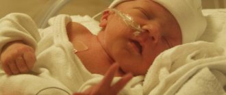
Rhabdomyoma in newborns
It is not always possible to detect a neoplasm in a newborn child at birth. Sometimes the disease can be detected only after several years of life. The pathology will form at the stage of fetal development, continuing to grow after birth.
It is sometimes possible to detect the disease in the fetus during a routine examination of fetal ECHO-CG. It is noted when there are disturbances in fetal development and hypoxia is detected.
The child suffers mainly from cardiac rhabdomyoma. The neoplasm is located in the area of the ventricle (left or right), at the site of the ventricular septum. It can also form in other departments. The danger of the situation lies in the growth of cells that can block the blood flow. And this can lead to the death of the child.
Sometimes there is an independent reduction of the tumor and the subsequent disappearance of the dangerous lesion. This happens in the absence of glycogen inside the tumor cavity. Therefore, doctors do not always rush into surgery. Surgical removal is performed in the presence of aggressively developing complications and chronic pathologies.
Rhabdomyoma diagnosed in parents recommends closely monitoring the child’s health. The newborn's heart is examined using the latest equipment. This is necessary to exclude the possible development of a neoplasm in a child. There are cases of heart disease being diagnosed due to a hereditary gene defect.
The baby may have tuberous sclerosis, a genetic disease that provokes the formation of a large number of benign nodes in different parts of the body.
Rhabdomyoma can cause serious complications - heart failure, cardiomyopathy, aortic stenosis and other pathologies. To prevent complications, it is recommended to carry out correct and timely therapy.
Signs of the disease
In the early stages, rhabdomyoma develops asymptomatically. The development of pathology can last several years. This is precisely what is associated with late diagnosis of the disease. After the tumor reaches a large size, the first signs appear in the victim. The clinical picture of the disease is associated with disturbances in the functioning of the heart.
Symptoms of rhabdomyoma in children:
- There are serious disturbances in the heart rhythm, which are manifested by tachycardia, bradycardia and other pathologies;
- Signs of edema appear in the soft tissues of the abdomen and limbs;
- The child is not gaining weight well – there is a weight deficit;
- Poor appetite or complete absence;
- The skin acquires a bluish tint;
- The child becomes capricious, fatigue sets in quickly;
- Mental and nervous disorders are present.
Damage to the ventricles causes heart block. The formation of a node in the area of the septum separating the ventricles is accompanied by prolonged fainting. An increase in formation causes cardiac arrest (asystole) or closure of the excretory duct, which causes acute heart failure.
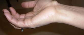
Signs of single neoplasms in the peritoneum, arms, legs, genitals, neck, thigh, tongue, maxillofacial area (MFA), and head are detected by visual inspection and palpation.
You can detect the pathology yourself or with a doctor during a routine examination. The formation develops without pain or other signs of discomfort. Outwardly they resemble small wen.
To clarify, you will need to undergo a detailed examination.
Diagnosis of the disease
Diagnosis takes place during fetal development, after the birth of a child and in the subsequent formation of a person. The lesion is examined using ECHO-CG, but the use of MRI provides an expanded study and a visual image in a 3D projection.
Based on the results obtained, the doctor will make an accurate diagnosis - identify the size, location, degree of damage to the heart muscle and the progression of the tumor.
To clarify, the doctor will take biological material for a biopsy. The diseased area is removed using cardiac catheterization. The procedure is unpleasant, and serious complications are possible after the procedure. Therefore, doctors do not always carry it out.
The study of other foci is carried out using the same techniques, but the removal of biological material is not difficult. Histology is performed after surgical removal of the entire area of rhabdomyoma.
Detection of a malignant form of pathology requires subsequent irradiation of the operated area with gamma rays.
Treatment of the disease
Rhabdomyoma is treated only with surgical excision. It must be removed together with the capsule in which the node was located. This will prevent or minimize the risk of relapse.
On the heart muscle, the operation is performed by complete excision and partial - open surgical access is used. After the operation, the biological material is sent to the laboratory for histology. Required to confirm the benign nature of the pathology.
For small sizes, a conservative course of therapy is used that does not affect blood circulation inside the heart. Medicines prescribed:
- To increase cardiac output;
- Myocardial support;
- Preventive drugs for heart failure.
After the course of therapy, the patient is under intensive medical supervision. This is required to prevent relapse. Tumor treatment always ends positively.
Source: https://onko.guru/dobro/rabdomioma.html
Symptoms of rhabdomyoma
If the formation is not diagnosed during the fetal development of the baby, then in the future it can only be discovered by chance, since for a long time the pathological process is asymptomatic. In most cases this is several years.
In general, the clinical picture of this disease will be characterized as follows:
- poor appetite, slow weight gain;
- cyanosis of the skin;
- weakness, lethargy, apathetic state of the child;
- moodiness, frequent crying and poor sleep for no apparent reason;
- swelling of the limbs;
- heart rhythm disturbance;
- unstable blood pressure;
- loss of consciousness for no apparent reason;
- delay in development, both mental and psychological.
In addition, the clinical picture of the pathology will be supplemented by corresponding signs, depending on the location of the formation - pain in the hip area and swelling, heart pain.
It should be understood that the growth of a tumor in the heart muscle is a rather dangerous process, since its excessive growth subsequently, if it is not removed, will lead to cardiac arrest.
Cardiac rhabdomyoma in newborns: types, causes, symptoms, treatment
Among tumor diseases there are those that are not malignant, but can threaten no less serious consequences. These include the disease described below.
Concept
A benign tumor formed by striated muscle tissue, rhabdomyoma, can have different localizations.
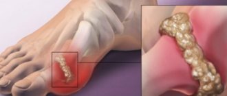
Usually this pathology is detected near large joints - knees, hips, but there are also formations of the heart (in children), tongue and throat (in adults) and genitals (only in women).
A specialist can make a preliminary diagnosis by examining a dense tumor up to 15 cm in size, which is not fused with the surrounding tissues, as it is encapsulated.
The section shows that it occupies the area of subcutaneous fat and muscle and is dark brown in color.
Experts note that this tumor can be independent or located inside other formations, for example, teratoma, and often consists of several nodules.
In childhood, this rare pathology mainly affects the heart (up to 60% of all myocardial tumors). The disease is detected at the age of 0-15 years, sometimes later.
In adults and adolescents, the neoplasm is most often detected in the retroauricular area, on the abdominal wall, tissues of the legs, and on the chest wall. The average age of diagnosis among adults is 36 years.
Types of disease
There are several classifications of pathology, the main one includes division by location:
- Cardiac tumors are found in the muscle of the heart.
- Extracardiac - have a different localization.
As for the types of tumors according to their histological examination, the following can be identified:
- Fetal. They can be polypoid, cellular, myxoid, differing from each other in the type of fiber arrangement and type of growth. They appear in humans from birth.
- Adults. Their structure is similar to that of physiologically normal striated tissue. A separate line is the tumor of the female genitalia.
All types of rhabdomyomas do not tend to grow invasively (damaging surrounding tissues) and are therefore considered benign. Some of them may recur after treatment, but this phenomenon is considered very rare.
Up to 10% of tumors can transform into malignant rhabdomyosarcomas.
Causes
The exact reasons remain a matter of debate. It is not clear exactly how normal muscle cells degenerate into a tumor, but it is assumed that this occurs under the influence of certain provoking factors.
These factors cause a failure in the production of myocytes (muscle cells) or the cells that form Purkinje fibers in the heart. But other researchers are of the opinion that such cells disrupt their structure already during ripening.
It is possible that both processes take place in the pathogenesis of the tumor, depending on which a fetal or adult formation appears.
What can influence the improper formation and development of myocytes in the tissues of the body? Presumably these factors are:
- burdened heredity;
- bad ecology;
- work in hazardous industries;
- poisoning, intoxication;
- hormonal imbalances;
- metabolic diseases, especially carbohydrate metabolism;
- mutation of genes during conception.
Cardiac rhabdomyoma in newborns
Despite the fact that this disease is not always detected immediately after birth (sometimes the diagnosis is made after 10-15 years), the prerequisites for its appearance are present immediately from birth.
Often, pathology is detected during fetal ECHO-CG, which is performed routinely or to search for causes of growth retardation and prerequisites for fetal hypoxia.
A benign heart tumor of this type is the most common location of rhabdomyoma in children. Most often it is located in the left or right ventricle or on the septum between them, but sometimes it is detected in other parts.
This benign tumor is very dangerous, because many of them are characterized by intracavitary increase in size, and therefore can block the blood flow path, which provokes the death of the child.
A favorable outcome is also possible, since almost half of such formations tend to spontaneously decrease (involution) and disappear without treatment when the glycogen supply in them is depleted. That is why indications for surgery for a heart tumor in a child are not always present, but only when complications and concomitant diseases develop.
If parents have rhabdomyoma, it is imperative to conduct a thorough examination of the heart in newborns. The fact is that there are familial forms of heart disease, which are caused by the presence of defective genes.
Complications of heart formation in children can include heart failure, aortic stenosis, cardiomyopathies and other severe pathologies, so treatment, if necessary, should be timely.
Symptoms
Late detection of the disease is often due to the fact that it does not give clinical signs, develops over years, but is not noticeable outwardly. Only when the tumor reaches a significant size will it manifest itself with the symptoms described below.
Of course, disruption of the heart will lead to a decrease in cardiac output and various disruptions in conduction.
The main manifestations of pathology that occurs in the heart area in children can be as follows:
- arrhythmias (bradycardia, tachycardia, extrasystole, etc.);
- swelling of the limbs and abdomen;
- poor weight gain;
- impaired appetite;
- skin cyanosis;
- frequent crying, weakness, capriciousness of the child;
- various types of deviations in mental development.
If the tumor is localized inside the ventricles, it most often causes the development of heart block. Serious conduction failures occur when it is located on the interventricular septum, and they are often accompanied by a prolonged loss of consciousness.
If the tumor continues to grow in size, it may result in asystole - cardiac arrest, and if it closes the ventricular outflow tract - acute heart failure with a poor outcome.
The symptom complex of single rhabdomyomas of another location - the abdominal wall, genitals, limbs, neck and head - is usually represented by visual or palpation detection independently or during a routine examination by a doctor.
Tumors do not cause pain or discomfort and are perceived by many as “fat tumors.” Only a doctor can make an accurate diagnosis after conducting appropriate tests.
Diagnostics
A heart tumor can be diagnosed in utero or after the birth of a child, as well as at any other age.
The main research method continues to be ECHO-CG (ultrasound of the heart), but in recent years, cardiac MRI has become a more accurate method of diagnosis.
These techniques allow you to make a diagnosis, assess the size and location of the formation, identify hemodynamic disorders in the heart, and also analyze the growth of the tumor.
An accurate diagnosis can only be made after a biopsy, despite the fact that instrumental methods are of great importance for a preliminary assessment of the scope of surgery (if necessary). A biopsy is taken by cardiac catheterization, but this procedure is fraught with various complications, so it is not always performed.
Tumors of other localizations are diagnosed using the same methods - ultrasound and MRI, but if the tumor is externally located, its biopsy will not be so difficult.
Often, histological analysis is carried out after complete removal of the tumor.
In case of erroneous diagnosis and subsequent detection of a malignant tumor, the patient subsequently undergoes irradiation of the operated area.
Treatment of rhabdomyoma
The only way to get rid of this disease is surgery. The tumor is removed along with the capsule, after which the risk of its recurrence is minimized.
Neoplasms on the heart are excised, as far as possible, completely or partially, using an open cardiac surgical approach. In the future, it is mandatory to wait for the histological report to confirm the benign nature of the pathology.
Conservative therapy is used if the tumor is small, does not cause concern and does not interfere with intracardiac circulation. In this case, drugs are used to increase cardiac output, to strengthen the myocardium and prevent heart failure.
Observational tactics at the beginning of rapid tumor growth should be replaced by surgery, and after it the person must be observed for a long time by a doctor for early detection of relapse.
Source: https://gidmed.com/onkologiya/lokalizatsiya-opuholej/serdechnaya-sistema/rabdomioma.html
What is rhabdomyoma
Rhabdomyoma is a benign type of formation, which is formed from striated tissue and can have different localization. However, cardiac rhabdomyoma most often occurs in children under 15 years of age. Rhabdomyoma of the tongue or nose is extremely rare.
Despite the fact that rhabdomyoma is a benign formation, it has a tendency to degenerate into malignant, therefore, after its removal, histological analysis is mandatory.
The clinical picture of such a pathological process does not appear immediately: in most cases, symptoms appear only after several years from the onset of the disease. However, it is possible to diagnose such a disorder in the fetus during intrauterine development.
It should be noted that in a fetus in the early stages of development, such a formation can lead to serious disorders, including Down syndrome. Diagnostically, rhabdomyoma is established only through laboratory and instrumental measures. Treatment is usually surgical: only removal of the tumor itself, as well as its capsule, will minimize the risk of relapse. In the early stages, if cardiac rhabdomyoma is diagnosed in newborns, conservative methods of therapy are used.
The forecast will be individual. According to ICD-10, this pathological process has a separate meaning - code D21.
Cardiac rhabdomyoma: in newborns, children, development prognosis
Cardiac rhabdomyoma is a neoplasm that develops from the striated muscle tissue of this organ. The tumor occurs in a benign form and is often diagnosed in children, but it also occurs in adults.
Causes
Factors contributing to the development of a benign heart tumor are the following:
- Genetic predisposition.
- Unfavorable environmental conditions in the region of residence.
- Work associated with hazardous production.
- Poisoning of the body.
- Failure of hormonal balance.
- Disturbance of metabolic processes in the body.
- Mutation of tissues during fetal development.
In most cases, cardiac rhabdomyomas are detected in newborns a few weeks after birth. But sometimes the pathology is discovered several years after birth.
Symptoms
Rhabdomyoma in the heart area may not show any signs for a long time. After all, this is a benign pathology that occurs gradually and grows slowly. But over time, the tumor begins to interfere with the organ’s ability to function fully, and therefore the patient is troubled by various symptoms.
These include the following:
- Heart rhythm disturbance.
- Swelling of tissues in the leg area.
- Increase in body weight.
- Decreased appetite.
- Blueness of the skin.
- Crankiness in a child.
- General weakness in the body.
- Development of mental disorders.
If the tumor is located in the ventricles, heart blockades often occur; if it is in the area of the interventricular septum, then prolonged fainting is observed. If treatment is not taken promptly, the tumor will continue to grow and may cause heart failure and cardiac arrest.
Therapeutic measures
Treatment is prescribed depending on the size of the tumor, the presence of concomitant diseases in the presence of rhabdomyoma, the age of the patient and other factors. Both conservative and surgical methods of therapy are possible.
Conservative treatment is prescribed if:
- The neoplasm is small in size.
- The patient has no symptoms.
- There is no disturbance of intracardiac circulation.
In this case, the doctor uses medications that increase cardiac output, strengthen the heart muscle, and prevent the occurrence of heart failure.
If the formation tends to grow rapidly and interferes with the functioning of the organ, the doctor prescribes surgery. It is carried out in an open way. The tumor is removed along with the capsule, which minimizes the likelihood of relapse.
Forecasts prevention
The prognosis for rhabdomyoma is favorable. The sooner measures are taken to eliminate the pathology, the easier it will be to cope with it without harm to health. But it is still better to prevent the development of a tumor disease at all.
To prevent the development of the disease in the mother’s child during pregnancy, the following is recommended:
- Avoid exposure to radiation and harmful substances.
- Stop smoking and drinking alcohol.
- Monitor hormonal balance.
- Do not overload the body.
- Eat properly.
- Walk more in the fresh air.
- Take vitamins if they were prescribed by your doctor.
To prevent rhabdomyoma, adults should also follow all of the above recommendations. Thus, the most common location of rhabdomyoma is the heart. The disease most often affects children, especially newborns. The pathology is benign, but treatment must be carried out.
Source: https://opake.ru/dobrokachestvennaya-opuhol/rabdomioma-serdca/
Causes of rhabdomyoma
The exact causes of this disease have not yet been established. However, clinicians suggest that the development of the disease in the area of soft tissue, heart or hip (other types of localization are not excluded) may be due to the following factors:
- mutation of genes during the conception of a child or its intrauterine development;
- bad heredity;
- metabolic disorders, especially when it comes to carbohydrates;
- work in production;
- hormonal disorders;
- poisoning with heavy metals, chemicals or poisons;
- living in an extremely unfavorable environmental environment.
It is also possible that rhabdomyoma may develop due to the bad habits of the mother: smoking, drinking alcohol during pregnancy.
Treatment of rhabdomyoma
Elimination of pathology by conservative methods is possible only in the early stages. In most cases, surgery is used - the tumor is removed along with the capsule to prevent relapse of the disease.
If the histological analysis shows the malignant nature of the formation, an additional examination is carried out, on the basis of which the doctor will prescribe subsequent treatment.
Even after surgical excision of the formation, its recurrence cannot be ruled out. Therefore, patients with such a diagnosis should be registered with an appropriate specialist and systematically undergo a preventive examination.
The prognosis will be positive if the formation is benign and not multiple. In other cases, everything will depend on the form and severity of the oncological process.
As for prevention methods, in this case there are none, since the nature of the development of such a pathology has not been established. Pregnant women need to regularly undergo ultrasound examinations for timely diagnosis of pathology, and parents regularly take their child for preventive medical examinations.
What to do?
If you think you have Rhabdomyoma
and the symptoms characteristic of this disease, then doctors can help you: pediatrician, cardiologist.
Be healthy!
Source
Did you like the article? Share with friends on social networks: