The size of the tumor can be several millimeters or several centimeters (cystic formations above 25 cm are very rare).
A cyst is a pathological phenomenon that may not manifest itself for a long time, so the disease is most often diagnosed in the later stages. In women, this pathology is detected 3-5 times more often than in the stronger sex. Mostly people of mature age (30-55 years) suffer from the disease.
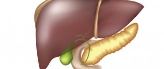
General information, liver cyst - what is it?
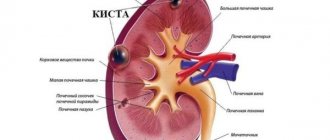
A liver cyst is a benign neoplasm that consists of a fluid-filled cavity with its own walls in the form of a capsule.
Cystic formations are diagnosed in 0.8-2% of people. Representatives of the weaker sex in the age category of 30-50 years are more often susceptible to the disease. The inside of the cyst is lined with cubic or columnar epithelium; inside there is liquid content. Unlike false cysts, true cysts have their own walls formed from liver tissue.
Inside the cavity, the contents may be transparent and colorless, but in rare cases a jelly-like mass of a greenish or brownish tint is determined. With the development of an infectious process inside, the contents become purulent, and with hemorrhages - hemorrhagic.
Cystic formations are detected in various segments and lobes of the liver and can reach very large sizes (more than 25 cm in diameter). The cyst can be located intraparenchymal or subcapsular. Code “liver cyst” according to ICD-10: cystic liver disease (congenital) (Q44.6), liver damage in diseases classified in other headings (K77*).
Pathogenesis
The formation of a parasitic cyst from the moment of infection includes 3 stages:
- Penetration of the parasite first into the blood and then into the hepatic system, forming a cyst capsule with small contents. At this stage, the liver is able to fully perform its functions, the immune system works as usual. The stage is completely asymptomatic.
- An increase in the size of the tumor, the formation of a specific stalk, which falls into the abdominal cavity. The tumor reaches such a size that it begins to compress the liver and nearby organs, causing severe pain to the patient.
- Rapid progression of the process, which is accompanied by a pronounced inflammatory process around the cyst and subsequent suppuration. In rare cases, it is at this stage that the cyst ruptures with damage to the liver structures.
Classification
There are several classifications of liver cysts. Depending on the etiological factor, the cyst can be:
- parasitic;
- non-parasitic.
A parasitic liver cyst can be alveococcal or echinococcal. The first occurs as a result of damage to the hepatic system by alveococcus . An hydatid cyst is formed after a tapeworm enters the human body and invades the liver for further reproduction. The echonococcal form occurs as a result of the introduction of the parasite through the portal bloodstream.
Classification of non-parasitic cysts:
- Congenital (true). They occur most often, because are formed as a result of abnormal development of the bile ducts. True cysts in the absence of dynamic growth are considered harmless.
- Acquired (false). Their occurrence is often associated with mechanical trauma to the liver.
Division of true cysts:
- Solitary . Characteristically, it is the right lobe of the liver that is affected, and specifically its lower part. The solitary cyst of the right lobe of the liver has a peculiar pedicle, which allows it to hang into the abdominal cavity.
- Multiple liver cysts (polycystic) . They arise as a result of a genetic mutation. Multiple cysts are always located in the upper layers of the liver. Neoplasms spread diffusely throughout the liver and increase in size throughout a person’s life.
- Cystic fibrosis . It is considered the most dangerous form, because poses a real threat to the life of a child.
- Fibrosis affects not only the hepatic system, but also the portal vein. The pathology is characterized by the growth of a huge number of bile microcysts.
Based on size, neoplasms are divided into:
- small (volume up to 1 cm);
- medium (size from 1 to 3 cm);
- large (up to 10 cm);
- giant (more than 25 cm).
Cysts can also be complicated or uncomplicated.
Surgical treatment
Usually, surgical or surgical treatment is prescribed to treat the pathology.
Indications for surgery:
- bleeding;
- ruptures of the walls of the neoplasm;
- increased symptoms;
- large tumor size;
- the beginning of suppuration;
- violation of bile flow.
Surgery can be of different types:
- Radical, when the patient needs an organ transplant, for example, in the case of polycystic formation.
- Conditionally radical, when the affected parts of the liver and the cyst are removed.
- Palliative, when the contents of the cyst are removed, and its walls are glued together and removed from the organ.
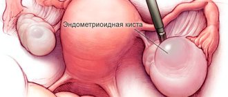
The last method is considered the most effective. However, after it is carried out, relapses may occur. Palliative intervention or percutaneous puncture under ultrasound guidance is performed. If the size of the cyst exceeds 6 cm, then puncture is ineffective.
If the cyst is parasitic, then it is treated with special antibacterial medications. However, such treatment is only possible if it is small in size. Otherwise, surgery is performed.
There are other methods of surgical intervention:
- Opening the tumor and draining it.
- Marsupialization - cleansing the formation and suturing its walls to the edges of the surgical incision.
- Fenestration is the opening of polycystic formations with excision of their walls.
Be sure to read:
Parasites in the human body - symptoms of presence and methods of treatment at home
A radical method of treating solitary tumors gives a favorable prognosis. The sooner you contact a specialist, the easier and faster the treatment will be.
Causes of liver cysts
There is no consensus on the causes of cysts in the hepatic system. Various reasons lead to the development of different types of neoplasms.
A true cyst occurs against the background of long-term inflammatory hyperplasia of the biliary tract, which is accompanied by obstruction. During intrauterine development, intralobular flows are not connected to the general biliary system, while the epithelium continues to produce fluid without interruption, which leads to its accumulation inside the cavity and the formation of a hydrated liver cyst. Bile is never found among the contents of the cyst , which indicates the absence of points of contact with the bile ducts, which function normally. Young solitary cysts contain clear liquid inside, while old ones contain green mucus.
Also, the causes of liver cysts may lie in the use of hormonal drugs, including oral contraceptives, during the treatment of which false cysts are formed.
False cysts are formed after liver injuries, inflammatory processes, and also as a result of surgical treatment of a liver abscess or other pathology.
Parasitic cystic formations are formed against the background of helminthic infestation, mainly by echinococci.
Diagnosis and treatment
Diagnosis of the disease is primarily done using imaging methods such as CT, ultrasound, and MRI. In some cases, the doctor can also detect the presence of a cyst by palpating the abdomen. Cystic echinococcosis is detected using an enzyme-linked immunosorbent assay aimed at detecting antibodies to echinococcus in the blood.
Most often, a liver cyst does not require treatment and only requires regular medical observation and an appropriate diet.
Symptomatic cysts that enlarge or become malignant are not treated but must be removed surgically. Sometimes drainage of the cyst fluid is also done, but this procedure has a temporary effect.
Symptoms of a cyst in the liver
Main signs of pathology:
- frequent belching;
- prolonged nausea;
- muscle weakness;
- shortness of breath with minimal physical activity;
- dull pain in the right hypochondrium with possible irradiation to the scapula, arm, and periumbilical region;
- prolonged diarrhea syndrome;
- discomfort in the right hypochondrium during shaking, traveling in transport;
- pain in the epigastrium during physical activity;
- excessively rapid fatigue;
- increased body temperature;
- decreased appetite;
- hyperhidrosis , sweating ;
- sudden weight loss for no particular reason;
- asymmetrical deformity of the abdomen.
Most often, a liver cyst does not manifest itself in any way, especially in cases where it is single or small in size. Formations are detected accidentally during ultrasound diagnostics or computed tomography for other reasons. Symptoms largely depend on the size of the cyst, its shape, nature, location and general condition of the patient.
One of the first signs of pathology is pain. The pain is localized in the right hypochondrium and can radiate to the left side, the central part of the abdomen and even under the shoulder blade. Patients describe the pain as dull and associate its occurrence with eating or physical activity. As the pathology progresses, the pain becomes constant and bothers the patient even at rest.
Symptoms indicating damage to 20% of the liver and reaching a cyst diameter of 70 mm:
- prolonged diarrhea ;
- increased gas formation ;
- quick satiety after eating a small amount of food;
- a significant increase in size of the hepatic system is detected by palpation and visually.
In some cases, nonspecific symptoms of pathology are detected:
- dyspnea;
- low-grade fever;
- increased sweating ;
- lack of appetite;
- fatigue.
With alveococcal and hydatid cysts, patients complain of:
- the appearance of a rash all over the body of unknown origin;
- yellowness of the skin and severe itching;
- increased body temperature;
- visual and palpable enlargement of the liver in size;
- irradiation of pain into the chest.
The following symptoms indicate possible perforation and bleeding from the cyst:
- chills, fever ;
- the appearance of sharp and intense pain in the abdomen;
- excessive tension in the abdominal muscles;
- pale skin;
- constipation;
- a drop in blood pressure with a parallel increase in heart rate;
- pronounced, profuse sweating .
If the symptoms described above are detected, it is recommended to immediately seek qualified medical help.
Symptoms of the disease, why a cyst is dangerous
Small cysts with a small diameter usually do not cause any clinical symptoms and are not diagnosed for many years. As the cysts grow in size, some patients may experience pain and discomfort in the liver area. Liver function in this pathology is usually not impaired, so cysts are not visible in any way in the laboratory.
Only 5% of patients with liver cysts have specific symptoms of the disease. Most often this is heaviness in the right hypochondrium, weakness, a feeling of fullness in the abdomen, bloating for no apparent reason, abnormal bowel movements, and increased gas formation. Some patients may experience a feeling of bitterness in the mouth and pain of varying intensity.
Sharp and sudden pain in the liver area, not relieved by taking antispasmodics, can occur with spontaneous rupture of cysts, but such complications are typical only for large formations and are rare.
The most striking clinical picture is the echinococcal type of cyst. Parasitic patients may experience febrile fever, severe skin itching, cough, severe pain in the liver, yellowing of the skin and sclera, weight loss, and bowel dysfunction. The appearance of these symptoms is a reason to immediately consult a doctor for examination and treatment.
Tests and diagnostics
Clinically, a liver cyst may not manifest itself in any way, and therefore is often a finding during a clinical examination.
First of all, it is necessary to determine the type of cyst, its exact location and size. It is important to timely determine the presence of an inflammatory process around the cyst.
Basic diagnostic methods:
- ultrasonography;
- CT, MRI;
- cyst puncture.
Computed tomography and magnetic resonance imaging help to clearly differentiate cystic neoplasms from other similar pathologies (tumor, hematoma, etc.). MRI is considered the most informative method that allows you to assess the overall picture of the disease. The study allows you to obtain a three-dimensional image of the organ and its inclusions.
Computed tomography provides layer-by-layer images, thanks to which it is possible to characterize the structural structure of the organ itself and the neoplasms in it. CT is used to identify small cysts located both on the surface of the liver and inside it. The study allows you to identify cysts at the earliest stages and timely diagnose polycystic disease. An abdominal CT scan to detect tumors involves intravenous administration of a contrast agent.
Ultrasound examination allows us to determine the morphology, size and type of tumor. It is considered the simplest, most accessible and widespread diagnostic method. To assess the benignity of the formation, a puncture of the cyst is performed to determine tumor markers .
If the cyst is parasitic in nature, then an additional blood test is performed to identify parasites in other organs and tissues.
It is important not only to diagnose the very fact of the presence of a cyst, but also to determine its size, nature, and the number of similar neoplasms in the organ. It is necessary to identify the true cause that led to the formation of the cyst in order to avoid a possible relapse of the disease. Timely and competent diagnosis allows you to start treatment at the earliest stages and avoid the development of complications that can have serious consequences for the health and life of the patient.
Diagnostics
To cure a disease, you need to diagnose it. Doctors usually prescribe the following tests:
- Ultrasound of the abdominal cavity or liver. The method is used to determine the number of tumors and their size. Using an ultrasound, the puncture site is determined to determine the contents of the cyst - fatty, with liquid or pus.
- CT and MRI. Examination methods are prescribed if it is necessary to determine the size and number of cysts, identify the causes of their appearance and possible complications.
- Laparoscopic examination. Laparoscopy is needed for accurate diagnosis, for example, when an ultrasound revealed a tumor similar to a cyst.
If the doctor suspects a parasitic origin of the cyst, he will order additional tests to determine the parasite. Such analyzes include ELISA, RIA, XRF and others.
Treatment of liver cysts with folk remedies
Herbal infusions and decoctions can provide powerful support to a weakened body affected by neoplasms. However, uncontrolled use of herbal infusions can have a negative effect on the entire body. Before starting treatment with folk remedies, it is recommended to consult your doctor about compatibility with the main drug therapy. Plants used in recipes:
- celandine (juice);
- burdock (root and juice);
- elecampane;
- tenacious bedstraw;
- yarrow;
- milk thistle;
- pine nut (shell).
In addition to medicinal herbs, traditional medicine recommends consuming quail eggs and kombucha infusion.
Folk recipes
Folk remedies can be used as an additional method of therapy. But it is recommended to use them only after consultation with your doctor. Uncontrolled consumption of folk remedies can cause a deterioration in the patient’s general condition and the development of dangerous complications.
The most effective folk remedies are based on:
- burdock leaves;
- celandine juice;
- pine nut shells.
Traditional recipes are effective only in tandem with traditional medicine methods. Under no circumstances should they be the only method of treatment.
Diet for liver cysts
Diet for liver cysts
- Efficacy: no data
- Terms: 1-6 months/lifetime
- Cost of products: 1500-1600 rubles. in Week
Diet for liver inflammation
- Efficacy: therapeutic effect after 10 days
- Timing: constantly
- Cost of products: 1300-1400 rubles per week
Dietary nutrition for cystic neoplasms in the liver involves avoiding some foods and including others in the diet.
It is prohibited to use:
- sweets;
- carbonated drinks;
- spices, seasonings;
- coffee;
- fried, salted, smoked and spicy dishes.
Permitted for use:
- greenery;
- fish;
- vegetables and fruits;
- cellulose;
- fermented milk products;
- sea buckthorn and rose hips.
The diet involves eating foods that have undergone deep heat treatment. The diet must contain easily digestible protein (at least 120 g per day) and a minimum amount of fat (no more than 80 g). Daily calorie content should not exceed 3000 Kcal. The meal plan must be fractional.
Complications and consequences of liver cysts
Patients are concerned about what consequences a cyst in the liver may develop in the near future.
After complete resection of a solitary cyst, the likelihood of relapse is very low if you follow the diet recommended by the gastroenterologist-hepatologist and all recommendations.
After puncture of a cyst, the risk of its reoccurrence or the appearance of a similar neoplasm nearby is very high.
In the absence of adequate therapy, complications such as:
- cyst rupture;
- bleeding from a cyst;
- inflammation of the tissue around the cyst;
- liver failure;
- entry of parasites into the abdominal cavity.
Treatment
Many people, when cysts are discovered, think about how to treat this pathology. Most cysts do not require treatment and resolve on their own over time. If the cysts are small and isolated, the patient only needs to undergo periodic examinations to determine their size and growth dynamics. If the cyst increases in size or if it spontaneously ruptures, surgical treatment may be offered.
One of the surgical treatment options is puncture removal of fluid from the cyst, as a result of which it “collapses” and regresses. However, this procedure may have only a temporary effect, which depends on the etiology of its occurrence. If the causative factor is not removed in time, the fluid will again begin to flow into the cyst and cause its growth. If the cyst is accessible, laparoscopic surgery is possible. The life prognosis is usually favorable.
Drug therapy is not used due to its low effectiveness and lack of pronounced clinical effect.