| Aplastic anemia | |
| ICD-10 | 60.-61. |
| ICD-9 | 284284 |
| OMIM | 609135 |
| DiseasesDB | 866 |
| MedlinePlus | 000554 |
| eMedicine | med/162 |
| MeSH | D000741 |
Aplastic anemia
is a disease of the hematopoietic system, classified as myelodysplasia and expressed in a sharp inhibition or cessation of growth and maturation of all three cell lines in the bone marrow, or so-called panmyelophthisis.
Aplastic anemia is characterized by severe pancytopenia - anemia, leukopenia, thrombocytopenia and lymphopenia.
Medical history
This disease was first described by Paul Ehrlich in 1888 in a 21-year-old woman. The term "aplastic anemia" was coined by Chauford in 1904. Aplastic anemia is one of the most severe disorders of hematopoiesis. Without treatment, patients with severe forms of aplastic anemia die within a few months. With timely adequate treatment, the prognosis is quite good. For a long period of time, aplastic (hypoplastic) anemia was considered as a syndrome combining pathological conditions of the bone marrow occurring with severe hypoplasia of hematopoiesis. Currently, a disease called “aplastic anemia” is isolated as an independent nosological unit - and it is clearly distinguished from hematopoietic hypoplasia syndrome, which is a manifestation of a number of known independent diseases of the bone marrow.
Classification of aplastic anemia
| Form of pathology | What is the difference? |
| By duration | |
| Acute | No more than one month |
| Subacute | From one month to six months |
| Chronic | More than six months |
| According to the severity of selective aplasia | |
| Moderate | Granulocytes less than 0.0x10⁹/l, platelets less than 20.0x10⁹/l. |
| Heavy | Granulocytes less than 0.5x10⁹/l, platelets less than 20.0x10⁹/l. According to diagnostic results, bone marrow cellularity is less than a third of normal. |
| Very heavy | Granulocytes more than 0.5x10⁹/l, platelets more than 20.0x10⁹/l. |
Pathogenesis
Aplastic anemia can develop when exposed to a number of myelotoxic factors: ionizing radiation, chemicals - benzene, gold salts, arsenic; medicines - chloramphenicol (chloramphenicol), phenylbutazone (butadione), chlorpromazine (aminazine), meprobamate, dilantin, antimetabolites (6-mercaptopurine, methotrexate), alkylating agents (cyclophosphamide, chlorbutine) and some other drugs. The myelotoxic effect from the influence of some factors (ionizing radiation, antimetabolites) always occurs at a sufficiently large dose, while others manifest themselves individually. The reason for individual sensitivity, in particular to certain drugs, is not always clear, but may be associated with genetic defects of hematopoietic cells. This applies, for example, to chloramphenicol and phenylbutazone, which cause suppression (depending on the dose) of erythropoiesis with a frequency of 1:24,000 and 1:40,000, respectively, in individuals taking them. The hereditary nature of the individual sensitivity of erythropoietic cells to these drugs is confirmed by the development of bone marrow aplasia in different members of the same family and in identical twins. In other cases, a connection between drug-induced inhibition of hematopoiesis and immune mechanisms is likely due to the appearance of antibodies to erythrocyte precursors. Cases of the occurrence of aplastic anemia after acute viral hepatitis (possibly due to the ability of the hepatitis virus to change the karyotype of cells, which was traced in a culture of leukocytes), infection with the Epstein-Barr virus, and parvovirus have been described.
There is also a hereditary form of aplastic anemia - Fanconi anemia.
In more than half of patients, it is not possible to identify any causative factors - this is the so-called idiopathic aplastic anemia. The mechanisms underlying idiopathic anemia are unclear. A possible autoimmune mechanism is associated with the effect of autoantibodies on bone marrow cells with the participation of immune lymphocytes. It has been shown that lymphocytes (T-suppressors) from patients inhibit the formation of erythrocyte colonies in the donor's bone marrow and can disrupt the differentiation and proliferation of hematopoietic precursors.
It is also assumed that the basis of aplastic anemia may be a lesion (internal defect) of the stem cell, as evidenced by the restoration of hematopoiesis in patients after transplantation of allogeneic bone marrow containing normal stem cells. There are experimental data indicating the importance for the development of the aplastic process and microenvironmental disorders - the primary defect of bone marrow stromal cells. However, the essence of these cellular defects remains unclear, as well as their primacy. It is possible that in different forms of aplastic anemia the pathogenetic mechanisms are different.
Diagnostics
The picture of peripheral blood is represented by tricytopenia. The decrease in hemoglobin is significant and can reach a critical level of 20 - 30 g/l. The color index is usually equal to one, but in some cases there may be hyperchromia and macrocytosis of erythrocytes. The number of reticulocytes is sharply reduced. Severe leukopenia (agranulocytosis) is characteristic. The absolute content of lymphocytes is unchanged or reduced. The platelet count is always reduced, and in some cases it is not possible to detect them at all. In most cases, ESR increases (up to 40 - 60 mm/hour).
The clinical picture of the disease allows us to form a primary idea of the pathology of the blood system. The starting point of the diagnostic search is a clinical blood test with counting the number of reticulocytes and platelets. Detection of bi- or tricytopenia during peripheral blood examination serves as the basis for performing a morphological examination of the bone marrow.
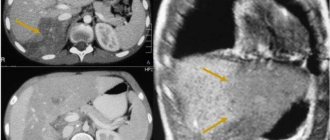
The diagnosis of AA is established on the basis of a typical histological picture of the bone marrow obtained by trepanobiopsy of the iliac crest. To obtain a high-quality (informative) biopsy, industrially produced trephines (Sherwood medical) are used.
Histological examination of the bone marrow reveals a large amount of adipose tissue, the content of which can reach 90%. Among the dominant adipose tissue there are stromal and lymphoid elements. Hematogenous cells are extremely poorly represented: erythroid and granulocytic precursors are found in small numbers. Megakaryocytes are absent.
Treatment
Treatment of aplastic anemia is a very difficult task.
- Treatment with glucocorticoids is effective if the disease is caused by autoimmune mechanisms, the appearance of antibodies against blood cells.
- Treatment with anabolic drugs stimulates hematopoiesis.
- Androgen treatment has an anabolic effect and stimulates erythropoiesis.
- Treatment with cytostatics (immunosuppressants) is prescribed only if there is no effect from other treatment methods in patients with an autoimmune form, including partial red cell aplasia.
- Splenectomy
- Treatment with antilymphocyte globulin is recommended if splenectomy and other treatments fail.
- Treatment with cyclosporine. Cyclosporin A (sandimmune) has an immunosuppressant effect, selectively inhibits the transcription of the interleukin-2 gene in T-lymphocytes, suppresses the production of interferon gamma and tumor necrosis factor alpha.
- Bone marrow transplantation.
The main and only pathogenetic method of treating aplastic anemia, which allows us to count on saving the patient’s life, is bone marrow transplantation from a compatible donor. If it is impossible to select a donor, palliative therapy is carried out. The immunosuppressant cyclosporine A is used as a basic drug. In patients with mild aplastic anemia, the use of this drug allows one to count on success in some cases. In addition, the use of cyclosporine A is advisable from the standpoint that glucocorticoids, androgens and antilymphocyte globulin can improve the state of hematopoiesis in patients with mild aplastic anemia, but, however, the increased risk of subsequently developing clonal bone marrow diseases should be taken into account. The use of cyclosporine A minimizes this risk. It should also be noted that some patients with mild aplastic anemia who have overcome the 6-month survival threshold may experience spontaneous improvement even if they have not received any immunosuppressive therapy. The effect of immunosuppressive therapy in patients with severe and extremely severe aplastic anemia is questionable.
- Treatment with stimulating factors or myeloid growth factors are glycoproteins that stimulate the proliferation and differentiation of various types of hematopoietic progenitor cells.
- Indications for red blood cell transfusion include severe anemia, signs of brain hypoxia, and hemodynamic disturbances.
All patients with aplastic anemia require replacement transfusion therapy with red blood cells and/or platelets. The volume of transfusion therapy is determined by peripheral blood parameters and clinical manifestations of the disease. In addition, antibacterial and mycostatic therapy is carried out to prevent or treat infectious complications.
Historical reference
In the pre-transfusion era, untreated aplastic anemia often resulted in the death of the patient. Thanks to the development of transfusion medicine, patients who do not respond to drug therapy have a chance to extend their lives by several years. At the same time, they may develop refractoriness (immunity) to allogeneic platelets due to the appearance of alloantibodies. Ultimately, persistent neutropenia increases the risk of bacterial or, more commonly, fungal diseases.
Forecast
Remission can be achieved in approximately half of patients. The prognosis is slightly better in children than in adults. The presence of a large amount of fat in the bone marrow does not mean the process is irreversible. There are cases when such patients experience complete remission and complete repair of bone marrow hematopoiesis. The prognosis is better when the content of reticulocytes is increased, when there is a more polymorphic picture in the bone marrow, when there is a slight increase in the size of the spleen and at least a small but clear effect of corticosteroid hormones. In these cases, splenectomy often has a good effect until complete recovery. In some patients, aplastic syndrome is the onset of acute leukemia. Sometimes signs of hemoblastosis are revealed only several years after the onset of the disease.
| This article lacks links to sources of information. Information must be verifiable, otherwise it may be questioned and deleted. You may edit this article to include links to authoritative sources. This mark was set on October 26, 2020 . |
Aplastic anemia: symptoms
The disease begins acutely, it is accompanied by a feeling of severe weakness and fatigue. The patient's skin and visible mucous membranes look pale, and he himself suffers from the following clinical manifestations:
- noise in ears;
- the appearance of shortness of breath even with insignificant efforts;
- unpleasant tingling in the chest area.
When the number of platelets per unit volume of blood decreases, hemorrhagic syndrome appears:
- even after slight compression or shock to the skin, bruises and hemorrhages appear on it;
- a rash in the form of small dots can be seen on the body, arms and legs;
- bleeding gums are observed;
- spontaneous nosebleeds;
- heavy menstruation (in women).
A decrease in the number of leukocytes per unit volume of blood is characterized by the regular frequent development of infectious diseases of the skin and structures of the urinary system, inflammatory processes of the oral mucosa, and pneumonia.
The congenital form of anemia develops in children under ten years of age and is accompanied by a number of other disorders:
- underdevelopment of the skull and brain;
- reduction in the size and weight of the kidneys (hypoplasia);
- intense coloring of certain areas of the skin - hyperpigmentation;
- severe hearing loss and speech impairment due to it.