Conservative treatment methods.
They are used only in cases of incomplete fusion and in the absence of inflammatory processes.
- Correct hygiene procedures. Washing in the morning, in the evening, and also after each bowel movement, with warm clean water only using hands without various flavors and antiseptics. Do not constantly use soap and sponge.
- The genitals should always be clean and dry.
- Refusal to use wet sanitary napkins, as well as powders and various waxed creams.
- Replacing colored synthetic underwear with colorless cotton.
- Bathing the baby in water containing a solution of potassium permanganate, St. John's wort or chamomile.
- Daily short-term massage with light pressing semicircular movements using baby cream.
This kind of treatment is, first of all, the prevention of fusion of the labia minora in little girls. Following these recommendations will allow you to get rid of small synechiae in a week or two. However, remember that even if you have never encountered synechiae, girls under 6 years of age need periodic visits to a pediatric gynecologist - at least once a year! Well, if there have already been fusions - at least once a month, because relapses of synechiae are a very common occurrence.
Big problems baby
Why does the baby become capricious when about to pee? Anything can happen, of course. But, if this happens again and again, most likely the problem is not trivial and it is called synechiae. This means that the baby’s labia minora “stick together”, that is, the entrance to the vagina is blocked by a membranous membrane.
The larger its size, the more difficult it is to write. Synechiae most often form in children from infancy to three years, but sometimes it happens that the process is delayed until adolescence, until the girl’s body begins to produce a sufficient amount of the hormone estrogen. What can trigger the disease?
Paradoxical as it may seem, our “obsession” with hygiene. If you wash your baby several times a day, instead of benefit there will be one harm: the protective water-fat layer is destroyed and the mucous membrane dries out.
Of course, this does not mean at all that the less often such procedures are performed, the less danger there is. The contact of bacteria on the delicate mucous membrane can also provoke inflammation, leading to synechiae. Therefore, you need to do everything correctly: wash the baby with warm running water from front to back and use less of any “detergent” products.
Note! The risk of synechia formation increases if during pregnancy the mother suffered from severe toxicosis, gestosis, or the baby received an intrauterine infection while passing through the birth canal. Premature babies are also susceptible to this disease.
Diagnosis of vulvovaginitis in girls
For the diagnosis of vulvovaginitis, anamnesis (concomitant diseases, provoking factors - foreign body, masturbation) and complaints are important. On examination, swelling and hyperemia of the vulva is noted, which can spread to the skin of the thighs. With a long chronic course of the disease, hyperemia is replaced by pigmentation. The mucous membrane of the vaginal vestibule can become macerated, erosions and small ulcers appear. Discharge from the genital tract is serous-purulent, purulent, with a foreign body in the vagina - mixed with blood.
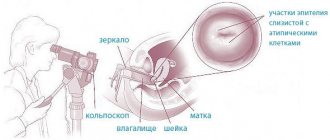
Additional research methods help in diagnosis. Vaginoscopy determines the presence and extent of damage to the vagina and cervix, as well as a foreign body. Swelling and hyperemia of the vaginal wall and the vaginal portion of the cervix, pinpoint hemorrhages, and erosions are noted. Microscopy of a native smear and a Gram-stained smear reveals an increased number of leukocytes in the field of view, gonococci, trichomonas, and fungi. At the time of examination, you can culture the vaginal discharge to determine the flora and sensitivity to antibiotics. The specific nature of vulvovaginitis in girls is revealed by PCR. Helminthic infestation is confirmed by examining stool for worm eggs and scrapings from the perianal area for enterobiasis.
Clinical signs of vulvovaginitis are determined mainly by the causative agent of the disease.
Trichomonas vulvovaginitis in girls is manifested by copious liquid discharge of a whitish or greenish-yellow color. They often foam and irritate the skin of the external genitalia, thighs, and perineum. The disease is accompanied by severe itching of the vulva, as well as symptoms of urethritis. The discharge may contain blood.
With a mycotic lesion, the vulva is hyperemic, swollen, with whitish overlays, under which, when removed with a spatula, areas of bright hyperemia are found. Vaginal discharge looks like a cheesy mass. The disease is often accompanied by symptoms of urethritis and cystitis.
Chlamydial vulvovaginitis in girls is in most cases chronic, with frequent relapses and complaints of periodic itching of the vulva. There may be a burning sensation when urinating. The vulva is moderately hyperemic. Vaginoscopy reveals cervicitis, petechial hemorrhages, and cervical erosion. The discharge is often scanty, mucous, and rarely purulent.
Urea- and mycoplasma vulvovaginitis in girls does not have a specific clinic. Typically, patients are bothered by serous-purulent discharge from the genital tract, often in combination with urethritis. The role of mycoplasma infection in the occurrence of vaginal inflammation has been questioned by many authors.
Herpetic vulvovaginitis in girls manifests itself as small blisters on the hyperemic vulva. The blisters contain clear and then, when a secondary infection occurs, purulent fluid. After 5-7 days, the blisters open with the formation of erosions and ulcers, which are covered with a scab. At the onset of the disease, burning, pain and itching in the vulva area are expressed. Common symptoms include headache, chills, and fever.
Gonorrheal vulvovaginitis in girls can be torpid, recurrent and even asymptomatic, although the most typical onset is acute. The lesion is multifocal, usually involving the vagina (in 100% of cases), the urethra (60%), less often the rectum (0.5%).
After a 1-3-day incubation period, abundant purulent discharge and diffuse hyperemia of the external genitalia, perineum, skin of the inner thighs, and perianal folds appear. Girls complain of pain when urinating, tenesmus. Discharge from the genital tract is purulent, thick, greenish in color, sticks to the mucous membrane, and leaves crusts on the skin when it dries.
Diphtheria vulvovaginitis in girls causes pain in the external genitalia, during urination, infiltration, severe swelling and hyperemia of the vulva with a bluish tint. During vaginoscopy, gray films are found on the vaginal mucosa, after removal of which bleeding erosions remain. Ulcers with necrotic changes and a yellowish coating are possible. The inguinal lymph nodes are enlarged and painful. Discharge from the genital tract is slight, serous or bloody-purulent, with films. Local changes are accompanied by symptoms of general intoxication and fever.
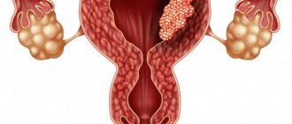
Treatment of intrauterine synechiae
Treatment and prognosis of the disease depend on the nature of intrauterine adhesions. Depending on the structure of the tissue, synechiae are light (in the form of films), medium (muscle fibers) and heavy (formed from connective tissue). Adhesions can occupy either a relatively small area of the uterine cavity or grow throughout its entire body.
If the patient complains and suspects the presence of intrauterine synechiae, the following methods are used for diagnosis:
- Hysteroscopy. The procedure allows you to install white strands of different lengths and densities.
- Ultrasound of the uterus and its appendages.
- X-ray of the uterine cavity.
When diagnosing synechiae of the uterine cavity, the results of a gynecological examination are taken into account, which allows one to determine the size of the uterus and its appendages, their mobility and soreness.
The main method of treating intrauterine synechiae is their dissection and removal using a hysteroscope, electric knife or laser. The procedure is carried out under ultrasound control. In the case of severe adhesions, more serious surgical intervention is required to separate them.
Unfortunately, excision of intrauterine synechiae does not always guarantee complete cure. Very often, relapses and the appearance of new adhesions are possible. To restore the normal structure and functioning of the endometrium, women are prescribed a course of hormonal therapy. The drugs are taken for 6 months to prevent the appearance of new synechiae of the uterine cavity. Treatment is supplemented with antibiotics if the cause of the development of adhesions was inflammatory processes in the body.
Most women are concerned about how quickly pregnancy can occur after treatment of uterine cavity synechiae. The normal menstrual cycle and the ability to fertilize resume quite quickly, but it is necessary to take into account the degree of endometrial restoration. Pregnancy can be planned only by eliminating infectious diseases and restoring the hormonal function of the body.
Etiology
The cause of vulvovaginitis in girls can be specific (gonococci, mycobacterium tuberculosis, corynebacterium diphtheria, trichomonas) and nonspecific (opportunistic aerobes and anaerobes, chlamydia, fungi, viruses, protozoa) infection. Vulvovaginitis can develop after the introduction of a foreign body, with helminthic infestation, masturbation, or impaired reactivity of the body due to secondary infection.
The routes of transmission of a specific infection are different. At an early age, the household route of transmission of infection predominates (through household items, public places, and violation of hygiene rules). Sexually active teenage girls may become infected through sexual contact.
Symptoms and causes of fusion
Synechia is the adhesion of the labia minora and the labia majora to each other, the fusion of the labia minora to the labia majora. This disease is characterized by the formation of adhesions and requires special treatment. Girls under 2 years of age are susceptible to this pathology. Symptoms of synechia:
- a sudden appearance of bright redness on the child’s labia minora, the presence of small rashes;
- the child is capricious when changing clothes and washing himself due to painful sensations;
- wet panties from constantly leaking urine.
The main reasons for the appearance of pathology:
- hormonal imbalance, lack of the female hormone estrogen, the cause of this disorder may be gynecological diseases of the mother during pregnancy;
- infectious lesions of the genitourinary system;
- too frequent washing with soap, which disrupts the pH balance and dries out the mucous membrane. As a result of dryness, adhesions of the labia are formed;
- uncomfortable underwear or diapers that contribute to chafing and irritation of intimate organs;
- untimely change of dirty diapers;
- development of vulvovaginitis, this disease can appear in young children due to neglect of hygiene rules;
- allergies to cosmetics, washing powders, fabrics. Similar reactions can cause fusion of organs;
- Diaper rash that occurs due to constant wearing of diapers when the baby’s skin does not breathe is a common cause of adhesions of the intimate lips.
Also, the cause of synechia can be any kind of pregnancy complications (infectious diseases of the urinary tract or gynecological organs of the mother). The fusion of the labia minora can form a closed vaginal cavity, in which all bacterial secretions will accumulate inside and give rise to new diseases.