Timely prevention of this pathology, which has social and economic significance on a national scale, is one of the priority problems of an integrated approach, which also includes osteopathic correction of dysfunctions of the hepatobiliary system. An osteopath (chiropractor), using soft visceral techniques, improves the functioning of the liver, gallbladder and bile ducts, thereby normalizing the qualitative composition of bile and its further passage in the body.
Gallstone disease (GSD) is a disease caused by the formation of stones in the gallbladder or bile ducts, as well as possible obstruction of the ducts due to stone blockage. In Europe and America, over the age of 50 years, about 1/3 of women and about 1/4 of men suffer from cholelithiasis. There is a clear connection between prevalence and gender.
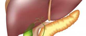
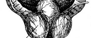
Anatomy and topography of the gallbladder
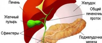
The left and right hepatic ducts, when merging at the point of exit from the liver lobes, form the common hepatic duct (3-4 cm long). The common bile duct is located lateral to the common hepatic artery and anterior to the portal vein.
The common bile duct has four parts:
- supraduodenal (from the confluence of the common hepatic duct with the cystic duct to the outer edge of the duodenum);
- retroduodenal (from the outer edge of the duodenum to the head of the pancreas);
- pancreatic (passing behind the head of the pancreas or through its parenchyma);
- intramural (passing through the thickness of the wall of the duodenum).
A duct opens into the duodenum at the papilla of Vater.
Options for connecting the common bile and pancreatic ducts:
- approach the duodenum as a single duct
- ducts join in the wall of the duodenum
- The common bile and pancreatic ducts empty into the duodenum separately
The sphincter of Oddi of the common bile duct is located at the site where the duct passes through the ampulla of the papilla of Vater; regulates the flow of bile into the duodenum.
Blood supply to the bile ducts:
The intrahepatic ducts receive blood directly from the hepatic arteries; The blood supply to the supraduodenal part of the common bile duct is variable. In most cases, blood flow is directed from the porta hepatis. The most significant vessels lie along the edges of the bile duct at 3 and 5 o'clock. The gallbladder is located in the vesicular fossa on the undersurface of the liver. It serves as a landmark for the border of the right lobe of the liver.
Anatomical parts of the gallbladder: bottom, body, Hartmann's pouch (located between the neck and body of the gallbladder - the part of the bladder located posteriorly). The wall of the gallbladder consists of smooth muscle cells and connective tissue. The lumen is lined with tall columnar epithelium.
Blood supply to the gallbladder:
Arterial blood enters the gallbladder through the gallbladder artery, a branch of the right hepatic artery (less commonly the hepatic artery itself); venous outflow from the gallbladder occurs mainly through the cystic vein, which flows into the portal vein. Lymph from the gallbladder flows both to the liver and to the lymph nodes of the porta hepatis. The cystic duct, common hepatic duct and cystic artery form Calot's triangle. The bile ducts have sphincters that regulate bile secretion: Lütkens' sphincter in the neck of the gallbladder, Mirisi's sphincter at the confluence of the cystic and common bile ducts.
Causes
The appearance of a gallbladder diverticulum in the body occurs in two ways:
- Congenital anomaly. This rarely happens. The pathology gradually developed during the period of intrauterine maturation of the fetus.
- Acquired defeat. This type of abnormal development is associated with the natural performance of organ functions.
A congenital, or true, diverticulum forms inside an organ. Outwardly, this does not manifest itself in any way, since the muscle layer has become denser. There are no symptoms or signs. The main location of the passages is the bottom or neck.
Gallbladder diverticulum
An ailment (acquired) acquired during life is localized in a place where there is a decrease in the tone of elastic fibers. During normal development of the gallbladder, fibers envelop the organ from the inside and create reliable protection from the negative effects of liver secretions.
The cause of diverticulum is a thin wall. Traces of perforation are visible. Protrusion outward occurs due to the appearance of a gallstone inside.
Of the acquired diverticula, Rokitansky-Aschoff sinuses are distinguished. This type of disease develops against the background of chronic inflammation. The pressure inside the organ gradually increases, and protrusions appear on the outer layer of the bladder.
Etiology
The formation of gallstones occurs in the gallbladder as a result of the deposition of dense particles of bile. Most of the stones (70%) consist of cholesterol, bilirubin and calcium salts. Stagnation of bile, increase in the concentration of bile salts. Stagnation of bile is promoted by pregnancy, a sedentary lifestyle, hypomotor dyskinesia of the biliary tract, and foods low in fat. An important factor is inflammation; inflammatory exudate contains large amounts of protein and calcium salts. Protein can become the core of the stone, and calcium, combined with bilirubin, forms the final appearance of the stone.
Cholesterol gallbladder stones: Most gallbladder stones form cholesterol by settling from supersaturated bile (especially at night, when the concentration in the gallbladder is highest). In women, the risk of gallstones is increased by the use of oral contraceptives, rapid weight loss, diabetes mellitus, and ileal resection. Cholesterol stones are large, have a smooth surface, are yellow in color, and are often lighter than water and bile. Ultrasound reveals the symptom of floating stones.
Pigmented gallbladder stones, composed predominantly of calcium bilirubinate, are found in patients with chronic hemolysis (eg, sickle cell disease or spherocytosis). Infection of bile with microorganisms that synthesize beta-glucuronidase also contributes to the formation of pigment stones, as it leads to an increase in the content of direct (unbound) bilirubin in the bile. Pigment stones have a smooth surface and are green or black in color.
Salt mixed stones (consisting of calcium bilirubinate) are more often formed against the background of inflammation of the biliary tract.
Calcium, combined with free bilirubin, settles in the form of stones (calcium salt of bilirubin). Normally, bile contains a glucuronidase antagonist, which prevents the formation of stones. When the gallbladder contracts, the stones migrate. Obstruction of the cystic duct with a stone leads to shutdown of the gallbladder and the occurrence of obstructive cholecystitis and hydrocele of the gallbladder.
Biliary tract diseases
Bile duct damage can occur for many reasons. The main one is unhealthy diet. Damage to the ducts, for example, during surgery, also plays a role in the development of diseases. This phenomenon is especially common during surgical removal of the gallbladder.
Blocked ducts
Blockage of the bile ducts occurs as a result of the appearance of a mechanical obstruction that interferes with the movement of bile. This phenomenon is a complication of some diseases. Blockage of the pathways may be complete or incomplete.
Symptoms of a blocked bile duct usually include discomfort and pain. Signs appear depending on how blocked the pathways are. The development of cholelithiasis is often observed, a disease in which stones form in the bladder. This is caused by a disorder of metabolic processes and stagnation of bile.
The disease may not show symptoms for a long time, but when the stones move into the ducts, inflammation develops and colic appears. When the bile ducts are clogged, pain of varying intensity appears. Often all this is accompanied by an increase in body temperature and vomiting. In the absence of therapy, liver failure develops, which often leads to death.
If therapy is not timely or is completely absent, complications develop. The walls of the ducts thicken, and the bile ducts narrow. In this case, the stone stops in the duct, which leads to the accumulation of bile and stretching of the walls of the organ.
In this case, the bile is directed back into the bladder, which stretches. As a result, the likelihood of it rupturing increases.
Duct stricture
Obstruction of the bile ducts may occur due to compression of the ducts from the outside. This phenomenon is often observed in the following pathologies:
- Cyst or tumor of various origins;
- Scar formation;
- Hepatitis or pancreatitis;
- Cirrhosis of the liver;
- Organ injury;
- Surgical intervention;
- Worm diseases.
This pathology is manifested by the following symptoms:
- Abdominal pain;
- Jaundice;
- Nausea accompanied by vomiting;
- Weight loss;
- Flatulence;
- Change in color of urine and stool.
Obstruction of the bile ducts can lead to serious consequences:
- Nutrient malabsorption disorder;
- Blood clotting disorder;
- Liver disorder;
- Abscess and sepsis.
Cholangitis and cholecystitis
Cholangitis is an inflammation of the bile ducts that develops due to infection entering them through the hematogenous or lymphogenous route, as well as from the gallbladder or intestines. Usually the pathology develops due to cholecystitis, but can act as an independent disease.
In this case, a person may experience abdominal pain, nausea with bouts of vomiting, chills and fever, increased sweating, and yellowing of the skin.
Acute cholecystitis is inflammation of the walls of the bladder, which is caused by infection. The disease is accompanied by colic, pain in the ribs, increased body temperature, the gallbladder increases in size, and bile changes composition and consistency. She cannot move freely along the ducts.
The disease can be life-threatening.
Dyskinesia
Dyskinesia is a disorder of organ motility, which is accompanied by a disorder in the movement of bile into the intestines. This contributes to indigestion. The disease has two forms:
- The hypermotor form manifests itself in active contraction of the muscular layer of the walls of the gallbladder and ducts, as a result of which they are strongly compressed, spasms occur, and pain radiates to the arm;
- The hypomotor form is characterized by weak muscle contraction, the movement of bile into the intestines slows down, and digestion is disrupted. A person develops a bitter taste in the mouth, heaviness in the stomach, and flatulence. In this case, the likelihood of stone formation increases and there is a risk of stone formation.
Dyskinesia has several causes:
- Genetic predisposition;
- Anatomical features of the organ;
- Diseases of the gastrointestinal tract;
- Wrong diet.
Therapy of the disease involves the use, first of all, of drugs for the treatment of neuroses.
Cholangiocarcinoma (cancer)
In some cases, especially in the presence of chronic diseases, thickening of the bile ducts occurs, which over time transforms into cancer. Symptoms in this case will manifest themselves differently, but are in many ways similar to the signs of cholecystitis.
A person develops jaundice, itchy skin in the area of the affected ducts, and fever. With the rapid growth of the tumor, severe pain appears and the person’s condition worsens.
In this case, surgical intervention is required in which the ducts are removed, sometimes including part of the liver. In the latter case, a donor organ transplant is performed.
Pathogenesis
There are 4 types of stones:
- cholesterol stones, containing about 95% cholesterol and some bilirubin lime;
- pigment stones, consisting mainly of bilirubin lime, cholesterol in them is less than 30%;
- mixed, cholesterol-pigment-limestones,
- limestones containing up to 50% calcium carbonate and a few other components.
Gallstone disease is a symptom complex that includes not only the formation of stones, but also the presence of typical biliary colic. The pathogenesis of the latter is the advancement of the stone, spasm and obstruction of the bile ducts. Stones located in the area of the bottom and body of the gallbladder usually do not appear clinically, i.e. are “mute” - 25-35% of people of both sexes after 65 years are such “carriers”.
What are called tumors of the bile ducts in the body, as well as the gallbladder?
Based on statistical data, we can safely say that of the total number of oncological formations, cancerous tumors of the gallbladder ducts account for no more than 5%, namely 4.3%. Only one percent of male patients are diagnosed with oncological tumors, and subsequently undergo surgery on the bile ducts and gallbladder. Women's statistics are less pleasant, because here the rates increase exactly three times, which confirms the fact that women are more susceptible to gallbladder cancer than men. Experts around the world try every year to reduce these numbers to zero. Israeli doctors have achieved particular results in this area. Thanks to the constantly increasing level of qualifications, cutting-edge equipment, the latest diagnostic and treatment methods, as well as an individual approach to each case, Israeli specialists have achieved worldwide respect and authority in the treatment of such diseases. A successful combination of practical and theoretical skills helps thousands of patients forget about their illnesses forever and live a full life.
Today, despite all the modern achievements of technological, scientific, medical and other progress, the etiology of this type of disease is unknown. Gallstone disease is found in almost 90% of all people with cancer. Adenocarcinomas account for almost 82% of all carcinomas present in the gallbladder of the average patient with a similar problem. Local invasiveness of growth into the liver tissue is very typical for this type of tumor. Metastases of these tumors penetrate through the lymphogenous route into the liver, pancreas and duodenal lymph nodes.
Read! Biliary (hepatic) colic
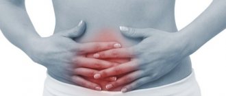
The clinical picture is as follows: patients complain of pain that is localized in the right upper abdomen, as well as frequent vomiting and nausea. In rare cases, it is possible to establish a diagnosis before surgery.
Clinical picture
Biliary colic is a syndrome characterized by sudden sharp pain in the right hypochondrium, radiating to the right collarbone, right arm, back, accompanied by nausea and vomiting. There may be bile in the vomit, hence the feeling of extreme bitterness in the mouth. With prolonged pain and obstruction, itching of the skin develops and jaundice appears a little later. Symptoms of peritoneal irritation are possible.
When the cystic duct is blocked, an inflammatory process and hydrocele of the gallbladder may develop. In the presence of inflammation, cholangitis and cholangiohepatitis can develop; with incomplete obstruction, secondary biliary cirrhosis of the liver can develop. When a stone is retained in the common bile duct, obstruction of the pancreatic duct is also possible with the formation of acute pancreatitis, including those associated with the reflux of bile into the pancreas.
When examining a patient, an enlarged gallbladder can be detected, but it may also be wrinkled and there may be practically no contents in it. As a rule, such patients have an enlarged liver, it is soft and painful on palpation.
A number of symptoms are characteristic. Ortner's symptom: pain when tapping along the edge of the right costal arch. Murphy's symptom: increased pain when pressing on the abdominal wall in the projection of the gallbladder during a deep breath. Kehr's symptom: the same on palpation at the point of the gallbladder (in the angle formed by the costal arch and the edge of the rectus abdominis muscle.). Zakharyin's symptom: the same with tapping at the point of intersection of the right rectus abdominis muscle with the costal arch. Mussy's symptom: pain when pressing between the legs of the right sternocleidomastoid muscle (phrenicus symptom is caused by irradiation of pain along the phrenic nerve, which is involved in the innervation of the liver capsule and gall bladder). Beckmann's sign: pain in the right supraorbital area. Yosh's sign: the same at the occipital point on the right. Mayo-Robson sign: pain when pressing in the area of the costovertebral angle.
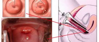
Diagnosis of cholelithiasis
Stones are detected by X-ray and ultrasound methods. Cholecystography, intravenous cholegraphy, and radionuclide scanning of the gallbladder are used.
If a tumor is suspected, with obstructive jaundice of unknown origin, concomitant liver damage - fibroduadenopancreatocholangiography, laparoscopy and laparoscopic cholecystocholangiography. Laboratory tests: high levels of bilirubin, increased bile acids, signs of inflammation in the blood. If the common bile duct is completely blocked, there is no urobilin in the urine, and a sharp increase in the secretion of bile acids is possible.