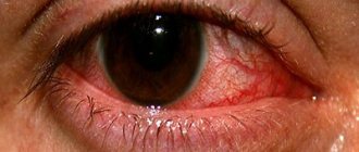
Rhinitis occurs at least once a year in every person. This condition causes significant discomfort, because it may be accompanied by symptoms such as congestion and discharge from the respiratory organ, pain and burning in the nose, swelling of the mucous membrane, etc. When the nose ceases to function fully, various disorders occur in the body. Thanks to this organ, a person can breathe, and it also humidifies, cleanses and warms the incoming air.
Thus, the nose is a very complex and at the same time delicate system, which often fails. There are many reasons for its malfunction. This may be a deficiency of vitamins, weak immunity and the entry of pathogenic microflora into the body. That is why the treatment of runny noses of different etiologies differs significantly. Therefore, it is important to know what diseases of the nose and sinuses there are.
Types of diseases of the nose and paranasal sinuses
The classification of rhinitis is quite extensive.
According to the etiological factor, there are four leading categories of diseases. The first group is pathologies with which a person was already born.
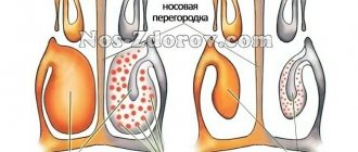
The most common birth defect is the curvature of the septum located in the respiratory organ.
But there are also more dangerous disorders that prevent the nose from working normally and require surgical intervention.
For example, fistulas and narrowing of the nasal passages provoke chronic nasal diseases.
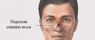
The second group is diseases caused by injuries to the nose. Damage can be of a different nature (open, displaced, mixed, etc.). Trauma to the organ is accompanied by severe swelling, which often leads to septal hematoma.
Attention! A runny nose is the most common disease in the world. According to statistics, 90% of the world's inhabitants complain of difficulties with nasal breathing at least once a year.
The third category of nasal diseases are infectious diseases caused by fungi, bacteria and viruses . These types of runny noses develop most often.
The fourth group is rhinitis provoked by irritating factors ( allergic ) . In this case, a runny nose can be caused by an allergen, medications or chemicals. Also, diseases associated with the nose are divided into chronic and acute according to the nature of their course. They are also distinguished by area of localization and shape.
Inflammation of the sinuses
Below we will consider in more detail the most common nasal diseases.
Acute rhinitis
Inflammation of the nose occurs when an infection penetrates into it, and the person’s immune system is weakened and cannot overcome it on its own. At the beginning of the disease, the nasal mucosa is susceptible to changes - it dries out and hyperemia appears. Then it swells and discharge appears. At the last stage of progression of a runny nose, purulent mucus is released from the nose.
If left untreated, acute inflammation spreads to the paranasal sinuses, Eustachian tube and lacrimal ducts. If the patient has high immunity, then the course of rhinitis will be mild and the unpleasant symptoms will go away in two to three days.
When the body's protective functions are weakened, the disease can last from 1 month or more. Treatment of acute rhinitis should be comprehensive. Its main goal is to eliminate signs of the disease . For this purpose, the patient is prescribed antiseptic aerosols, corticosteroids in the form of ointments and nasal rinsing with saline.
Chronic runny nose
It develops as a complication of acute rhinitis due to incorrect treatment or lack thereof. Other etiological factors are poor circulation, stagnation of purulent exudate in the sinuses and exposure to irritants. Characteristic signs of the disease:
- deterioration of sense of smell;
- snore;
- nasal congestion;
- headache;
- snot.
Basically, an exacerbation of the runny nose occurs in autumn and winter, and by spring the patient’s condition returns to normal. The course of chronic rhinitis leads to malocclusion in children, deformation of the skull, and changes in the development of the chest.
Attention! Long-term untreated rhinitis worsens hearing and contributes to the appearance of pharyngitis, tonsillitis and furunculosis.
Chronic diseases of the nasal mucosa are treated by eliminating the factors that provoke the disease, physiotherapy, climatic treatment and taking medications. Cauterizing and astringent agents ( silver nitrate, Protargol ) and drops that constrict blood vessels are used. If complex therapy does not bring the desired results, galvanocaustics are performed.
Ozena
Fetid rhinitis occurs due to atrophy of the mucous, bone and cartilage tissues of the nose. The clinical picture of the disease is viscous discharge with a putrid odor, the formation of crusts in the nose and lack of perception of odors. The causes of ozena have not been established. But there is an opinion that the disease develops due to congenital pathologies, after inflammatory diseases and infection of the nasal cavity.
Attention! In 80% of cases, the appearance of a foul runny nose is caused by Klebsiella.
Ozena therapy can be medicinal and surgical.
During drug treatment, the patient is prescribed antibiotics and nasal rinsing.
After removing the crusts, endonasal installations are made, and then ointments are placed in the nasal passages.
Also, fetid rhinitis can be effectively treated with physiotherapeutic methods.
During surgical treatment, operations are performed aimed at:
- narrowing of the nasal passages;
- stimulation of mucosal trophism;
- moisturizing mucous layers;
Allergic rhinitis
This type of runny nose can be seasonal, occurring during flowering of plants, and year-round, caused by household allergens. Based on the duration of its course, allergic rhinitis is divided into intermittent (up to 4 days) and persistent (occurs at least 4 times a year).
When irritants enter the nasal cavity, rhinorrhea, sneezing and difficulty breathing through the nose occur. Treatment consists of specific immunotherapy. Antihistamines and glucocorticosteroids are prescribed as medications.
Hematoma and abscess of the nasal septum: causes, symptoms, diagnosis, treatment - Healthy Family
A nasal septum hematoma is a limited collection of blood under the mucous membrane. The ICD-10 code is J34.0. It is formed in the process of trauma to the nose, accompanied by bleeding in the submucoperichondrial layer. Under the influence of secondary infection, suppuration may occur and an abscess may begin. In some situations, pathology occurs as a consequence of viral diseases.
Causes of pathology
The causes of the pathological process are:
- Vascular trauma due to septal surgery (septoplasty). At the postoperative stage, blood from vessels damaged during surgery accumulates under the mucosa on one or both sides.
- Nose bruises.
- Nose fractures.
Development mechanism
The first is characterized by unilateral accumulation of blood and a mild pathological process. It is also easier to treat. The bilateral form involves the accumulation of blood on both sides and often develops into an abscess.
In many situations, the bruise covers only the cartilaginous part of the septum. If there is a unilateral nasal hematoma, breathing will not change. The painful syndrome will be mild or completely absent.
Directly because of this, many patients discover the disease late.
At this time, the hematoma begins to fester and turns into an abscess, which is dangerous due to the curvature of the nasal septum and complications inside the skull.
Complications
- nose bleed;
- suppuration and abscess;
- purulent consequences of an abscess: an inflammatory process in the soft tissues of the face, the orbit (leading to the formation of ulcers), inside the skull. In addition, thrombosis of the cavernous sinus sometimes occurs, which is accompanied by intense pain in the head, convulsive and comatose states and other central nervous system disorders;
- curvature of the external nose;
- perforation of the nasal septum.
Diagnostics
The following diagnostic methods are used to identify pathology:
- Physical examination. During anterior rhinoscopy, a unilateral or bilateral thickening of the nasal septum of a reddish hue is determined. The nasal cavity in such situations is poorly or completely inaccessible for examination. Sometimes the protrusion is discovered when the tip of the nose is raised.
- Clinical study. When diagnosing peripheral blood in patients with abscess of the nasal septum, neutrophilic leukocytosis is detected.
- Instrumental diagnostics. Sometimes, in order to detect a hematoma, it is necessary to remove blood from the nasal cavity using an electric suction device and use a button probe to palpate the protrusion.
- Indications for consultation with highly specialized doctors. The complex course of the pathological process, the presence of pathogenetically interrelated diseases in the patient that require appropriate therapy (diabetes mellitus), is considered an indication for consultation with specialized doctors.
A comprehensive and thorough diagnosis allows you to select the appropriate therapy for the pathology.
Therapeutic measures
Treatment is carried out on an outpatient or inpatient basis. It depends on the patient’s well-being and the intensity of the hemorrhage. Therapeutic measures include:
- Aspiration of blood. The hematoma cavity is punctured, then aspiration is carried out using a syringe. Used for mild forms of pathology.
- Drainage. Used if puncture is ineffective. The surgery is performed under local anesthesia. To remove the blood clot, an incision is made into the mucous membrane above the hematoma. Then the dried blood is pumped out and drainage is installed into the formed cavity.
- Treatment with antibacterial agents. Used only for surgical therapy. The drugs of choice in this case are antimicrobial agents of the 2nd-3rd generation cephalosporin subgroup and aminopenicillins. In many situations, they are used in a course lasting up to one week.
After removing the dried blood and treating the affected area with antibiotics, special tubes are installed and packing is performed. Subsequent therapy involves injections of broad-spectrum antimicrobial agents.
They are used in accordance with the scheme throughout the week. Drugs from the subgroup of cephalosporins are effective against various pathogenic microorganisms, which often cause pathology.
Remedies for bruising and swelling
The use of external agents should be started as soon as possible. An exception may be heparin ointment; it is used only on the second day. The drugs are applied to the bruise four to five times a day. Creams and gels are used only in situations where there are no scratches, abrasions or other minor damage to the skin.
Medicines
It is possible to eliminate the manifestations of pathology through medications, which include troxerutin and heparin. They belong to a subgroup of anticoagulants that help thin the blood. The most common drugs:
- troxevasin;
- troxerutin;
- heparin ointment;
- hepatrombin.
To eliminate a bruise, medications containing vitamin K are also used. It helps improve the penetration of active elements into the skin cells and normalizes blood clotting.
Folk remedies
Folk remedies for getting rid of bruises should have a good absorbable effect.
They are considered an additional element to traditional therapy, and their use requires prior approval from a specialist.
Natural remedies that help reduce unpleasant symptoms include arnica and comfrey. Medicines intended for the treatment of hematomas often include leech extract.
Often, in order to eliminate the pathological process, badyagu is used. This freshwater algae has a beneficial effect on the affected area and gives a positive result in the shortest possible time. There is a large number of different pharmaceutical products, the basis of which is badyaga.
It is possible to make a compress based on it on your own at home. It is necessary to purchase dried plant raw materials. The powdery mass is diluted with hot water, 1 teaspoon of algae is taken for 2 hours of liquid.
Prognosis and prevention
One of the main preventive measures is to prevent nasal injuries. If such a problem occurs, then the following instructions must be followed as soon as possible:
- after a blow, bruise or fracture, you must immediately contact a specialist;
- on the way to the hospital, you need to keep ice near your nose (helps curb the formation of a hematoma);
- It is necessary to ensure that during transportation the patient is in a semi-sitting position.
Timely treatment of diseases of the ENT organs is important in preventing adverse consequences.
Prevention for patients at risk is key. These include:
- children;
- patients with diabetes mellitus;
- people with arterial hypertension.
If any injury to the nose occurs, they need to find out the doctor's recommendations. If surgery is performed in a timely manner, opening the hematoma and removing dried blood or pus, the prognosis will be positive.
When a secondary infection is added or a nasal abscess has formed, involvement of the quadrangular cartilage cannot be ruled out. As a result, the external nose becomes deformed.
If suppuration spreads further against the background of other diseases, the prognosis will depend on the effectiveness of therapy.
Sources: medscape.com, health.harvard.edu, medicalnewstoday.com.
Source:
Forecast
Timely opening of the nasal septum hematoma and removal of the contents allows for a favorable prognosis. With an abscess, cartilage may melt, which can lead to permanent deformation of the external nose (recession of the nasal dorsum).
Source:
Hematoma in the nose
The nose is sensitive to mechanical influences, therefore, with strong impacts, a nasal injury may occur, in the form of a hematoma of the nasal septum. In this case, damage to the vessels occurs, blood flows out of them, but it cannot go beyond the intact mucous membrane and accumulates under it. As a result, swelling and edema of the septum is formed.
Symptoms of a hematoma in the nose
At the time of injury, severe pain is felt, which then goes away. As the hematoma fills, the person feels a feeling of fullness and a painful condition, which intensifies when pressing on the nose.
The larger the size of the hematoma, the more difficult it becomes to breathe through the nose: in especially severe cases, with large bilateral hematomas, the nasal passages can be completely blocked.
Based on the symptoms, the traumatologist makes a diagnosis.
Treatment of nasal septum hematoma
When examining the nasal cavity, the surgeon sees a hematoma in the form of a soft-to-touch, bright red formation on the side of the septum. With this injury, it is very easy to eliminate the symptoms. It pierces the mucous membrane, and all the blood comes out. Emptying hematomas must be performed without fail.
Source: https://niikelsoramn.ru/profilaktika/gematoma-i-abstsess-nosovoj-peregorodki-prichiny-simptomy-diagnostika-lechenie.html
Sinusitis
Sinusitis
Occurs when the maxillary sinuses become inflamed. The acute form develops against the background of infectious diseases, as a complication of rhinitis and in inflammatory diseases of the teeth.
The chronic form appears if negative factors are added to the acute stage (deviated septum, thickening of the mucosa, narrowing of the sinus openings).
Symptoms of acute sinusitis:
- temperature;
- pain radiating to the forehead, teeth, root of the nose;
- chills;
- lacrimation;
- purulent discharge;
- nasal congestion.
Chronic sinusitis is accompanied by pain in the sinuses and head, malaise, loss of smell and nasal congestion. Inflammation is treated with conservative, physiotherapeutic methods or through puncture, drainage and treatment of the nasal cavity. The main goal of therapy is to eliminate swelling and inflammation and improve the outflow of pus.
Frontit
Inflammation of the paranasal frontal sinuses occurs for reasons similar to sinusitis. Symptoms and treatment of nasal diseases depend on its shape. During an acute inflammatory process, swelling, changes in skin color, and an increase in body temperature occur. Sometimes phlegmon, purulent fistula, and abscess develop. Chronic sinusitis is accompanied by swelling of the mucous membrane or polyps form in the nasal passage. In case of necrosis of the posterior wall, meningitis and abscess develop.
Acute frontal sinusitis is treated with conservative methods: lubricating and instilling the nasal mucosa with a solution of Naphthisin and Cocaine with adrenaline. Also does therapy mean?
- physiotherapeutic effects;
- taking Analgin and taking acetylsalicylic acid;
- carrying out inhalation or intramuscular administration of antibiotics.
Attention! Chronic frontal sinusitis is treated using traditional methods, but if there is no success, surgical intervention is performed.
Ethmoiditis
Develops during the inflammatory process of the mucous cells of the nose of the ethmoid labyrinth. Causes: infections, untreated frontal sinusitis or sinusitis, sepsis in newborns.
The disease is characterized by symptoms such as difficulty breathing, discomfort and a feeling of fullness in the nose, anosmia, and mucous secretion. Therapy involves the use of systemic antibiotics, antipyretics, painkillers and vasoconstrictors.
Sphenoiditis
Appears when the mucous layers of the sphenoid sinus become inflamed. Provoking factors are:
- dysfunction of the sphenoid sinus anastomosis;
- sinus defects (narrowness);
- penetration of foreign particles into the sinus;
- chronic inflammatory diseases of the respiratory organs (upper).
The leading symptom of sphenoiditis is headache of different localization. Sometimes ophthalmological problems develop (diplopia, decreased visual function). It is also possible to release purulent and mucous exudate and have a foul odor from the mouth.
Therapy consists of the use of vasoconstrictor, antibacterial and thinning drugs. The doctor also performs a procedure for enemizing the nasal cavity, which improves the outflow of secretions. The chronic form of the disease often requires surgical intervention.
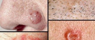
Diseases of the external part of the nose
This group of diseases includes lesions on the outer surface of the nose and adjacent areas. Such pathologies include:
- Erysipelas is an infectious disease accompanied by redness and swelling of the skin on the vestibule of the nose.
- Infection of the surface of the nose through damage.
- Boils.
- Infection in the nose - manifested by the formation of a purulent lump the size of a pea. With simultaneous inflammation of the glands and hair follicles, this phenomenon is called a carbuncle.
- Rhinophyma is an inflammation characterized by the proliferation of layers of skin on the tip and wings of the nose. Externally, the formations look like mounds that can hang down and stretch out.
Topographic anatomy
Region boundaries. The upper one is a horizontal line connecting the medial ends of the eyebrows, the lower one is a horizontal line drawn through the base of the skin of the nasal septum, and on the sides there are the nasobuccal and nasolabial folds. There is a distinction between the external region and the nasal cavity. External area of the nose (regio nasi externa). The skin of the outer area is thin, mobile on the back of the nose, and rich in sebaceous glands on the wings. Subcutaneous tissue is poorly expressed. Under the skin there are facial muscles: m. nasalis (on the lateral surface near the wings), m. depressor septi nasi (at the nasal septum above the upper lip). The branches of a. spread along the back of the nose. dorsalis nasi (terminal branch of the ophthalmic artery), and branches of a. angularis (terminal branch of the facial artery). Venous outflow is carried out through the veins of the same name. Sensitive innervation is provided from the first and second branches of the trigeminal nerve (n. infratrochlearis, ethmoidalis anterior, infraorbitalis). The facial muscles are internalized by the facial nerve. The bony basis of the external nose is made up of the nasal bones, supplemented by the lateral cartilages of the dorsum and wings of the nose. The cartilage of the nasal septum occupies the middle position.