Have you been struggling with GASTRITIS and ULCERS for many years without success?
“You will be amazed at how easy it is to cure gastritis and ulcers just by taking it every day...
Read more "
Many people experience pathological processes in different parts of the intestine, which are accompanied by a characteristic clinical picture. It is discomfort, pain and other symptoms that force them to go to hospitals for consultation. To make an accurate diagnosis, specialists prescribe hardware examinations to patients, in particular fluoroscopy with barium. This technique allows you to examine the condition of different parts of the organ using x-rays, as well as identify any abnormalities and pathologies.
What does it show
Barium examination of the intestines is considered one of the most informative diagnostic methods, allowing to identify the following pathologies in the early stages:
- polyps;
- ulcerative lesions;
- malignant neoplasms, etc.
X-rays of the large or small intestine are performed using special equipment that passes a dose of radiation through the body that is safe for human health.
The image reflected in the image, which is transmitted by X-rays, allows you to determine the following parameters of the organ:
- dimensions;
- motor functions;
- shape;
- location relative to other organs;
- contours;
- lumen diameter;
- pathological changes (for example, narrowing, obstruction, developmental abnormalities, polyps, ulcerative lesions, neoplasms, diverticula);
- degree of elasticity of the organ;
- the ability of the intestine to stretch.
Barium is used for intestinal X-rays, thanks to which specialists are able to correctly diagnose the disease, with minimal risk of causing injury to the mucous membranes of the organ. This medication is not absorbed into the blood and does not provoke the development of allergic reactions, therefore it is considered the safest drug for conducting a hardware examination of this type.
Indications
Indications for an X-ray of the intestine with barium are the following patient complaints:
- rapid weight loss for no apparent reason;
- disruption of defecation processes, for example, diarrhea began, in which the stool acquired a tarry structure and changed its usual color;
- the occurrence of pain in the abdominal area;
- persistent constipation;
- impaired intestinal motility;
- blood, mucous or purulent impurities are detected in the stool.
X-rays of the small or large intestine are prescribed to people in whom experts suspect the development of the following pathologies:
- Crohn's disease;
- diverticula;
- colitis;
- enteritis;
- polyps;
- tumor processes;
- abnormalities of organ development, etc.
Results of an X-ray examination of the intestines
What does a barium x-ray show? Thanks to the contrast and air, the doctor can see the relief and structure of the organ, its contours in the resulting images. Based on this, he makes a conclusion about the degree of its extensibility and elasticity, and the ability for peristaltic contraction. By studying the lumen, a specialist evaluates the patency of the intestine, the presence of fecal accumulations and formations in it.
Article on the topic: Why does it sting in the right side under the ribs: causes and symptoms
X-ray of the intestines with barium
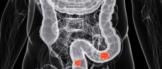
An X-ray of the intestine can detect the following pathologies.
- Intussusception. In this condition, a section of the intestine twists in such a way that the lumen becomes completely blocked. This leads to its obstruction. This pathology manifests itself very sharply and painfully. The patient should be immediately sent to the hospital.
- Malignant formations. When examining the intestine, they appear as a narrowed area and can also lead to closure of the lumen, but this occurs already at a late stage of cancer development.
- Intestinal obstruction. This disorder manifests itself in the form of vomiting and severe abdominal pain, since peristalsis is severely impaired. During an X-ray examination, the pathology manifests itself in the form of a lack of movement of contrast and air into other parts of the organ, different levels of accumulation of substances.
- Polyps. Despite the fact that these formations on the intestinal wall do not harm the body, they must be removed, as they can lead to a malignant process.
- Diverticula. These are protrusions of the intestinal wall. As a rule, the cause of their formation is high pressure in the intestines. Diverticula can get fecal matter, causing inflammation.
Peculiarities
Currently, when performing intestinal radiography, specialists use several techniques:
- Irrigoscopy is performed, in which a contrast agent is injected directly into the patient's rectum. This method is used when examining the large intestine. Specialists can also use double contrast tactics, in which the organ is filled with air or an inert gas in parallel with a special liquid.
- Barium fluid is used to examine the small intestine. The patient must drink a contrast agent before starting the diagnostic procedure.
- A technique called irriography is used. During the diagnostic procedure, an image of the large intestine filled with a contrast agent is displayed on the monitor screen.
Intestinal X-ray techniques
The intestine is a hollow organ, which on an x-ray is no different from neighboring tissues, since its ability to absorb x-rays is the same. That is why a contrast agent must be administered to study it:
- barium sulfate;
- sodium amidotrizoate;
- air.
Water-soluble sodium amidotrizoate is used when examining the intestines in newborns or when fistulas or perforation are suspected. Air is used when performing double contrasting.
The method of administration depends on the purpose of the study and the department to be studied.
Contraindications
There are a number of contraindications to X-ray examination of different parts of the intestine:
- the presence of bleeding in the internal organs;
- toxic megacolon;
- period of pregnancy and breastfeeding;
- presence of intestinal obstruction;
- ulcerative form of colitis;
- the occurrence of severe abdominal pain;
- perforation of the intestinal walls;
- diagnosed tachycardia and heart failure in severe form;
- unconsciousness of the patient;
- a recent intestinal biopsy (the prohibition is due to the fact that the contrast agent can provoke inflammation in the places from which biological material was taken).
Preparation rules
In order for the patient’s examination to show the most reliable result, he needs to properly prepare for it:
- You must stop drinking alcoholic beverages within a week.
- Three days before the scheduled x-ray, a person should exclude from his menu foods that can increase intestinal gas formation and provoke fermentation processes. This list should be supplemented with: fatty meats and fish, cabbage, milk, black bread, soda, legumes, raw vegetables, which contain large quantities of coarse fiber.
- If the patient has chronic constipation, he needs to drink as much fluid as possible these days, and also take medications that have a laxative effect. For example, Duphalac.
- The day before the hardware examination, the person must stop eating solid food. He is recommended to drink broths, fruit juices, and teas for 24 hours.
- In the evening, before the procedure, the patient must undergo a total cleansing of the intestines. He needs to take a laxative medication, and then give two enemas, between which a certain time interval must be observed - 2-2 hours.
- In the morning, on the day of the x-ray, the person must take another enema. The cleansing procedure is carried out until clean water begins to emerge from the anus.
If the patient cannot tolerate mechanical cleansing manipulations, he should prepare the intestines for examination using special medications:
- The patient should use special medications designed to prepare the intestines for surgical and diagnostic procedures. For example, Fortrans powder or Bsakodyl suppositories (this drug is available in pharmacy chains and in tablet form).
- If Bisacodyl tablets are used, then 2 of them should be taken the day before the examination, during the morning meal.
- After 3 hours, the patient should take any laxative medication.
- After 3 hours, take two Bisacodyl tablets.
- Before going to bed, take a Bisacodyl suppository.
- After waking up, on the day of the x-ray examination, the person must place another suppository.
X-ray of the intestines with barium: what does the procedure show?
Fluoroscopy allows you to obtain sufficiently complete information to make a correct diagnosis and prescribe an adequate course of treatment for the disease. In particular, the doctor receives an overview of the intestine being examined:
- its shape;
- external diameter;
- lumen
What else does fluoroscopy show? It allows the doctor to see the causes that disrupt the normal functioning of the intestines (erosions, ulcers, worms, tumors).
How to cleanse the intestines before an x-ray?
The effectiveness of fluoroscopy largely depends on proper preparation for it, which mainly comes down to cleansing the intestines. To properly cleanse the gastrointestinal tract, it is necessary to take the following steps.
- You need to start preparing three days in advance. All foods that can cause increased formation of gases in the intestines (legumes, raw vegetables, black bread, carbonated drinks) are excluded from the menu.
- When there is a tendency to constipation, it is recommended to take laxatives.
- The day before fluoroscopy is performed, solid food is excluded from the diet.
- The main bowel cleansing begins the day before fluoroscopy, in the evening. A laxative is drunk and two enemas are given. The break between enemas is two hours. It is important to administer the enema correctly. A description of the procedure for this procedure can be found on the Internet or obtained from a doctor.
- On the day of fluoroscopy, another enema is done to finally cleanse the intestines of its contents.
- People who cannot give enemas to prepare for fluoroscopy must take special medications (Bisacodyl, Fortance, etc.).
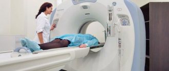
How do doctors do fluoroscopy?
Before proceeding with fluoroscopy, the patient takes off his outer clothing and all metal objects, and puts on a hospital gown.
Then a barium solution (0.5 liters) is drunk. With double contrast, it is drunk through a tube into which the device supplies air or an inert gas.
After taking the suspension, you have to wait until the contrast reaches the desired part of the small intestine.
Fluoroscopy is performed in various positions of the body to obtain a detailed picture.
During fluoroscopy of the colon, the condition of the rectum is first examined using a sigmoidoscope. After this, a barium solution (about one and a half to two liters) is carefully introduced into the large intestine. The patient periodically changes his body position so that the doctor can obtain more informative images.
After the fluoroscopy is completed, it is recommended to drink more fluids and take a laxative to quickly remove barium from the body.
Conducting research
Hardware examination of the intestines is carried out on an empty stomach. The patient will have to remove all metal objects and outer clothing.
Radiography will be carried out in several stages:
- Initially, the specialist will give the patient barium liquid, which needs to be drunk in a volume of 500 ml.
- After this, the person must wait two hours for the liquid to spread throughout all parts of the small intestine.
- Next, the specialist will begin taking x-rays at intervals of 45 minutes. In this case, the patient will have to take different positions.
- The last x-ray is taken after the patient has had a bowel movement in the designated area.
In the case when the double contrast technique is used, the person will have to drink barium liquid prepared by a specialist through a straw. In parallel, inert gas or air will be supplied through it. Due to the forced air, the patient may experience discomfort and severe distension in the intestines. He should take deep breaths at these moments. In this case, the study will be considered complete when the contrast agent reaches the appendix.
After the X-ray examination is completed, the patient can lead a normal life. Within a few days, his stool will have a whitish tint as the contrast agent is naturally excreted from the intestines. It is worth noting that barium mixture causes constipation in some people, so a specialist may recommend taking laxatives.
How is an X-ray of the small intestine done?
X-rays of the small intestine will help diagnose diseases.
The x-ray will take about half an hour. The patient will need to drink half a liter of solution, which will resemble milk in appearance and taste like lime.
When it is necessary to carry out double contrast, barium must be drunk using a tube that goes from the machine, which supplies air a little at a time.
After this, after 5 hours, you can start x-raying, since during this time the contrast will enter the small intestine. While the solution passes through the small intestine, it is necessary to take about 8 x-rays in different positions. This happens every 45 minutes.
Read: X-ray of the intestines: preparation for the procedure according to all the rules
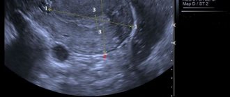
If the solution is not distributed evenly throughout the intestines, then the doctor, by palpating the abdominal wall, will be able to distribute the substance evenly. The procedure will come to an end when the substance completely fills the space between the small and large intestines - this is the ileocecal angle, and the solution fills the cecum.
The radiologist, seeing the uniformity of filling of the intestines with contrast, will be able to determine whether there are any pathologies. When the mucous membrane is enveloped in this contrast in the form of a speckled pattern, the intestines function as usual.
When the substance settles on the walls of the mucous membrane in the form of flakes, this indicates malabsorption syndrome or malabsorption.
When tumors are present inside the intestine, contrast filling will be uneven.