Kidney ultrasound is one of the safest diagnostic methods. Ultrasound can detect virtually any abnormal processes and diseases of the renal apparatus. Other prerogative aspects of the procedure include:
- lack of direct contact of medical instruments with the organ (non-invasive);
- absolute painlessness of the process;
- no contraindications to the examination (with the exception of damage to the epidermis in the area of the examined organs);
- insignificant time costs for the procedure;
- high information content.
The last point is largely ensured by proper preparation. To get objective results, you should know what to do before a kidney ultrasound. Separate examination of the kidneys is rarely performed. For the most part, an ultrasound of the abdominal cavity with kidneys or an ultrasound of the urinary system is prescribed. Based on this, preliminary preparation for the procedure is carried out.
The main recommendations for preparing for a kidney examination using ultrasound are aimed at correcting nutrition a few days before the procedure. For people who have difficulty with regular bowel movements and flatulence, preparation should begin 5-7 days in advance; for other patients, three days will be enough.
About food
Changing your eating habits will make it easier for your organs to function. An unloaded digestive system and kidneys are easier to study. This will help the doctor make the most accurate diagnosis possible. The diet is made up of products that are easily processed by the digestive tract.
You can eat the following ready-made meals and products:
- porridges cooked in water (milk and cream bases are excluded);
- chicken, turkey, rabbit, lean fish, cooked by stewing or boiling;
- cheese and cottage cheese with low fat content;
- boiled or stewed vegetables;
- boiled eggs;
- light, non-rich soups.
The diet before a kidney ultrasound is aimed, first of all, at avoiding foods that cause flatulence. The accumulation of gases in the intestinal cavity is an obstacle to the free passage of ultrasound, and distorts the image of the digestive organs and urinary system on the monitor of the ultrasound machine.
Before the study, you should exclude from your diet:
- legume source of vegetable protein (peas, lentils, chickpeas, beans);
- pickled, raw, pickled cabbage;
- whole fresh milk;
- yeast baked goods;
- fruits: pears, apples, grapes;
- raw vegetables: radishes, radishes, cucumbers, tomatoes;
- sweet dessert dishes;
- chocolate;
- sausage, frankfurters;
- whole grain flour products, including bread.
The consumption of heavy foods is also subject to restrictions. Not recommended: fatty pork, fish and meat prepared by smoking, potato dishes, canned foods, including marinades and pickles, fatty mayonnaise-based sauces and ketchups, seeds, dried fruits, any nuts.
Dietary nutrition for 2–6 days will allow the doctor to assess in detail the condition of the abdominal cavity and kidneys, and speed up the examination process itself. Meals include small portions at 3-4 hour intervals.
The culinary method of processing food by frying is not recommended. Meat, vegetables, fish should be boiled, stewed or baked in foil, and steam cooking should also be used. Herbal and vitamin decoctions will be useful.
The main question of the preparatory period is whether it is possible to eat before an ultrasound? In the case when an ultrasound examination of the kidneys is prescribed in conjunction with ultrasound diagnostics of the abdominal cavity, it is necessary to maintain a fasting regime of 8 to 12 hours. During this time, the gastrointestinal tract will have time to process food. In this case, the evening meal should be light and consist of easily digestible dishes.
This can be any water-based porridge, boiled or steamed fish of not very fatty varieties (pollock, hake), stewed vegetables (excluding cabbage). Is it possible to eat before an ultrasound of the kidneys, when a separate examination of these organs is performed? If the ultrasound is scheduled for the afternoon, a breakfast that is easy on the stomach is allowed. When diagnosing in the morning, it is better to refuse breakfast.
For preschool children, the food restriction before diagnosis is at least five hours. Breastfed babies are examined no later than half an hour after feeding. To avoid problems with the gastrointestinal tract, you should not overeat after the procedure. A large meal after a diet can cause dyspepsia (painful and difficult digestion) and difficulties with bowel movements.
Limiting certain foods is necessary to prevent gas formation in the intestines.
About the liquid
The drinking regime during the preparatory period should be plentiful and regular. You are allowed to drink water, green or weak black tea, diluted fruit drinks and juices without pulp. It is prohibited to consume sweet soda and kvass containing yeast, as these drinks provoke gas formation. Immediately before diagnosis, the bladder should be full.
This recommendation is especially relevant if examination of this organ is planned along with the kidneys. It would be a good idea to prepare water in advance so that you can drink it immediately before the ultrasound diagnostic room.
How much water should you drink for the procedure to show optimal results? Doctors are of the opinion that one liter will be enough. The main thing is not to empty your bladder, otherwise drinking beforehand loses its meaning.
If the digestive tract is stable, before the study it is enough to take carminatives for two days: activated carbon (1 tablet per 10–15 kg of weight) or Espumisan (capsule three times a day).
For patients with digestive problems (regular bloating, constipation), it is recommended to add an enema procedure to the medications the evening before the ultrasound (enema is prohibited on the day of diagnosis). You can do a regular enema, 2 liters in volume, or use the modern Microlax microenema. If it is not possible to give an enema, then you should resort to the help of mild laxatives (Guttalax, Fitolysin).
For the smooth functioning of the digestive tract, it would be useful to use enzyme medications that improve digestion. Mezim, Pancreatin, Festal should be taken one tablet during breakfast, lunch and dinner. Correctly fulfilled preparation conditions enable the doctor to easily diagnose diseases of the kidneys and other organs responsible for the process of urine excretion.
If the doctor suspects the presence of not only renal pathologies, but also a deviation from the norm in the functioning of adjacent vessels, a special type of diagnosis is prescribed - ultrasound with Doppler (duplex). Double examination is more effective. It allows you to identify changes directly in the organ, the speed of blood flow and the cleanliness of blood vessels (the presence of blood clots, atherosclerotic growths on the vascular walls, expansion or narrowing of the vascular system of the kidneys).
The doctor will tell the patient about the necessary preparation before the test
Ultrasound evaluates the general anatomical structure of the organ in the same way. In the postoperative period, ultrasound is performed to monitor the results of the intervention. Duplex scanning of the kidneys is not performed if examination of other organs of the digestive system is scheduled on the same day, as well as if there are lesions on the epidermis in the area of the organs being examined. If Doppler sonography was prescribed, compliance with preparatory measures for the procedure remains unchanged.
You should not eat before vascular examination, regardless of whether the procedure is morning or evening. Duplex allows you to identify diseases in the initial period of their development, when the patient may not be aware of their presence. Ultrasound is performed on an outpatient basis. Treatment is prescribed by a therapist or nephrologist in accordance with the examination results. If the disease cannot be determined by ultrasound, the doctor refers the patient to tomographic diagnostics of the kidneys (MRI and CT).
How to prepare for an ultrasound of the kidneys so that the ultrasound doctor can examine them well and make an accurate diagnosis - we will analyze the answer to this question in detail in the article. In this case, it is the absence of any interference from the intestines and other structures of the abdominal cavity that will help the sonologist to form an adequate picture of the condition of this most important paired organ. This study can be carried out as a routine annual examination - against the background of complete health or existing chronic pathology.
Preparation during pregnancy
The kidneys of a woman who is “in position” are subject to triple stress . Most often, ultrasound is prescribed for preventive purposes to exclude pathologies in mother and child.
Preparing for pregnant women involves following a diet. The differences are that you cannot take laxatives, adsorbents or do an enema. All this is harmful to the fetus.
An hour before the procedure, you need to empty your bladder and drink a liter of water.
How to properly prepare for a kidney ultrasound
There are also the following indications for this ultrasound diagnostic:
- Pain or discomfort in the lumbar region (simultaneously with a general urine test).
- A detected increase in blood pressure.
- Changes in the color, quantity and nature of urine.
- For diseases of the endocrine system.
- Fever, chills, weakness, lower back pain radiating to the groin and thigh.
- After a kidney transplant.
- After a lumbar injury.
- Detection of large amounts of any salts in the urine.
- In children - as a routine examination to detect congenital anomalies of organ development.
- Pain associated with urination.
The same indications apply to the urinary system. Ultrasound of the adrenal glands may be prescribed in the following cases:
- arterial hypertension
- obesity
- muscle weakness
- infertility
- rule out adrenal tumor
- other manifestations of increased or, conversely, decreased adrenal function.
Ultrasound of the adrenal glands and kidneys should be postponed if there are open wounds in the abdominal area and sterile dressings are applied. Also, this study should be performed later than 24 hours after an X-ray examination of the intestine with barium.
Preparation for ultrasound diagnostics of the kidneys and urinary system
The doctor who will conduct the study should tell you how to prepare for a kidney ultrasound.
He knows exactly what causes the most difficulties in organ imaging and can explain how to avoid it.
Below is a standardized protocol for preparing for a kidney and adrenal test in the form of answers and questions.
Patient reviews
Irina, 37 years old, Noyabrsk
I have chronic pyelonephritis. I do ultrasounds a couple of times a year. The procedure is painless, there is no discomfort. It helps to see the condition of the kidneys, whether there is inflammation or not.
Preparing for a kidney ultrasound is not burdensome. I recently had my organs examined using this method, stones were found, and a diet and medications were prescribed. I hope for a speedy recovery.
Olga, 33 years old, Ekaterinburg
I agree that the procedure is good. But what a queue we have! Sometimes you have to wait almost a month to get an ultrasound! And you can skip the line only for a fee.
Natalya, 29 years old, Saratov
I would like to say a big thank you to the specialists - uzologists. Last year I went to a nephrologist with lower back pain. They sent me for an ultrasound examination. They found sand and inflammation of the bladder.
Thanks to the correct diagnosis, appropriate treatment began immediately. Now I feel healthy, my tests are good. Low bow to all good specialists.
Honored Doctor of the Russian Federation L. Z. Ginzburg will tell you what preparation is needed for an ultrasound examination of the kidneys. in the video below:
When should you do a kidney ultrasound?
The basis of kidney ultrasound is the use of a high-frequency sound signal, which helps to clearly visualize the organ. The examination does not cause any pain and is completely safe. The correctness of the conclusions about the results obtained depends on the experience and level of professionalism of the doctor, so it is better to undergo the procedure in trusted medical institutions.
Ultrasound is performed for various diseases of the urinary system, in the presence of oncological processes, after kidney organ transplantation. Doctors recommend that healthy people undergo an ultrasound examination once a year for prevention. Main indications for kidney ultrasound:
- infectious diseases (cystitis, pyelonephritis);
- poor urine test;
- enuresis;
- renal colic;
- sharp pain in the lower back.
Ultrasound of the kidneys and bladder in children
Using this study, the structure, size and anatomy of the organs of the children's urinary system are assessed. Kidney ultrasound in children reveals diseases such as:
- congenital anomalies of the urinary system and the vessels supplying it;
- sand, stones;
- abscesses;
- cysts;
- tumors;
- enlargement of the renal pelvis;
- various inflammations.
Pediatricians prescribe an ultrasound for a child if a urine test reveals a large amount of urates or oxalates, if there is discomfort and pain during urination, or if blood is found in the urine. The procedure is prescribed for a newborn child if a neoplasm or compaction is felt in the retroperitoneal space, and there is a suspicion of an anomaly in the development of internal organs. Doctors often advise performing an ultrasound on a child if there are pathologies of the urinary organs in his close relatives.
Ultrasound of the kidneys during pregnancy
Ultrasound examination is not mandatory for women while pregnant, but is done frequently. During pregnancy, kidney pathologies are common, because the urinary organs work more intensely, the load increases, which provokes inflammation and exacerbation of chronic pathologies. Ultrasound of the kidneys in pregnant women is indicated in the following cases:
- abnormal urine test;
- the presence of any renal pathologies;
- back injuries;
- endocrine diseases;
- increased blood pressure;
- blood or unusual color of urine;
- violation of the act of urination;
- lower back pain.
Ultrasound of the kidneys in pregnant women
Ultrasound examination is a harmless and painless procedure for all people, including pregnant women.
Unfortunately, during pregnancy, chronic diseases worsen, especially kidney diseases, since they bear a double load and are displaced under the influence of the fetus.
It happens that problems with this organ of the urinary system arise for the first time during pregnancy. In both cases, the doctor will prescribe an examination, including an ultrasound.
Preparation for kidney ultrasound in pregnant women is the same as in all patients: diet for 3 days, full bladder. This medical procedure does not pose any danger to the unborn baby.
Pregnant women are prohibited from doing enemas, taking most types of sorbents, and laxatives. This leads to increased uterine tone and fetal development disorders.
How to prepare for a kidney ultrasound
In order for the procedure to be effective, you should carefully prepare for it. Ultrasound penetrates perfectly through fluid in the body, but will not be able to pass if there is air in it. For this reason, preparation for an ultrasound of the bladder and kidneys begins with the removal of gas accumulating in the abdomen. To do this, three days before the test you need to keep a special diet, and then drink activated charcoal. On the day of the procedure, it is advisable to cleanse the intestines with an enema.
Is it possible to eat before a kidney ultrasound?
To prepare for the examination, you need to exclude from your diet foods such as baked goods, cabbage, potatoes, raw vegetables/fruits, dairy products, chocolate, and sweets a couple of days before the examination. Is it possible to eat before an ultrasound of the kidneys and abdominal cavity? Immediately before the procedure, it is forbidden to eat food for 8 hours. Kidney ultrasound is performed on an empty stomach. When the examination is scheduled in the afternoon (in the second half), you can eat in the morning before 11 o’clock, but only foods allowed by the diet.
Do I need to drink water before a kidney ultrasound?
If an ultrasound is performed exclusively on an empty stomach, then the amount of liquid you drink before the procedure may not be limited. Should I drink water before an ultrasound scan of the kidneys and adrenal glands? Immediately before the test, you are allowed to drink. If the patient’s bladder will be examined at the same time, then an hour before the procedure the doctor will advise you to prepare specially, that is, drink 1-1.5 liters of a non-carbonated drink. You can drink liquids right before the treatment room. Water, compote, tea or fruit juice are better suited for these purposes.
Preparing for the study
Specific preparation for ultrasound of the kidneys is related to their location in the body. The kidneys are located behind the intestines, in the retroperitoneum. Intestinal contents, especially gases, can make diagnosis difficult. Before the procedure, the patient is recommended to follow a special diet.
When prescribing an examination, the doctor explains in detail how to prepare for a kidney ultrasound at home. As a rule, this is a three-day preliminary diet that excludes foods that contribute to gas formation. It is recommended not to eat:
- green vegetables, especially cabbage;
- rye bread, fresh pastries;
- legumes;
- fruits;
- whole milk.
Sweets, carbonated drinks, and alcohol are also prohibited. You can eat porridge, light soups, fermented milk products (if there is no individual reaction of bloating). The bread is only white, slightly dried. As a rule, the patient is invited to the study on an empty stomach. If the ultrasound is scheduled for the afternoon, then you can eat a light breakfast in the morning. Sometimes, in addition to the diet, the doctor may prescribe activated charcoal, or espumizan.
If dietary restrictions are contraindicated for health reasons (for example, diabetics), then how to prepare for a kidney ultrasound should be discussed specifically with a specialist.
Normally, fluid intake is prescribed an hour before the procedure. Diagnosis is carried out with a full bladder, so you cannot urinate.
Types of kidney ultrasound
Nowadays, ultrasound examination of the kidneys is carried out using different methods, which helps doctors detect tumors and inflammation at an early stage. Urological practice uses the following diagnostic options:
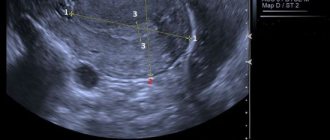
- Dopplerography or Color Doppler mapping (CDC). It is carried out to study the renal vessels. The technology of the method is based on fluctuations in the frequency of sound waves, which change after a collision with blood (a moving object). As a result, the doctor receives information about the presence of inflamed vessels and the nature of blood flow in the renal tubules. This method is based on the Doppler Effect.
- Ultrasonography (echography). This type of study determines disturbances in topography, detects stones and tumors, and reveals renal parenchymal changes. It is based on the principle of reflecting high-frequency waves from tissues, muscles and other dense structures of the organ. During the session, the specialist receives complete structural information about the organ being examined.
The essence of the procedure
Kidney ultrasound is divided into 2 types:
- Ultrasound echography - the procedure is based on the principle of sound reflection from tissues of different densities, which allows for a thorough examination of the renal parenchyma, timely detection of emerging tumors, conglomerates and topographic abnormalities;
- Doppler ultrasound is a method based on the Doppler effect and helps in the study of blood circulation in the renal vessels.
Ultrasound is practically the only safe technique that is so effective and informative. The procedure does not use radiation and does not have any negative effects when the skin comes into contact with the sensors.
How to do an ultrasound of the kidneys
Examination of the urinary system is carried out standing, sitting, lying or on the side. The sonologist applies a hypoallergenic water-based gel to the patient’s skin to ensure full contact of the body surface with the sensor. This increases the transmission level of ultrasound waves. First, the ultrasound procedure of the kidneys and adrenal glands is performed in the lumbar direction, then oblique and transverse sections are studied. In this case, the specialist moves the sensor to the side and front of the abdomen, and the patient turns alternately to the right and left side. The technique helps you see:
- location, size, shape of organs;
- condition of the parenchyma, renal pelvis, calyces, sinuses.
To determine the mobility of organs and improve their visualization, the doctor asks the patient to breathe and/or hold his breath after changing position. The necessary sections are visible much better when you inhale. The procedure is performed in a standing position if nephrosis is suspected. A lateral or sitting ultrasound examination is performed to view the renal vessels. The duration of the examination is no more than half an hour.
Kidney size is normal according to ultrasound
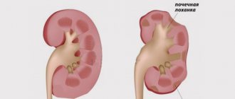
Interpretation of the results is done only by the doctor. The specialist in the conclusion indicates the number of organs, their location, size, shape, mobility, describes the condition of the ureters, adrenal glands, and tissue structure. Ultrasound diagnosis of the kidneys is considered normal if the contours of the organ are smooth in the photo, the fibrous capsule is clearly defined, and the tissues have a homogeneous structure. The renal pelvis should not be dilated, the organs should be located at the level of the first and second vertebrae, and the thickness of the parenchyma should be 15-25 cm.
Adult kidney size is normal
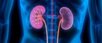
The left kidney should be located higher than the right. Some mobility of up to 2 cm in a vertical position is allowed. The shape of healthy organs should be bean-shaped (bean grain), and the size should be constant, but a slight difference between them of up to 1 cm is allowed. Normal kidneys according to ultrasound in adult men and women: width 5-6 cm, length 10-12 cm, thickness 4- 5 cm. 1 organ weighs up to 200 grams. An increase in parameters may indicate the presence of inflammatory processes or diseases such as hydronephrosis or pyelonephritis. Reduction in size occurs with hypoplasia.
Normal kidneys in children
The price of an ultrasound examination is no different for an adult or a child, but their standards are different. For a normal determination of the size of paired organs, it is necessary to conduct a correlation analysis between the body weight, age, height and gender of the child. There are certain tables that a specialist should take into account when deciphering the diagnosis.
It is difficult to determine the normal size of kidneys using ultrasound in children, because each child develops differently. You can navigate development based on average statistical indicators. For example, the size of the kidneys in a newborn baby is 4.9 cm. From three months to one year, the organs increase to 6.2 cm. Then, until the age of 19, they should normally grow by 1.3 cm every 5 years.
What does a kidney ultrasound show?
The range of pathologies of the urinary system is very wide. After an ultrasound examination, interpretation of a kidney ultrasound may show the following diseases:
- Pyelonephritis. Infection of the renal pelvis, which eventually spreads to the parenchyma. The disease occurs in acute or chronic form.
- Urolithiasis disease. The pathology is characterized by the presence of stones in the pelvis, bladder or along the ureter.
- Kidney block. Stopping the flow of urine due to swelling or inflammation in any part of the urinary system. This condition can be caused by a stone, blood clot, or injury.
- Renal vein thrombosis. Complete or partial blockage occurs due to a blood clot, increased echogenicity of the parenchyma, increased size of the organ, or the presence of fluid in the tissues.
- Damage to the urinary system. These include many diseases for which no treatment was carried out. The condition can also occur after injury.
- Prostatitis. The disease affects the strong half of humanity. Inflammation of the prostate gland is accompanied by severe pain in the perineum or lower back, difficulty urinating, and discomfort during sexual intercourse.
Proper preparation for a kidney ultrasound will help to obtain the most reliable results, and this is important if there are problems with the paired organ and a specific selection of a treatment regimen is required. It is not at all difficult to properly prepare for the procedure; the main thing is to strictly follow the basic rules, and also not to ignore the recommendations of the treating doctor.
Advantages
Ultrasound has been used in medical practice for approximately 40 years, but is still considered a young diagnostic method for imaging organs using ultrasound waves.
The ultrasound examination apparatus consists of a console with a screen. The console is connected to a probe (detector), which sends sound waves to the organ being studied. Returning to the probe, the converted waves display the examined areas of the body on the display. Thus, the probe, like bats or an echo sounder on a ship, “listens” to its modified echo.
To ensure complete but painless connection of the probe head to the body, the study area is lubricated with a special gel, which prevents the formation of air bubbles that can distort the image.
It is not difficult to do an ultrasound. The doctor will initially ask the patient to lie on his back and then change positions twice to get an idea of the condition of the organ on both sides. Almost always, to complete the picture you will need to hold your breath for a moment. An ultrasound examination of the kidneys is done within a few minutes, usually no more than half an hour. At the end of the procedure, excess gel is removed with a napkin. Interpretation of the ultrasound of the kidneys - the sonologist’s report will be ready while the patient gets himself in order.
The advantages of this diagnostic method are manifold:
- preparation for the procedure does not require much effort and time;
- the method is non-invasive;
- the process is safe and painless;
- no side effects;
- it is possible to repeat the study after some time;
- it is possible to use ultrasonography for pregnant women (X-rays are not used);
- the procedure is more accessible and cheaper than other imaging methods;
- Using ultrasound, tissues that are not qualitatively recorded by other methods are studied;
- ease of visualization of tumors, their shapes and sizes;
- After the ultrasound, the patient returns to daily activities without any restrictions.
Investigations complement the diagnosis and should not be underestimated. The only disadvantages of the procedure include the inaccuracy of visualization of individual organs and the examination of obese patients.
When is a kidney ultrasound prescribed?
Ultrasound of the obstructive system and kidneys is prescribed when a person has complaints and suspicions of dysfunction of these organs:
- pain and discomfort in the kidney area;
- increased body temperature;
- swelling of the body;
- changes in the color and composition of urine;
- discomfort and pain during urination.
Pregnant women at certain stages of pregnancy, as well as young children who have a history of renal pathologies or are suspected of developing them, are required to be examined.
The essence of the procedure
Ultrasound of internal organs is an informative and high-quality diagnostic study that has a number of advantages, including:
- accuracy and information content of the results;
- no contraindications or side effects;
- easy tolerability and painlessness;
- speed of implementation;
- absence of pain and discomfort.
A special gel promotes more efficient delivery of ultrasound waves.
The procedure is performed with equal success on men, women, children and the elderly. A person is exposed to high-frequency ultrasonic waves, which, upon reaching the desired organ, are reflected from it, transformed, and display the image on the monitor. A special gel, which is applied to the skin before the examination, helps to obtain the most accurate results from examining internal organs. Thanks to it, the waves penetrate better inside and transmit more accurate and clear images to the monitor. The results will be deciphered by a doctor who is specially trained and knows all the parameters necessary to make an accurate diagnosis.The examination is done in 2 ways:
- Doppler ultrasound. Allows you to identify and evaluate pathologies in the blood circulation and the condition of the kidney vessels.
- Ultrasound echography. Thanks to this method, it will be possible to evaluate the structure of the organ, identify the presence of foreign stones and neoplasms of various etiologies.
Carrying out an ultrasound of internal organs will be difficult if the patient:
- is overweight;
- suffers from flatulence and excessive gas formation;
- the day before I had undergone a diagnostic test during which barium was injected into the intestines as a contrast agent.
Types of ultrasound diagnostics
There are two types of ultrasound:
- “normal” - echography;
- Doppler ultrasound.
Using echography, the structure, location and size of the kidneys are examined. Information is transmitted to the screen in black and white. The specialist conducting the diagnosis, based on the picture on the computer screen, has the ability to accurately assess the pathology or normal size of the organs. However, it is impossible to study renal blood flow with “conventional” ultrasound.
Doppler ultrasound is prescribed when ultrasound of the renal vessels is necessary. The method is based on the ability of a sound wave (of a certain frequency) to be reflected from red blood cells. Moreover, the reflection shows not only the size and speed of blood flow, but also the structure of the blood vessels. The information is presented in the form of a graph – a Dopplerogram. Ultrasound of kidney vessels allows you to diagnose blood clots, blockages, aneurysms (stretching), narrowing of blood arteries and veins.
How to properly prepare for a kidney ultrasound?
Preparing for a kidney ultrasound is not at all difficult, it will not require significant effort from the patient, but you need to take the recommendations seriously, because if a dangerous disease develops in the body, it is important to undergo a high-quality examination and determine the diagnosis in a short time.
You should not drink beer a few days before the procedure.
To prepare for the study for adults in 2-4 days you need to:
- Normalize nutrition and exclude from the diet any food that provokes the formation and accumulation of gases in the rectum. You cannot eat or drink: white bread and baked goods;
- potato;
- whole milk;
- white cabbage;
- strong alcoholic drinks, beer, sweet soda.
- white or black coal;
It is better to take wipes with you to remove any remaining gel from the skin.
During the procedure, the laboratory technician uses a special gel, which ensures the penetration of ultrasonic waves into the abdominal cavity, making the results of the study as accurate as possible. After the procedure, the gel must be wiped off, as it leaves stains on clothes. Not all diagnostic institutions are provided with special napkins, so for your comfort you should take your own napkins or towel with you.
Features during pregnancy
Pregnant women often experience problems with the kidneys, since during this period the paired organ is exposed to repeated stress, so ultrasound diagnostics are performed to monitor its functioning. During pregnancy, it is also worth preparing responsibly for the procedure. The use of special absorbent drugs, as well as enemas, is contraindicated. It is important to normalize your diet and avoid foods that cause flatulence. The diet before an ultrasound scan of the kidneys also includes adherence to the drinking regime, how much liquid to drink is agreed with the doctor. It is better to carry out the procedure on an empty stomach, because if the doctor sees abnormalities in other abdominal organs, the absence of food in the intestines will make it possible to obtain a more accurate result.