Pathology in the genitourinary system associated with narrowing of the urinary canal is quite common. This disorder is called ureteral stricture. The pathological process can affect the organ either completely or partially. Due to disruption of the urinary system, urine is either not released at all, or it happens slowly. There are acquired and congenital strictures.
Description of the pathology
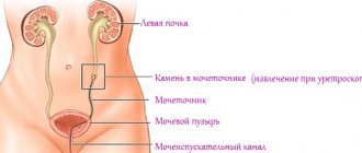
The ureter is a hollow tubular organ that connects the kidney to the bladder (in most mammals).
It starts from a narrowed area of the renal pelvis, where urine formed in the kidney flows. Its outlet end ends in the wall of the bladder.
For a healthy person, narrowing of the ureter of an anatomical or physiological nature is considered an acceptable norm. This phenomenon occurs due to the elastic properties of its wall. However, if stenosis or stricture occurs, the changes begin to take on a fibrosclerotic form. As a result of this pathological process, a violation of the submucosal membrane, as well as the muscular and outer walls of the ureter, occurs. Some muscle elements die off and are replaced by scar tissue, which is unable to perform any functions because it is atrophied.
Diagnostics
A doctor, when a patient comes to him with complaints indicating a stricture of the ureter, collects an anamnesis, including information about previous diseases and operations. This will allow you to correctly prescribe treatment later.
After clarifying the existing symptoms, the time and nature of their manifestation, the doctor refers the patient to a laboratory test, which includes urine and blood tests.
Blood analysis
A general analysis of both blood and urine indicates an increase in the number of leukocytes and ESR, which makes it possible to diagnose inflammatory processes. A biochemical blood test allows you to determine the level of kidney function.
Urinary duct stricture can be detected by ultrasound.
The pelvicalyceal system of the kidneys is clearly expanded, which indicates an incomplete urinary process, accompanied by congestion.
Urine cannot be excreted fully due to obstacles along the way, one of which is a stricture.
When performing excretory urography, the doctor completely visualizes the existing narrowing of the ureter.
However, in the most difficult cases, when narrowing is observed almost along the entire length of the ureter, or it has bends, excretory urography will not be able to unambiguously confirm the presence of scar changes in its walls.
In these cases, they resort to retrograde ureterography.
Having introduced a contrast agent, the doctor observes to what level it moves freely along the ureter, at what stage its movement stops.
Another diagnostic way to examine the ureter to identify pathological changes in it is to examine the organ using a ureteroscope.
Organ dysfunction
The lumen of the urinary duct at the site of ureteral stricture decreases, which disrupts the normal functioning of the organ. Urine cannot be completely eliminated from the body and begins to accumulate in the bladder, eventually causing increased pressure on the ureter. Subsequently, it stretches and elongates. In some cases, it comes to curvature of the ureter. In the absence of proper treatment, the pathology affects the kidneys.
Ureteral stricture can develop in any area of the organ. Most often, the pathology is localized in the space between the bladder and ureter. In addition, there are cases of identifying a stricture between the ureter and the pelvis.
Types of strictures
Narrowing of the ureter can take different forms depending on the area of localization of the pathology, as well as on the nature of the disease. First of all, a distinction is made between acquired and congenital stenosis. The latter appears during the intrauterine development of the unborn child.
The pathological process may occur due to thickening of the walls in certain places. Congenital ureteral strictures appear as a consequence of certain disorders during fetal development, namely:
- An inflection that occurs due to the curved shape of the ureter.
- The appearance of a membrane in the ureteral valve, which provokes the accumulation of urine in the bladder.
- Ureterocele. This is a disease that is characterized by a narrowing of the lumen in the lower part, while the ureter expands, and in some cases falls into the cavity of the bladder.
- Squeezing of blood vessels.
- The formation of diverticula, which provoke protrusion of the lower part of the ureter.
An acquired form of narrowing of the ureter can occur under the influence of various factors, depending on the person’s state of health. Depending on the area where the stricture was localized, right-sided and left-sided stenosis are distinguished. It also happens that both sides of the ureter are affected. Also, stenosis can be localized both in the upper part of the ureter and in its lower part, where the transition to the renal pelvis occurs. If the pathological process develops in the middle section, then both the upper and lower parts of the organ are affected.
Provoking factors
Factors that can cause the development of ureteral stricture are:
- Formation of kidney stones. This falls under the category of internal injuries. Urolithiasis causes inflammation, and the mucous membranes are easily damaged by the stones, leading to scarring.
- External injuries to the lumbar spine. As a result, a hematoma appears behind the peritoneum, which later becomes the basis for a stricture.
- Injury sustained during surgery.
- Radiation therapy and radiation injury.
- Tuberculosis, inflammation in the ureter.
The causes of ureteral stricture should be determined by a doctor.
In addition, in some cases, pathology appears as a consequence of gunshot or knife wounds. Also, self-treatment of sexually transmitted diseases can lead to injury to the ureter. Men are more susceptible to injury and overuse and are more likely to develop strictures. If any of the listed factors are excluded, the doctor makes a conclusion about the congenital form of the disease.
The ICD-10 code for ureteral stricture is N13.5.
Stricture of the pelvis and ureter
Stricture of the ureteropelvic segment (UPS) is any pathological narrowing of the lumen of the upper urinary tract that impairs its patency. Due to impaired urine outflow, the pathology leads to the development of other diseases of the urinary system.
REASONS FOR THE DEVELOPMENT OF LUS STRICTURE
Strictures of the pelvis and ureter can be congenital or acquired. Acquired ones arise for a number of reasons:
- Past inflammatory diseases of the upper urinary tract
- Due to chronic cystitis with reflux of urine into the ureter
- Damage to the wall of the ureter due to stones
- As a complication of specific infections, for example, tuberculosis
- As a consequence of radiation therapy
- As a complication of oncological processes
- Consequence of abdominal operations and manipulations
HOW DOES LMS STRICTURAL MANIFEST?
The clinical picture consists of symptoms of urinary outflow disturbance and its consequences. Typically patients feel:
- Lower back pain
- Renal colic
- Decreased urine output
- Cloudy urine mixed with blood
DIAGNOSTICS OF STRICTURE OF THE LUS
For complete treatment, it is necessary to accurately determine the cause of the disease and then eliminate it. For this purpose, urologists at the WMT clinic carry out comprehensive diagnostics:
- General inspection
- History taking
- Ultrasound diagnostics
- CT with contrast and mandatory delayed phase
- MRI
TREATMENT OF STRICTURE OF LMS
The only effective method of treatment is surgery. There are a large number of options for PUJ strictures, so for an effective result it is necessary to choose the optimal type of operation. Urologists at the WMT clinic perform surgical treatment of PUJ strictures of any complexity. In the absence of damage and impaired renal function, specialists perform reconstructive operations aimed at removing the problem area and restoring urinary outflow:
- Anastomosis (artificial connection of two ends of a hollow organ)
- Boari operation - anastomosis between the ureter and bladder
- Replacement flapplasty
- Plastic surgery of the ureter with a fragment of intestine
In case of severe damage to the renal tissue with loss of kidney function, urological surgeons remove the kidney and ureter.
MAKE AN APPOINTMENT
Book a consultation with a urologist at the WMT clinic by calling 8
or leave a request on the website.
Appointment with a Urologist
Please leave your phone number. We will select the most convenient time and make an appointment for you.* - required fields are marked with an asterisk Cost of services
Symptoms
As a rule, symptoms and severe pain accompany bilateral stenosis. Unilateral stenosis, on the contrary, mostly occurs in a latent form. For this reason, it is almost impossible to diagnose the disease at an early stage of its development. With bilateral damage, the following symptoms are observed:
- Increased pressure in the arteries.
- Pain syndrome in the lumbar region.
- Nausea and vomiting.
- Convulsive syndrome.
- Passage of a small amount of urine.
- Pain during urination.
- An increase in body temperature, which indicates an inflammatory process in the body.
- Presence of blood in the urine.
The symptoms of ureteral stricture are very unpleasant.
In the absence of proper treatment, the pathological process can progress and spread to adjacent organs, including the kidneys. Due to incomplete excretion of urine from the body, the risk of stagnation increases, which will ultimately lead to urolithiasis, pyelonephritis, hydronephrosis, as well as chronic kidney failure. It is important to promptly identify the pathology and receive qualified medical care.
Symptoms of the disease
The ureters are paired organs, but this does not mean that both organs can be susceptible to the same disease. Bilateral ureteral stricture is not common but is much easier to identify. Unilateral stenosis can occur without obvious pain; something is wrong only by indirect signs.
Important! Problems with urination should be a warning sign, especially for men. Even if urinary stagnation is not a major concern, it will ultimately lead to the development of chronic kidney disease.
What symptoms indicate a possible abnormal narrowing of the ureter? Most often, patients indicate:
- pain when urinating;
- the need to strain your stomach on the toilet;
- decrease in urine output;
- splashing stream of urine in men;
- dull, not entirely clear pain in the lumbar region;
- sharp pain along with chills, fever, nausea and vomiting;
- bloody discharge from the ureter;
- the presence of blood particles in the urine;
- weakness, temperature;
- muscle cramps;
- yellowness of the skin;
- cloudy urine with a strange, pungent odor;
- increased blood pressure;
- general intoxication.
The diagnosis is established as a result of ultrasound of the genitourinary system, x-ray with contrast agent, MRI. Urography makes it possible to visualize the location of the kidneys and urinary tract, determine all possible narrowings, their extent and how they affect the functioning of the genitourinary system.
In leading clinics, it is possible to conduct three-dimensional angiography with a diuretic load, which allows you to simultaneously see the abnormally dilated section of the ureter above the stricture, and assess the condition of the renal vessels.
Important! Numerous studies are necessary to clarify the full picture of the disease and all possible treatment methods, which in most cases are only surgical.
Diagnostic methods
To get a complete clinical picture, it is necessary to schedule a detailed examination of the patient. Diagnostic procedures include blood and urine tests, and ultrasound of the genitourinary system. In addition, the patient is prescribed computed tomography and magnetic resonance imaging. Endoscopic examination is contraindicated if the patient has an inflammatory process in the vagina, uterus, urethra or prostate gland.
The most informative and common method of research for ureteral stricture is urethrography. The procedure is an X-ray examination using contrast. This technique makes it possible to identify those areas in which there is stagnation, as well as localize the presence and position of narrowed areas. The contrast is injected directly into the urethra or intravenously.
Preparation for urography
Urography is considered an effective, safe diagnostic method. The study is prescribed if there is a suspicion of kidney pathologies, bladder diseases, problems with filtration and urine output.
The basic rules for preparing for urography will be the following:
- 3 days before the procedure, the patient must refuse food that causes excessive gas formation.
- A test must be carried out to detect an allergy to a radiocontrast substance.
- Meals should be taken no later than 8 hours before the test; you should not drink too much liquid during the day.
- You can't eat in the morning.
- In the office, you need to remove metal products and jewelry, and, as directed by the doctor, empty your bladder.
- If there is anxiety shortly before urography, you can take a sedative (calming) drug.
Therapy
After a thorough examination and clarification of the diagnosis, the patient is prescribed the necessary treatment. The main goal of therapy is to normalize urine excretion. The treatment regimen is selected based on the results of the studies. It is also important to consider the general condition of the kidneys and genitourinary system. Another important factor in determining treatment is the size of the stricture.
Ureteral stricture cannot be treated at home or with traditional medicine. Contrary to popular belief, you should not heat the affected area, as this may make the pain more intense.
One of the effective treatment methods is plastic surgery in urology centers. This is a rather complex procedure with a long rehabilitation period, so it is prescribed only as a last resort. The operation is not suitable for every patient, as it has a number of contraindications.
Another treatment method is bougienage of the ureter. The procedure involves using a metal rod that is inserted into the ureter and dilates it. The procedure is very painful, and the effect is short-lived. Bougienage is rarely used.
How are strictures (stenoses) of the ureter treated?
Treatment is primarily aimed at restoring the flow of urine. There are several ways to do this, and they are completely different. The choice of one option or another depends on the results of the examination, the condition of the organs, and the size of the stricture.
Important! No traditional medicine methods work for strictures. Do not self-medicate; a traditional hot water bottle is contraindicated in this case. It can cause more urine production, which will lead to more pain.
A summary of possible methods:
- Complete plastic surgery – in the most severe cases. This is a serious operation with a difficult rehabilitation period; there is a wide range of contraindications for it.
- Bougienage is an outpatient procedure in which the narrowing is moved apart using a bougie - a smooth metal rod. The procedure is quite painful and gives a short-term effect, so it is used extremely rarely in modern hospitals.
- Plastic replacement is a surgical intervention for small strictures up to 10-20 mm long. During the operation, the damaged area is excised and replaced with tissue from the patient himself.
- Optical urethrotomy is a method in which the abnormal narrowed area is dissected using a cystoscope. Suitable for strictures up to 5 mm. A special spring is implanted into the lumen of the ureter. In this case, the mucous membrane, gradually regenerating, acquires an anatomically correct shape.
Plastic replacement method
Urology centers also use the plastic replacement method. This method is suitable for the treatment of small strictures, the size of which does not exceed 20 mm. The operation involves making an incision and replacing the scars with tissue from the patient. In addition, optical urethrotomy using a cystoscope is used. Any intervention to treat stenosis must be agreed with the attending physician and supervised by a qualified surgeon.
The pathology is quite serious and cannot be treated with medications or traditional methods. If surgery is not performed, complications may occur that affect the kidneys and other organs.
Health care
When organizing treatment measures, special attention is initially paid to restoring the normal outflow of urine.
This is very important for the patient’s health, since congestion provokes serious diseases leading to kidney failure.
Self-treatment of any kind is strictly prohibited. Under no circumstances should you apply warming compresses or warm up the pain areas with heating pads.
When warming up, urine begins to form intensively, and due to its difficult exit, it cannot find an outlet and stagnates in the kidneys, leading to even more serious problems, provoking pain of even greater intensity.
Only a doctor can carry out treatment using one of the methods that, in his opinion, is most appropriate according to the results of a diagnostic examination.
There are four methods in total, including bougienage, replacement and complete plastic surgery, and urethrotomy.
Bougienage is an outpatient procedure. Bougienage involves inserting a metal rod with a smooth surface into the ureter, with the help of which the walls of the ureter are moved apart, eliminating the narrowing.
Bougienage is a procedure accompanied by significant pain, and therefore it is very rarely used.
In addition, treatment involves surgical intervention involving plastic replacement of the damaged area of the ureter with tissues of another organ of the patient himself.
This operation is performed only if the area where the stricture is observed does not exceed 20 mm. For even smaller sizes (up to 5 mm), an optical urethrotomy is performed, during which the damaged area is excised and a special spring is implanted into it.
As it recovers in the postoperative period, thanks to the regenerative properties of the tissue, the ureter restores its physiological shape. Complete plastic surgery is performed only in the most severe cases.
promoipochki.ru
Prevention and prognosis
Stenosis develops rapidly, especially when it is preceded by injury. A hematoma forms in the affected area, which must be detected and drained. If first aid is carried out correctly, the formation of strictures is excluded. Any, even minor, injury to the lower back requires contacting a specialist for examination and examination. It is important to avoid injury to the pelvic area when playing sports. It is important to use special protective shields that can soften the blow.
The sooner surgery is performed after detection of strictures, the better for the patient and the less likely it is to develop complications. In addition, this will shorten the recovery period, and the operation itself will not be so painful. An important point in proper rehabilitation is compliance with all instructions of the attending physician.
Prevention of stenosis
Constrictions do not occur extremely quickly in most cases. In case of trauma, the phenomenon is preceded by extensive retroperitoneal hematomas, which should be drained in a timely manner. Thanks to the professional actions of doctors, in this case, stenosis will not develop and there will be no complications.
Note! The main prevention of ureteral strictures is the timely and correct treatment of all injuries in the lumbar region. A qualified consultation with a urologist in case of blows to the lower back is mandatory, do not neglect it!
What should athletes do if they have such blunt injuries that can be considered a professional phenomenon? Winter sports are dangerous. Many sports disciplines have developed special shields that soften the impact of collisions and impacts.
The prognosis of the disease is positive if the patient complies with all instructions during postoperative rehabilitation. The patient should not assume that ureteral stricture is a one-time occurrence. In most cases, everything depends on the person himself, what kind of life he has, how he views his own safety. Take care of yourself!
But perhaps it would be more correct to treat not the effect, but the cause?
We recommend reading the story of Olga Kirovtseva, how she cured her stomach... Read the article >>
ozhivote.ru
Complications
If the above conditions are not met, complications arise that can affect the functioning of the genitourinary system and other organs. The operation can also have consequences if the patient’s tissues have not grown together correctly or have not taken root at all.
Lack of treatment can lead to the development of pathologies such as cysts or kidney failure, as well as hydronephrosis, when the renal pelvis expands. In some cases, cystitis and kidney stones appear against the background of strictures.
Treatment
Treatment depends entirely on what causes the bend.
If this is associated with prolapse of the kidneys, then the patient is recommended to wear a bandage that helps maintain the internal organs, including the kidneys, in the correct position.
Physiotherapy
To strengthen the abdominal muscles, a special complex of physical therapy has been developed. It is also recommended to adhere to the rules of a balanced diet and stop the diet that has caused depletion of the fatty renal capsule.
In order not to bring the bend to a critical state during the period of conservative treatment, it is recommended to avoid lifting heavy objects.
If the bend was caused by other reasons, then in most cases drug treatment will be unsuccessful, so they resort to surgical intervention followed by conservative treatment of those complications that have already been caused by the bend of the ureter.
In particular, pyelonephritis, urolithiasis, and arterial hypertension are subject to such treatment.
In some cases, surgical intervention is also performed for nephroptosis.
During the operation, doctors secure the kidney in the place intended for it by nature. As a result, effective blood supply to the organ is restored, accordingly, at the junction with the ureter, the kink is eliminated, and the outflow of urine is normalized.
After such a serious operation, it is important to maintain bed rest so that the kidney can be successfully fixed in the position in which the doctors fixed it.
Due to the displacement of the kidney beyond the physiological norm, a kink of the ureter occurs. This dangerous pathology is characterized by a change in the free outflow of urine, inflammation of the urinary system and creates suitable conditions for the addition of secondary infections. The lack of adequate therapeutic therapy leads to changes in the renal blood supply, the occurrence of pyelonephritis and arterial hypertension.