Indications for coronary angiography Contraindications Preparation for the procedure Methodology for coronary angiography Interpretation of results Complications
As you know, the heart has blood vessels that supply blood and oxygen to the myocardium (heart muscle). These vessels are called coronary (coronary, or proper cardiac). Their normal functioning is very important for the proper functioning of the heart muscle, on which, in turn, the well-being of the whole organism depends. If the lumen of blood vessels is blocked by a blood clot or atherosclerotic plaque, acute or chronic hypoxia of the heart tissue (lack of oxygen) occurs, leading to necrosis (tissue death). As a result, diseases such as coronary heart disease (CHD) and myocardial infarction develop. In most cases, such diseases are easily diagnosed based on the results of a clinical examination, ECG and ultrasound of the heart.
But it is not always possible to determine the presence of coronary artery pathology on the basis of these data alone and to develop specific treatment tactics. Sometimes a doctor needs to literally “look” into a person’s heart in order to understand what pathological processes occur in this important organ. Is this feasible?
The capabilities of modern medicine are constantly expanding. A hundred years ago, doctors could not even imagine that someday they would be able to see the heart of a living person from the inside, see how it beats, evaluate how its internal structures and the vessels that supply it with blood work. Currently, all this has become possible thanks to special equipment and high-tech research methods. One such method is coronary angiography (CAG).
Coronary angiography (or coronary angiography) is an instrumental method for diagnosing cardiovascular diseases, which is carried out by introducing a radiopaque substance into the heart’s own circulatory system, as a result of which the doctor receives X-ray images of the coronary arteries with subsequent assessment of their patency. This study allows us to identify the degree of disturbance of blood flow in these arteries, arising due to a blood clot, atherosclerotic overlay, vascular spasm (for example, as with Prinzmetal’s angina), clarify the presence of myocardial ischemia, and also determine the doctor’s further actions in terms of cardiac surgical treatment - the need for stenting arteries or coronary artery bypass grafting (CABG).
Indications for coronary angiography
The main indications for this diagnostic method are the following: - acute myocardial infarction in patients for whom stenting is considered necessary by the doctor (within the first 12 hours from the onset of clinical manifestations); - severe stable angina pectoris 3 - 4 FC (functional class); - stable angina with signs of severe ischemia with minor physical exertion; - variant Prinzmetal angina; — lack of effect from the drug therapy, in this case the question of the advisability of stenting or CABG is decided; — previous myocardial infarction, accompanied by fatal rhythm disturbances (ventricular fibrillation, complete AV block, etc.) or clinical death; - high risk of sudden cardiac death; - impossibility of performing an ECG or ultrasound of the heart with exercise (low exercise tolerance, as well as for patients with low ejection fraction according to ultrasound); - before surgery on heart valves in patients over forty years old, as well as for pain in the chest and in the heart area; - clarification of the diagnosis according to clinical or professional indications - in cases where the results of other examination methods are doubtful; - relapses of angina or myocardial infarction within 9–12 months after stenting and CABG, respectively.
Indications for use
Coronography of the heart is used:
- For chest pain and shortness of breath, often indicating a narrowing of the blood vessels of the heart;
- In cases where treatment with medications does not bring results, and the symptoms of the disease intensify;
- Before undergoing heart valve replacement surgery (to identify narrowing of the heart vessels);
- After bypass surgery to evaluate the results of surgery;
- If there is a suspicion of congenital defects of the heart vessels;
- For diseases of the heart vessels;
- In case open heart surgery is planned;
- For heart failure;
- For serious chest injuries;
- On the eve of surgery, which is associated with a risk of heart problems.
Contraindications for coronary angiography
There are no absolute contraindications for this method. Among the relative contraindications, the following can be noted: acute infectious diseases, anemia (decrease in hemoglobin in the blood), pathology of the blood coagulation system with a possible risk of prolonged bleeding, stroke, acute or chronic diseases of other organs (acute surgical or gynecological pathology, decompensation of diabetes mellitus, bronchial asthma etc.).
For each patient, indications and contraindications are determined by a cardiologist, cardiac surgeon, and, if necessary, doctors of other specialties strictly individually.
Preparing for the study
Before coronary angiography, it is very important to follow the drinking and eating regime. The study is carried out strictly on an empty stomach (the last meal 6–8 hours before), since vomiting may develop during the intravenous administration of contrast and aspiration (entry into the respiratory tract) of vomit. Two to three hours before the examination, you are allowed to drink not a very large amount of clean drinking water for the proper functioning of the kidneys, since they are the ones who will have to remove the contrast agent from the body.
In the case of a planned examination, when a patient is sent from a clinic or cardiology hospital, he should have the following examination methods on hand: a general urine test, a complete clinical blood test with the determination of platelets, prothrombin index, blood clotting time and other indicators of the blood coagulation system, biochemical blood test, tests for HIV, syphilis, hepatitis B and C, ECG results, echocardiography (ultrasound of the heart).
If the patient is brought in for examination on an emergency basis (by an ambulance, from the cardiology or intensive care unit with suspected myocardial infarction), these examinations can be carried out urgently if necessary.
How is coronary angiography performed?
Coronary angiography is an invasive diagnostic method, that is, during the research process, it is introduced into the tissues and organs of the human body. Performed on a scheduled or emergency basis. During a routine examination, the patient is hospitalized several days before in the cardiology or cardiac surgery department of the hospital, where the necessary diagnostic methods described above are carried out at the discretion of the attending physician.
Before the nurse takes the patient on a gurney to the X-ray surgery room, he is given premedication - the administration of painkillers and sedatives (ketorol, Relanium intramuscularly or intravenously). Next, the patient is transferred to a table in the office, the puncture site of the radial artery (on the wrist) or femoral artery (in the groin) is anesthetized using subcutaneous anesthesia with lidocaine or other anesthetics, then the puncture (puncture of the skin and artery) is started directly. After access to the artery (most often the radial one), an introducer is inserted into it - a sterile disposable tube with a valve that prevents blood from entering it and a side port for administering contrast. A conductor is inserted through the introducer, reaching the aorta with the coronary sinuses through the radial artery. Next, a catheter is inserted into the conductor and installed at the ostia of the right and left coronary arteries; an X-ray contrast agent is introduced through this catheter, which allows you to see the shadow of the artery on the screen, since the arteries and heart absorb X-rays without contrast. In this case, an X-ray image is taken, which allows one to evaluate the coronary artery in different projections (the artery does not lie in the same plane).
The contrast results are displayed on the installation screen and then saved in the computer for further evaluation and interpretation of the results. After successful contrast, the catheter is either removed or doctors decide whether emergency balloon angioplasty or a stent is inserted into the narrowed artery.
After the procedure is completed, a pressure bandage is applied to the wrist, which does not require further dressings, and the patient is taken to the ward. The entire procedure takes about 15 - 30 minutes, without causing pain in the patient, not counting the puncture site.
After a routine examination, the patient remains in the cardiology department for several days to assess the general condition and decide on further treatment methods. If necessary, hospitalization time can be increased in accordance with the need for cardiac surgery.
In the case of an examination carried out for emergency reasons, the patient is transferred to the cardiac intensive care unit for further observation and treatment.
How is the procedure done?
We will consider the progress of the entire procedure of coronary angiography of the heart vessels “from the patient’s side.”
Hospitalization and preparation
The patient enters the department in the evening or comes in the morning at the appointed time for the study. He should have blood tests on hand (the doctor will specify which ones), electrocardiography and the results of an ultrasound of the heart.
In the reception department or in the ward, the patient will receive an information consent, which must be signed (unless you change your mind about undergoing the study). Coronary angiography is performed on an empty stomach, the duration of the entire procedure is from 30 minutes to 2 hours. The patient is discharged the next day. In the morning before discharge, all tests will be taken.
This procedure can be carried out in two ways (we are talking about a standard planned diagnostic method): through the vessels of the arm and through the femoral artery.
Methods for inserting a catheter for coronary angiography of the heart vessels
Before coronary angiography, an injection (premedication) will be given to relieve nervous tension.
Usually the patient is conscious during the study and communicates with the doctor. In rare cases, it is necessary to put the patient into a state of medicated sleep - then an anesthesiologist will be present for the study.
What happens in the operating room itself?
- In both cases, local anesthesia (lidocaine and other means) is initially given.
- A vessel is pierced on the thigh or arm, and a catheter or tube is inserted into the vessel. Initially, you need to reach the ostia of the coronary artery (this is where the coronary artery exits the aorta).
The surgeon inserts a tube into the patient's right arm vessel. Click on the photo to see it clearly
- The doctor inserts a catheter directly into the mouth of the coronary arteries. At the other end (where they entered through the skin), a syringe with contrast is attached to the catheter. So they introduce him. The contrast fills the heart arteries and is washed away by the blood flow. The entire procedure is video recorded. The doctor monitors the progress of the process on the screen. The monitor can be rotated so that the patient can also see his own arteries. You will be able to talk with the doctor.
The surgeon injects contrast from a syringe through the catheter. Click on the photo to see it clearly
The doctor watches the progress of the process on the screen
- After the procedure is completed, the doctor applies physical pressure with his hands to the puncture area. This is necessary to stop the bleeding.
- Then a sterile pressure (very tight) bandage is applied and the patient is transferred to the ward.
After the procedure, the surgeon applies a tight bandage to the patient. Click on the photo to see it clearly
After coronary angiography
The patient is not recommended to get out of bed for 5 to 10 hours. This difference is understandable - after all, some patients take medications that thin the blood. And not in all cases it is possible to cancel them before the procedure.
You can eat immediately after the procedure. A surgeon will come into the room to discuss all the nuances of the study.
The recording of the coronary angiography procedure is carefully and repeatedly studied and analyzed by doctors. A copy of the video will be given to you immediately in the operating room.
Interpretation of coronary angiography results
The assessment of data obtained during coronary angiography is carried out by an x-ray surgeon, a cardiac surgeon and a cardiologist. Depending on the degree of narrowing of the coronary arteries, the following terms are distinguished:
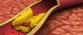
- occlusion - complete blockage of an artery by an atherosclerotic plaque or thrombus - the lumen of the artery according to the results of coronary angiography is narrowed by more than 90%; - stenosis - partial narrowing of the lumen of the artery by 30 - 90% - there are ostial stenosis (at the mouth of the artery or no more than three millimeters from its beginning), local stenosis (over 1 - 3 mm of the artery), extended stenosis (over a significant area artery narrowing of its lumen); — arterial aneurysm (protrusion of the wall, which interferes with normal blood flow and is fraught with rupture of the wall with bleeding); - arterial calcification (deposition of calcium salts, usually in combination with atherosclerotic plaques in the artery wall, which also causes narrowing and disruption of blood flow through this artery).
The figure shows partial obstruction of the coronary artery.
The results obtained are important for doctors from the point of view of the need for surgical treatment. For example, if the degree of narrowing of the artery lumen is more than 75%, the patient is indicated for cardiac surgical reperfusion (restoration of blood flow) of the myocardium.
What is coronary angiography?
Coronary vessels are the thinnest arteries that provide the flow of oxygen and blood to the myocardium. This is the only supply route to the heart, and it is very vulnerable and prone to damage. When the lumen of the coronary vessels is blocked and reduced, serious heart pathologies occur - ischemia, myocardial infarction, atherosclerosis.
Coronary angiography is the most accurate and reliable way to diagnose coronary artery disease
Coronary angiography refers to a radiopaque instrumental study, the essence of which is that a certain substance is injected into the vessel cavity, completely filling the lumens and allowing one to see the condition of the artery on an X-ray image.
What is the method of coronary angiography of the heart used for?
- To assess the condition of the coronary vessels;
- To diagnose abnormalities in the structure of arteries;
- To identify the source of blockage or spasm;
- To study the state of blood flow to the heart.
The coronary angiography method allows timely detection and diagnosis of pathologies of the heart and arteries, preventing the development of dangerous heart diseases.
Important! Coronary angiography is called the “gold standard” for diagnosing vascular pathologies of the heart, because the procedure helps to most accurately identify birth defects and objectively assess the condition of blood vessels from the inside.
Coronary angiography can be performed using several methods, depending on the indications for the study:
- Intravascular ultrasound examination (general coronary angiography) – all arteries of the coronary bed are diagnosed. It is used in rare cases - more often in scientific research. It is carried out invasively using a catheter, at the end of which an ultrasound sensor is installed.
- Selective interventional coronary angiography - a contrast agent is injected through a catheter only into those vessels that need to be examined. In the CIS, this method of diagnosing heart vessels is more often used.
- Coronary angiography is the most informative and clinically significant research method, allowing one to evaluate not only the lumens in the vessels, but also the affected areas in the walls of the vessels. The advantage of the method is less invasiveness (the catheter is inserted into a vein), short duration of the diagnostic procedure, and no need for hospitalization. The downside is the high cost of implementation, which makes the method inaccessible to most segments of the population.
Each of the methods has its own indications for use, and any of the listed diagnostic procedures has both disadvantages and advantages compared to others. Therefore, the method of coronary angiography of the heart vessels is selected by the doctor individually based on the clinical picture and condition of the patient.
Complications of coronary angiography
Since this study is invasive, and especially carried out on the heart, there is a risk of complications, which statistically develop in two cases out of a hundred. Mortality during coronary angiography is less than 1%. But still, in extremely rare cases, it is possible to develop ventricular fibrillation, thrombosis of the coronary arteries with the development of extensive myocardial infarction, stroke, radial artery thrombosis, infectious inflammation at the puncture site, acute renal failure as a reaction to the removal of contrast through the kidneys, an allergic reaction to the contrast agent , up to the development of anaphylactic shock.
Prevention of the development of complications is a careful collection of anamnesis regarding kidney diseases, anaphylactoid (apllergic) reactions, especially to iodine preparations, as well as timely administration of anticoagulants (heparin, fraxiparin, warfarin).
As a long-term complication, statistics show that low-dose radiation from cardiac imaging studies increases a patient's risk of cancer by an average of 3%.
General practitioner Sazykina O.Yu.
What changes do they see during the study?
| Type of coronary angiography | Changes |
| Planned | Narrowing or ulceration of the artery due to atherosclerotic changes Blood clots in the vessels of the heart Pathological spasm of the coronary arteries Anatomical changes in the vascular network (passage of vessels in the thickness of the myocardium, malformations of their development) |
| Urgent | The same disorders as during a routine study, but with signs of initial decompensation of blood flow in the heart muscle (significant stenoses, spasms or almost complete blockage of the lumen of the vessel by a thrombus) The presence of bypass blood supply and its functionality in maintaining normal blood flow to the affected area of the heart muscle |
| Emergency | Vessel with complete or almost complete blockage of the lumen Length of artery change Myocardial area with impaired blood supply |
Urgent and emergency coronary angiography in most cases includes not only diagnosis, but also surgical treatment of the resulting disturbance of blood flow in the heart muscle (installation of a stent in the artery or expansion of its lumen with a balloon).
To install a stent, a balloon is inflated, which straightens it inside the artery and it remains in this position. The balloon is removed.


