A kidney cyst is a benign cavity formation on one or both kidneys, which has a capsule of connective tissue and is filled with various contents. It has an oval or round shape, and most often occurs on one side of the paired organ. People of all ages can face a problem such as a kidney cyst and identifying its cause. They most often develop in men. This pathology is the most common renal tumor, accounting for 70% of all clinical cases. The size of the formation can reach more than 10 centimeters.
Classification of neoplasms
Types of formations.
Increase. Types of kidney cysts and their classification depend on the origin, location and quantity. A cyst, like a kidney pathology, can be congenital. It occurs as a result of improper development of the organ in the prenatal period. It is also caused by a genetic predisposition and is a hereditary disease. In this case, the onset of the disease occurs in childhood.
Congenital kidney tumors
Congenital neoplasms include:
- Solitary - filled with serous fluid, which may contain impurities of blood and pus. Usually located in the right or left kidney, they have one cavity.
- Multicystic disease affects one of the organs. In severe forms, it spreads to almost an entire organ, which loses its function. In some cases, unaffected healthy areas are preserved.
- Polycystic cysts are cysts that affect the tissues of both kidneys, causing them to take on the appearance of bunches of grapes. It is often a congenital disease caused by a genetic predisposition.
- Multicystic medulla - the formation of many cysts with expansion of the collecting ducts of the organ. It is a congenital disease.
- Dermoid is a congenital disease in which a neoplasm on the right or left kidney is filled with ectoderm elements (teeth, fat, hair, bone inclusions).
- Renal cystic formations - their appearance occurs in hereditary diseases (tuberculosis syndrome, Hippel-Lindau syndrome, Zellweger syndrome, Meckel syndrome).
Causes of acquired tumors
Acquired kidney cystosis occurs due to pathologies of this organ present in the body:
- tuberculosis;
- heart attack;
- pyelonephritis;
- cystic kidney dysplasia;
- parasitic infections (echinococcus);
- tumors;
- glomerulonephritis;
- medullary necrosis.
Localization, structure, contents of cysts
When classifying neoplasms by number, the following are distinguished:
- Single - there may be one cyst in one of the organs, or one formation in each organ.
- Multiple – several neoplasms can be located in one or both organs without interfering with their functioning. Multiple cysts can cause kidney failure in the case of polycystic disease.
According to their location in the tissues of the organ, they are distinguished:
- Subcapsular cysts are located under the renal capsule.
- Intraparenchymal kidney cyst - located in the parenchyma.
- Peripelvic cyst - located near the renal sinus. The most common are peripelvic cysts of the left kidneys.
- Cortical cyst - affects the cortex.
According to its structure, the neoplasm can be:
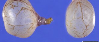
- Unicameral - a simple kidney cyst, has one cavity. Simple kidney cysts have a low tendency to malignancy.
- Multi-chamber - a complex kidney cyst, has several cavities separated by septa, and thus forming several neoplasms. A multilocular renal cyst may have calcifications, and there is a risk of it becoming malignant.
Single-chamber Multi-chamber
One example of a multilocular cyst is an atypical renal cyst. It has a broken structure and multiple partitions. It is often caused by injuries, infectious and parasitic diseases.
Based on the nature of the contents, cysts in the kidneys are divided into:
- serous - filled with a transparent green-yellow liquid;
- hemorrhagic – filled with blood, occurring after a traumatic impact, or as a consequence of a heart attack;
- purulent - suppurated formations that arise due to infection;
- calcified – formations containing calcifications.
We invite you to watch an informational video on the topic of our article:
Features of the therapeutic diet
When a kidney cyst appears, various treatment tactics can be chosen, be it observation, surgical or drug treatment. But in any case, it is complemented by a dietary table.
Food should be healthy
Basic principles of nutrition:
- Reducing salt intake, especially when, against the background of the presence of a cyst, there are disturbances in the functioning of the organ, kidney failure develops. In some cases, you have to give up salt completely.
- Refusal of a number of foods and dishes. The ban applies to salty and fried, spicy, as well as any alcohol, especially such a popular drink as beer. In addition, you will have to give up coffee and seafood.
- Strict adherence to the drinking regime. In cases where, against the background of a cystic formation of the kidney, severe swelling, high blood pressure, and also signs characteristic of heart failure such as shortness of breath appear, it is necessary to control the volume of fluid. The more severe the symptoms, the more stringent control is necessary, and vice versa.
- Limiting the presence of protein. The protein component of foods has a negative effect in the development of various kidney ailments. With a significant proportion of protein in food, the formation of a number of products during nitrogen metabolism, including uric acid and guanidine, is observed. All of them are characterized by severe toxicity, therefore, to minimize the load on the organ, the proportion of protein foods in the diet should be minimal.
It is not possible to cure a cyst if you follow a special diet. But diet is the most important component of complex treatment, which helps to increase the effectiveness of other methods.
Classification of renal cysts according to Bosniak
| Category | Character traits | Further tactics |
| 1 | A simple benign thin-walled cyst without septations, calcifications or solid component. The contents have liquid density and are not contrasted. | No further observation is required. The formations are benign. |
| 2 | Benign cyst. May contain several thin partitions. There may be small or slightly thickened calcifications in the walls and septa. The same group includes formations of uniform high density with a size | |
| 3 | Cysts may have many thin septa. There may be minimal uniform thickening, as well as “apparent contrast” of the walls or partitions. They may contain calcifications, incl. wide and nodular, but no measurable accumulation of contrast occurs. The contours are usually clear. This category also includes non-contrast-accumulating high-density lesions measuring > or = 3 cm and located entirely within the kidney. | Dynamic observation. A small part of the formations are malignant. |
| 4 | Cystic formations with uniformly or unevenly thickened walls or septa with measurable contrast uptake. | Surgery or follow-up. Over 50% of neoplasms are malignant. |
| 5 | Obviously malignant cystic formations that have all the characteristics of category 3 and, in addition, contain soft tissue components that accumulate contrast. | Surgery is recommended. Mostly malignant tumors. |
To view the table, move left and right. ↔
Categories of kidney cysts
Cysts during diagnosis
Renal cyst is also divided into categories:
- The category of the first group is a benign formation of a common type. Determined by ultrasound, the tumor is clearly visible on the monitor.
- The category of the second group is a neoplasm with the presence of a septum; minor changes in shape and size are possible. The changes are: infected, calcified and hyperdense.
- The category of the third group is a tumor predisposed to malignancy. In this case, the membranous formation and membrane thicken significantly and are therefore poorly detected. This pathology requires surgical intervention.
Treatment regimen for cystitis in women: causes of the disease, types, diagnosis, types of therapy
Causes of formation of cysts on the kidneys
The causes and nature of the formation of cysts on the kidneys are not precisely known. In 5% of cases, the pathology is congenital. There are various causes that lead to the development of such pathology as a cyst of the right or left kidney:
- the presence of various diseases (pyelonephritis, infectious kidney diseases, prostate adenoma, hypertension, urolithiasis, tuberculosis);
- hypothermia;
- injuries;
- age over 60 years;
- inflammatory processes;
- heredity;
- hypertonic disease.
Causes
Why such pathologies arise is not fully known. Presumably the reasons are:
- sand in the kidneys
- pyelonephritis
- renal pneumonia
- drug intoxication of the body
- organ trauma
- stones in the kidneys
- renal infarction, venous type
- ischemic heart disease leading to heart attack
- damage to the renal capsule
- development of renal hematoma
In some cases, the tumor is a congenital abnormality or develops later due to genetic inheritance.
Symptoms and signs
For a long time, the kidney cyst does not show symptoms, and treatment does not occur.
It can be detected on an ultrasound. The manifestation of signs of pathology occurs when the formation grows in size, which can cause pressure on surrounding organs. A cyst in the kidney manifests itself with the following symptoms:
- Pain in the lumbar region (a cyst of the left kidney leads to pain on the left side, and accordingly a formation on the right side causes pain on the right);
- high blood pressure (renal hypertension);
- difficult urine output;
- impaired blood circulation;
- blood in the urine;
- feeling of heaviness in the hypochondrium;
- increased levels of the hormone renin;
- problems with urination (frequent urge, decreased urine volume).
When an infection develops, a cystic formation on the right or left kidney is manifested by the following symptoms:
- fever, chills, increased body temperature;
- cloudy urine;
- abdominal pain that is sharp.
Possible complications
The main danger to humans comes not only from a cyst in the kidney area, but also from complications resulting from its presence. It could be:
- enlargement of the kidney against the background of delayed urine excretion from it;
- cyst suppuration;
- peritonitis due to cyst rupture;
- arterial hypertension;
- chronic renal failure;
- pyelonephritis of various forms;
- hemorrhage into the cyst cavity;
- anemia.
A kidney cyst is a disease that requires complex treatment. It is important to make every effort to minimize the likelihood of complications. In some cases, death is possible. Seeing a doctor and getting a diagnosis will help prevent a number of consequences.
Diagnostic methods
In order to detect tumors on the kidney and make a diagnosis, a comprehensive examination is required. The initial examination includes history taking and palpation. Upon palpation, an increase in the size of the organ and bumps on its surface are determined.
Additional research is required to make an accurate diagnosis. Laboratory tests are prescribed: general and biochemical blood tests, urine tests.
Violations are indicated by such results as a high erythrocyte sedimentation rate, an increased number of red and white blood cells, altered creatinine values, and a reduced specific gravity of urine.
In addition to analyses, it is necessary to use the following examination methods:
- Ultrasonography - allows you to determine the location, size, number of tumors, and their relationship with surrounding organs. On ultrasound, the tumor is displayed with a pseudo-enhanced echo signal in the area behind the mass.
- Computed tomography and magnetic resonance imaging are necessary before surgical treatment to obtain the most accurate data on location and size.
- Biopsy is an analysis of tumor tissue to determine whether it is benign or malignant.
- Angiography is a method of contrast study of blood vessels.
- Nephroscintigraphy is a radioisotope study that helps assess the functional activity of an organ.
- Urography is an x-ray examination using a contrast agent.
Biopsy Urography Nephroscintigraphy
Angiography Computed tomography
Treatment of the disease
Faced with such a phenomenon as a cyst on the kidney, the question arises - what to do to make it resolve? In most cases, kidney cysts do not require treatment. If the tumor is less than 5 centimeters in size, medical supervision and regular examinations are required to monitor the dynamics of the development of the tumor. If complications such as chronic or acute pyelonephritis, arterial hypertension, or urolithiasis occur, symptoms are treated conservatively.
Drug treatment for kidney cysts is aimed at relieving symptoms and eliminating the causes of the disease.
The attending physician prescribes medications to normalize blood pressure, destroy bacterial infections, relieve pain in the lumbar region, and normalize the outflow of urine. If the cause of the pathology is echinococcus, the patient is recommended to take anthelmintic drugs, but only as a preparatory measure before surgery. The attending physician must explain in detail how and with what to treat a kidney cyst. Cysts of any origin do not have the ability to resolve, however, they can be cured.
Surgery
A complicated cyst of the right or left kidney requires surgical intervention. Indications for surgery are large size of the formation (more than 5 cm), pressure on surrounding organs and tissues, persistent pain, infection of the organ, circulatory disorders, bleeding, suspicion of malignancy of the formation, its rupture.
A kidney cyst requires treatment such as percutaneous puncture. The procedure takes place with the participation of monitoring on an ultrasound machine. Drainage is inserted into the cavity of the formation, the contents are pumped out. Then the procedure for sclerosing the kidney cyst is performed, for which a sclerosing substance is injected into the collapsed walls of the cyst, which prevents its re-development. This procedure is quite dangerous, since there is a possibility of contents entering the organ, resulting in infection and possible sepsis. Also, with this method of treatment, relapse is possible.
Sclerosing therapy.
The most common method is laparoscopic resection, in which 3 incisions are made on the abdominal wall through which the laparoscope and other instruments are inserted. Then the formation is excised and removed. This method has the advantage of being less traumatic and preserving the qualities of a radical operation.
Nephrectomy.
Open surgery is recommended in case of suppuration of the formation, as well as in case of its rupture. The process of malignancy is also an indication for open surgery. Other indications include loss of parenchyma, development of renal hypertension, and some cases of urolithiasis. The surgical operation involves several types of intervention: enucleation (enucleation), excision, organ resection, nephrectomy (organ removal).
Rehabilitation period
After any type of surgical intervention, a person may experience various complications: damage to blood vessels, the abdominal cavity system, infection, urinary leakage, bleeding. As a preventive measure, patients are recommended to adhere to a diet that involves reducing the amount of salt consumed, controlling the amount of liquid they drink, and reducing fried, spicy, salty foods, alcoholic beverages, seafood, chocolate, and protein foods in the diet. Kidney tea is a widely known traditional medicine method.
Brief description of cystic neoplasms
Kidney cysts
Kidney cysts are benign tumors in the kidneys that are capsule-shaped and filled with fluid. The formation is often small in size: it can be of a simple type or complex (multi-chamber). According to medical statistics, it was found that the maximum size of a cyst is no more than ten centimeters in diameter.
It is almost impossible to diagnose the disease at an early stage, since the etiology of this pathology and the causes of its occurrence have not yet been fully studied. Most often, there is serous fluid inside the formation. If the tumor is in an advanced state, then pus, kidney fluid or bloody streaks may appear. Cystic tumors form together with other tumors that appear on the walls of the cystic formations. The tumor can be either acquired or congenital.
There are cases of polycystic kidney disease - this is the formation of multiple cysts on the parenchyma. Division occurs quite quickly, so treatment is prescribed immediately.
How dangerous is the disease and what are the complications?
Cystic formations in the kidneys often do not show symptoms and remain undiagnosed for a long time. The danger of cysts depends on their location and size. They can lead to the occurrence of such diseases in humans as chronic and acute pyelonephritis, urolithiasis, hydronephrosis, and arterial hypertension. In rare cases, the pathology can lead to kidney failure. Large cystic formations lead to compression of blood vessels, renal pelvis, and ureters. This can lead to organ atrophy. Backflow of urine and dissemination of kidney toxins throughout the body may also occur.
If left untreated, suppuration may occur, followed by rupture of the capsule, and hemorrhage. Suppuration leads to the development of an abscess and infection of the body as a whole. The formations also often recur, so regular examination is necessary.
You should not expect that a tumor on any kidney will be able to resolve. A simple kidney cyst, as well as a multi-chamber formation, require medical supervision and therapy.
We invite you to watch an informational video from Elena Malysheva’s program:
Signs involved in classification
Neoplasms in the kidney area are divided according to certain features identified when patients undergo examination.
These symptoms allow you to prescribe treatment, a period of examination, and observation by a specialist to monitor the progress of the disease. Determining the correct classification allows you to eliminate features characteristic of a particular species. In the Bosniak classification of kidney cysts, the following symptoms are distinguished:
- Category 1 and 2 – “ignoring”. It does not manifest itself in any way and does not cause complications in the patient; observation by a specialist is postponed, but until the next period.
- 2F view – “examine and observe”. The presented type already has a greater impact on the complication of the body, so consult a doctor and get examined. It will reveal the reasons for the exacerbation. After identifying the causes, it is necessary to constantly monitor how the disease manifests itself and its effect on the body as a whole.
- Type 3 – “removal”. It is necessary to prescribe therapy to the patient and monitor the reaction to procedures and medications. In case of serious complications, surgical intervention is required as complications are discovered.
There are a number of signs that distinguish a complex form from a simple one and prevent its expansion.
Signs of identifying a complex and simple disease:
- calcification;
- septa in the cavity of the cystic formation. This sign is observed and an increase in the partitions or their decrease is detected;
- increased density of the capsule, partitions;
- multi-chamber;
- accumulation of contrast agent. This point indicates the complexity of the neoplasm and allows you to determine the form of its treatment;
- thickening of septa, capsule cystic formation;
- nodular seals in the capsule and its septa.
Depending on the signs and complexity of the formation, treatment and examination are prescribed.