Pancytopenia is a hematological concept that refers to the reduction of all peripheral blood cell types. Due to the sharp reduction in the number of all types of blood cells in the clinical picture, pancytopenia is divided into three main syndromes:
- hemorrhagic,
- infectious
- and anemia.
Pancytopenia can affect susceptible people of all ages, genders, and races. For pancytopenia, chemotherapy and cytotoxic drugs are constantly needed.
General concepts
The disease is not common, so most people are not aware of this phenomenon. Pancytopenia is a state of deficiency of all blood cells, the development of which is caused by disturbances in the structure of blood cell precursors at the stage of hematopoiesis. The level of red blood cells, white blood cells and platelets decreases due to the fact that stem cells cannot function adequately.
The disease develops in two forms:
- idiopathic, caused by internal factors;
- secondary, which is caused by external influence.
The pathological condition in approximately 50% of cases develops as an autoimmune one. That is, the body independently fights its own blood cells, mistaking them for foreign agents. The idiopathic form may occur against the background of the addition of an additional external factor, which accelerates the unfavorable outcome.
Pancytopenia is a disease that can be inherited, developing even when the baby is in the womb. A child is born with problems with the functioning of the hematopoietic organs. Bone marrow hypoplasia can also be congenital, as a result of which the system cannot cope with its functions.
Lab tests
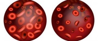
Pancytopenia is usually diagnosed by a complete blood count (CBC).
In pancytopenia, the CBC will show insufficient levels of all types of blood cells, including:
- The red blood cell count is less than 4.2 million cells per cc in women or less than 4.7 million cells per cc in men (this may also be described by low hemoglobin levels).
- The leukocyte count is less than 4,000 leukocytes per cubic cm (normally, a person has from 4,000 to 10,000 leukocytes per cubic cm).
- Platelet count is less than 150,000 cells per cubic cm (normally from 150,000 to 400,000 cells per cubic cm).
Etiology and risk factors of the disease
Among the main reasons causing the development of pathology are genetic mutations, long-term use of medications, radioactive or toxic effects. The idiopathic form is characterized by the fact that the cause cannot be determined, but there have been cases of the disease developing after bearing a child.
Establishing the cause is important for a favorable outcome of the disease. Eliminating external negative influences in this disease is 50% success.
In addition to the main reasons, there are a number of factors that contribute to the development of pathology:
- working with benzene or arsenic;
- Fanconi anemia;
- viral infections;
- chemotherapy;
- antibiotics and immunosuppressants;
- autoimmune diseases such as lupus;
- radiation therapy.
Fanconi anemia is one of the etiological factors of pancytopenia. Develops against the background of congenital anomalies and is considered an autosomal recessive disease. Anemia is characterized by the presence of defects in the regions of proteins responsible for DNA repair, as well as fragility of chromosomes.
Symptoms of Peyronie's disease
The symptoms of Peyronie's disease are as follows:
- erectile deformation of the penis;
- pain during erection;
- formation of a palpable plaque or “bumps” on the penis
There are different types of clinical course of Peyronie's disease.
Symptoms of Peyronie's disease may be absent and manifest only by the presence of “neoplasms” of the penis, which can be detected by palpation. The clinical course of Peyronie's disease may include severe pain and deformation of the penis during erection. In some cases, especially with the circular nature of the lesion, there is a significant shortening of the penis, and sometimes Peyronie's disease is clinically manifested only by erectile disorders.
Clinical picture of the disease
The pathological condition manifests itself in the form of three syndromes. Each of them is characteristic of signs of a decrease in the amount of one type of blood cells. Leukopenia is manifested by a picture of acute sepsis and the appearance of ulcerative-necrotic processes on the skin and mucous membranes.
Erythropenia and a marked decrease in hemoglobin provoke the development of symptoms of anemia. This condition is characterized by:
- pale skin;
- spots before the eyes;
- dizziness;
- noise in ears;
- weakness and fatigue;
- adynamia.
Patients complain of shortness of breath and increased heart rate. Auscultation allows you to determine the dullness of heart sounds, systolic murmur over the entire surface of the cardiac projection, and the sound of a spinning top over the jugular vein. The worsening of the patient's condition causes changes in the electrocardiogram, which indicate the development of heart failure.
Thrombocytopenia is manifested by frequent bleeding (nasal, gastrointestinal, uterine), bleeding gums, the appearance of petechiae and hemorrhages on the skin. Acute manifestations of the disease last about a month and often lead to death. Chronic pancytopenia resolves more favorably and has less severe symptoms
The course of the disease is considered very complex. Pancytopenia, complications of which are more common in older people, is accompanied by infections and bleeding.
Current [edit | edit code]
Two phases of the disease can be distinguished: the acute inflammation phase and the fibrotic stage.
The stage of acute inflammation can be manifested by pain in the penis at rest and painful erection, as well as the appearance of “soft” plaques and curvature of the penis.
During the fibrotic stage, a dense palpable plaque is formed, which can subsequently calcify, this indicates stabilization of the disease.
Over time, deterioration is observed in 30-50% of patients, stabilization - in 47-67%. Spontaneous improvement occurs in only 3-13% of patients. Improvement is possible at an early stage of the disease. After the formation, and even more so the calcification of a fibrotic plaque, spontaneous improvement is very rare.
Diagnostic measures
The first case of contacting a specialist requires an examination, collecting an anamnesis of the patient’s life and illness. Next, a general blood test is prescribed. If the following indicators are present, you can think about the development of pancytopenia and carry out further diagnostic measures:
- leukocytes <2 x 10⁹/l;
- platelets <150 x 109/L;
- red blood cells <3.5 x 10⁹/l.
A bone marrow puncture test is prescribed, which determines the presence of abnormal cells and myelokaryocytes. The presence of anomalies is determined, including bone marrow hypoplasia. Hemolytic tests are also performed.
Diagnosis of omphalitis
A specialist can easily make a diagnosis even from the first symptoms of omphalitis, but to clarify the form, tests will be required, the main one of which is a laboratory examination of blood counts. If you have omphalitis, you can contact a therapist or surgeon. To exclude complications, additional diagnostic methods may be prescribed:
- Ultrasound of the abdominal cavity - to ensure that there are no purulent foci on the abdominal wall;
- x-ray of the peritoneum;
- Ultrasound of soft tissues.
Hospitalization for adults is required in rare cases - only when there is a complication of the underlying disease that caused omphalitis.
Treatment of pancytopenia
Therapeutic stages depend on the clinical case, the patient's condition and the etiological factor that caused the disease. The following treatments may be used:
- Blood transfusion to balance the balance of formed elements.
- Bone marrow or stem cell transplantation. It is most often performed on young and middle-aged patients.
- In the case of autoimmune pancytopenia, the use of immunosuppressants is prescribed: Cytoxan, Methylprednisolone, Neoral, Sandimune.
- Agents that stimulate bone marrow function: Neupogen, Neulasta, Prokrit, Leukin.
pregnancy
Anti-platelet autoantibodies in a pregnant woman with ITPOM will attack the patient's own platelets as well as the placenta and react to fetal platelets. Therefore, ITP is a significant cause of fetal and neonatal immune thrombocytopenia. About 10% of newborns affected by ITP will have a platelet count of neonatal alloimmune thrombocytopenia (NAIT).
No testing laboratory can reliably predict if neonatal thrombocytopenia will occur. The risk of neonatal thrombocytopenia increases with:
- Mother with history of splenectomy for ITPA
- Mothers who have had previous children are affected by ITP
- gestational platelet count (maternal) less than 100,000/µl
It is recommended that pregnant women with thrombocytopenia or a previous diagnosis of ITP should be tested for serum antiplatelet antibodies. A woman with symptomatic thrombocytopenia and an identifiable antiplatelet antibody should be started on therapy for their ITPA, which may include steroids or IVIG. Fetal blood tests to determine platelet counts are usually not performed as ITP-induced thrombocytopenia in the fetus is usually less severe than NAIT. Platelet transfusions may be performed in newborns, depending on the degree of thrombocytopenia. It is recommended that newborns be auscultated with serial platelet counts within the first few days after birth.
Diagnosis of Werlhof's disease is carried out on the basis of clinical manifestations and laboratory tests.
Main hematological markers characteristic of pathology:
- platelet level below 150x109/l;
- increased bleeding time according to Duke (more than 1.5-2 minutes);
- decreased retraction (speed of passage) of a blood clot.
In addition, endothelial tests are carried out - tourniquet, pinch, prick. During a crisis, they turn out to be positive (as a result of exposure, petechiae appear in increased numbers). Bone marrow examination demonstrates a normal or increased number of megakaryocytes.
Idiopathic thrombocytopenic purpura is differentiated from leukemia, hemophilia, hemorrhagic vasculitis, gastrointestinal bleeding, dysovarian bleeding (in girls) and other diseases.
Medicinal plants
Complex therapy of the disease includes the use of herbal medicine. The level of red blood cells and hemoglobin can be increased with the help of berries. Vitaminized teas are made from rowan, black currant, rose hip, strawberry, and apricot berries. Rutabaga, gooseberries, cherries and grapes are rich in iron, the consumption of which will not only balance the balance of blood elements, but also strengthen the immune system.
A number of traditional methods for treating pancytopenia include honey. It is combined with the following products:
- with red wine and aloe juice;
- garlic;
- oats;
- lemon, dried apricots, raisins, prunes and walnuts.
Great importance is attached to infusions and decoctions of medicinal herbs: stinging nettle, birch, wormwood, clover, dandelion, orchis, sweet clover, and buckwheat.
Prevention
Measures to prevent the development of pathology:
- healthy lifestyle,
- rejection of bad habits,
- proper nutrition,
- protecting the body from negative exogenous factors,
- compliance with safety precautions when working with toxic substances,
- wearing respirators and other protective equipment,
- control over medication intake,
- timely treatment of infectious diseases,
- annual medical examination with mandatory laboratory examination.
Pancytopenia is a severe pathology that, without adequate therapy, can cause death in patients. It is important to prevent complications from developing. To do this, it is necessary to organize careful care and constant monitoring of the patient. Long-term maintenance therapy makes the prognosis of the pathology favorable. The outcome of pancytopenia depends on the severity of symptoms and the time of initiation of treatment. Such a disease is easier to prevent than to eliminate. That is why you should be attentive to your health, and if the first signs of illness appear, consult a doctor.