What is gallstone disease?
Gallstone disease (GSD) is a disease characterized by the formation of stones in the gallbladder and its ducts due to disruption of certain metabolic processes. Another name for the disease is cholelithiasis.
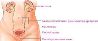
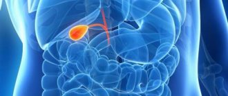
The gallbladder is an organ adjacent to the liver and acts as a reservoir for liquid bile produced by the liver. Gallstones, or stones, can be found both in the gallbladder itself and in its ducts, as well as in the liver and the trunk of the hepatic duct. They vary in composition and can have different sizes and shapes. Gallstone disease often provokes the development of cholecystitis (inflammation of the gallbladder), as the stones irritate its walls.
Gallstones in the gallbladder are formed from cholesterol crystals or calcium pigment-lime salts (in more rare cases). Biliary colic occurs when one of the stones blocks the duct that carries bile from the bladder to the small intestine.
The formation of gallstones is a fairly common disease, affecting about 10% of the adult population in Russia, Western Europe and the USA, and in the age group over 70 years old this figure reaches 30%.
In the second half of the twentieth century, the frequency of surgical interventions performed on the gallbladder exceeded the frequency of surgical operations to remove appendicitis.
Gallstone disease mainly occurs among the population of industrialized countries, where people eat large amounts of food rich in animal proteins and fats. According to statistics, cholelithiasis is diagnosed in women 3-8 times more often than in men.
Diet
At the first symptoms of gallstone disease and even after recovery, it is necessary to adhere to dietary nutrition. Eliminate from your diet
- fatty, spicy, canned, pickled foods;
- fatty meat (pork, lamb, duck);
- fatty fish;
- tea, coffee, alcohol;
- chocolate;
- eggs;
- sausages;
- legumes, mushrooms.
Build your diet from the following products:
- cereals (rice, buckwheat);
- vegetable broth soups;
- lean meat (rabbit, turkey, veal, chicken);
- dairy and fermented milk products;
- vegetables;
- fruits.
After treatment for gallstones is completed, continue to follow the diet. Otherwise, new stone formations are possible.
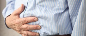
Symptoms of gallstones
In most cases, cholelithiasis is asymptomatic and does not have any clinical manifestations for several (usually five to ten) years. The appearance of symptoms depends on the number of stones, their size and location.
The main signs of cholelithiasis are:
- Paroxysmal drilling or stabbing pain in the liver and right hypochondrium;
- Nausea, in some cases vomiting;
- Bitter taste in the mouth due to the flow of bile into the stomach, belching of air;
- Flatulence, problems with stool (constipation, diarrhea), discoloration of stool;
- Weakness, general malaise;
- Temperature increase;
- Jaundice.
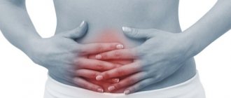
Hepatic (biliary) colic usually develops after eating fatty, heavy foods, spicy and fried foods, alcohol, as well as under conditions of increased physical or stress. Pain begins on the right under the ribs and can radiate to the right arm (shoulder and forearm), shoulder blade, lower back, and right half of the neck. Sometimes the pain can spread beyond the breastbone, which is similar to an angina attack.
Pain appears due to spasm of the muscles of the gallbladder and its ducts, which occurs in response to irritation of the walls of the bladder with stones, or due to excessive stretching of the walls of the bladder as a result of excess bile accumulated in it.
Severe pain is also observed when stones move along the bile ducts and stones block the lumen of the bile duct. Complete blockage leads to enlargement of the liver and stretching of its capsule, which causes constant dull pain and a feeling of heaviness in the right hypochondrium. In this case, obstructive jaundice develops (the skin and sclera of the eyes become yellow), which is accompanied by discoloration of the stool. Other symptoms of a completely blocked duct may include high temperature, excessive sweating, fever, and cramps.
Sometimes biliary colic goes away on its own after the stone passes through the bile duct into the small intestine. Usually the attack lasts no more than 6 hours. To relieve pain, you can apply a heating pad to the area of the right hypochondrium. If the stone is too large and cannot leave the bile duct on its own, further outflow of bile becomes impossible and the pain intensifies, immediate surgical intervention is required.
A common symptom of cholelithiasis is vomiting mixed with bile, which does not bring a feeling of relief, since it is a reflex response to irritation of certain areas of the duodenum.
An increase in temperature to subfebrile levels (not higher than 37°-37.5°C) indicates the addition of an infection and the development of an inflammatory process in the gallbladder. The development of cholecystitis is accompanied by decreased appetite and increased fatigue.
The first symptoms of gallbladder problems that should not be ignored:
Causes of formation of gallstones
Healthy bile has a liquid consistency and does not form stones. Factors that provoke their formation include:
- Increased cholesterol levels in bile, which changes its properties;
- Impaired outflow and stagnation of bile;
- Infection in the gallbladder and subsequent development of cholecystitis.
The main reason for the formation of stones is a violation of the composition of bile - the balance between cholesterol and bile acids. Bile with excess cholesterol and deficiency of bile acids is called lithogenic.
Increased cholesterol content in bile is caused by the following reasons:
- Excessive consumption of foods with high cholesterol levels (animal fats);
- Liver dysfunction, when the production of bile acids decreases;
- The presence of obesity, which is observed in approximately 2/3 of patients;
- Long-term use of oral contraceptives containing estrogens (in women);
- The presence of other diseases such as diabetes mellitus, hemolytic anemia, liver cirrhosis, allergies, Crohn's disease and other autoimmune conditions.
With a decrease in the contractile function of the gallbladder, cholesterol flakes settle, from which clots are subsequently formed - cholesterol stones.
The reasons for the obstructed outflow of bile and its stagnation are the following factors:
- The presence of certain diseases: dyskinesia (impaired contractile function) of the biliary tract, flatulence (increased pressure in the gastrointestinal tract impedes the flow of bile), as well as a history of surgical interventions on the gastrointestinal tract (vagotomy, etc.);
- Sedentary lifestyle;
- Pregnancy (pressure of the uterus on the peritoneal organs also prevents the outflow of bile);
- Improper diet with significant gaps between meals, as well as fasting and sudden weight loss.
In addition to functional genesis (dyskinesia), stagnation of bile can be caused by mechanical reasons, i.e., the existence of obstacles to its movement: these include adhesions, tumors, swelling of the bladder walls, kinking or narrowing of the bile duct, as well as congenital anomalies: cysts of the main bile duct, diverticula (protrusion of the walls) of the duodenum.
And finally, the third reason is infection of the gallbladder, which occurs ascending from the intestine or through the blood and lymph flow and as a result leads to cholecystitis (inflammation of the mucous membrane of the bladder walls) and cholangitis (inflammation of the bile ducts). Chronic cholecystitis and cholelithiasis are interdependent conditions when one of the diseases supports, accelerates and complicates the course of the other.
There are two types of stone formation:
- Primary stones begin to form in intact bile ducts and do not cause any clinical symptoms for a long time.
- Secondary stone formation occurs against the background of disturbances in the outflow of bile: cholestasis (decreased volume of bile entering the duodenum), biliary hypertension (increased pressure in the common bile duct, which leads to its expansion); due to blockage of primary bile duct stones. The formation of cicatricial stenoses and lumen in the biliary tract leads to the entry of ascending infection from the lower gastrointestinal tract into the gallbladder.
Thus, disturbances in the structural composition of bile play a decisive role in the appearance of primary stones. The formation of secondary stones is the result of cholestasis and infection of the gallbladder. Primary stones are formed mainly in the gallbladder due to stagnation and thick consistency of bile. Secondary stones can form both in the bladder itself and in the bile and intrahepatic ducts.
How big are gallstones?
The gallbladder is a hollow organ located under the liver and is designed to store bile. Bile is continuously produced by the liver, concentrated in the gallbladder and periodically enters the duodenum through the bile ducts. Bile is directly involved in the digestive process and consists of bile acids, pigments, cholesterol and phospholipids. With prolonged stagnation of bile, cholesterol precipitates, which gradually leads to the formation of so-called “sand”, the particles of which increase in size over time and join together to form larger stones.
According to their structure, gallstones are divided into homogeneous and complex (consisting of a core, body and cortex). The core usually consists of bilirubin. Homogeneous stones usually consist of clumps of mucus, pure cholesterol and foreign objects (fruit pits, etc.).
Based on their chemical composition, stones are classified into cholesterol, calcareous, pigment and mixed stones. Stones consisting of one component are relatively rare. Most stones have a mixed composition with a predominance of cholesterol. Stones with a predominance of pigments usually contain a significant proportion of lime salts, which is why they are called pigment-lime. The structure of the stones can be crystalline or layered, the consistency is solid or waxy. In most cases, the gallbladder of one patient contains stones with different composition and structure.
The sizes of the stones vary widely, from a few millimeters to several centimeters, and can reach the size of a hazelnut or a chicken egg. Sometimes one stone occupies the entire cavity of the distended gallbladder and weighs up to 70-80 grams. Gallstones can also be of any shape.
Stones with a diameter of 1-2 mm can pass through the bile ducts; in the presence of larger stones, the consequences and symptoms described above occur. In medicine, a fact has been recorded when one gallbladder contained about 7,000 stones.
Possible complications
- Acute cholecystitis;
- Blockage of the bile ducts with subsequent infection and the development of chronic cholecystitis and pancreatitis;
- Perforation (rupture) of the gallbladder and its consequences in the form of peritonitis;
- Entry of large stones into the intestines and intestinal obstruction;
- The risk of cancer occurring in the gallbladder.
Prevention
Prevention of gallstones consists of the following rules:
- proper nutrition;
- physical activity;
- normal weight.
Eat vegetables and fruits rich in vitamins and nutrients, avoid unhealthy foods with chemical additives, fatty, spicy foods that damage the liver.
Improve your metabolism with regular physical activity. You will not only improve your body health, but also lose weight. Normal weight is a prerequisite for the normal functioning of all organs, especially the gastrointestinal tract.
Gallstones detected in time can be removed more quickly and painlessly. Therefore, do not ignore the symptoms of malaise and digestive disorders.
Diagnosis of cholelithiasis
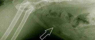
The presence of stones in the gall bladder is determined based on ultrasound examination. Large stones can be identified by touch. Using ultrasound, the number, size and location of stones are determined, and the condition of the gallbladder is diagnosed (for example, thickening of its walls indicates an inflammatory process).
If making a diagnosis is difficult, more complex methods are used, which include oral cholecystography (x-ray examination after oral administration of drugs that contrast bile), retrograde cholangiopancreatography (x-ray examination with endoscopy and injection of contrast into the bile ducts).
Non-surgical stone removal methods
An accurate diagnosis will tell the doctor which method will be the most effective and what the patient will have to do after treatment. Gallstone disease can be diagnosed in the early stages, in which case preventive and drug treatment is suitable. It is possible to remove stones by taking special medications, but treatment may last more than one month. The patient is prescribed a medicine containing bile acids, and with their help the stones in the gall bladder are dissolved. Ultrasonic crushing is carried out under almost the same conditions. Under ultrasound pressure, the stone in the gall bladder is crushed into pieces of 12-13 mm and come out naturally. The condition for treatment in this way is a completely healthy biliary part of the body. There is also a technique that involves destroying the stone in the gallbladder with a laser. With this treatment, consequences are possible, so additional studies of the body are carried out before starting it. Not only the doctor, but also the patient, whose consent will be required, will choose exactly how to treat. If it is not possible to be cured using one of the listed methods, an operation is performed and surgeons remove the organ completely. It is also done after other treatment methods have not given the desired results or the patient’s condition is rapidly deteriorating.
Treatment methods for gallstones
Modern conservative treatment, which allows preserving the organ and its ducts, includes three main methods: dissolving stones with drugs, crushing stones using ultrasound or laser, and percutaneous cholelitholysis (invasive method).
Drug dissolution of stones (oral litholytic therapy)
The dissolution of stones is carried out with the drugs Ursosan (ursodeoxycholic acid) and Henofalk (chenodeoxycholic acid). These drugs lower the level of cholesterol in bile and increase the content of bile acids in it.
Litholytic therapy is indicated in the following cases:
- The stones are of cholesterol nature. The chemical composition of stones can be determined using duodenal intubation or oral cholecystography;
- The stones are small in size (from 5 to 15 mm) and fill no more than 1/2 of the gallbladder;
- The contractile function of the gallbladder is normal, the patency of the bile ducts is good;
- The patient can take acids regularly for a long time.
At the same time, you should stop taking other drugs that provoke stone formation: estrogens, which are part of contraceptives; antacids, which are used for ulcers to reduce acidity and will interfere with the absorption of acids; cholestyramine, designed to bind and remove cholesterol.
Most gastrointestinal and kidney diseases are contraindications to this method. Doses and duration of administration are prescribed by the doctor on an individual basis. The course of treatment lasts from 6 to 24 months (minimum) and is carried out under ultrasound control. The effectiveness of therapy depends on the dose of medication and the size of the stones and is 40-80%. At the same time, you need to lead a correct lifestyle and take preventive measures to prevent the formation of new stones.
This method is characterized by a high relapse rate after completion of treatment (up to 70%), since after stopping the drugs, the level of cholesterol in the bile rises again. Therefore, as a preventative measure, you will have to continue to take low (maintenance) doses of these drugs.
On the subject: Preparations for dissolving gallstones
Ultrasound extracorporeal lithotripsy
This method is based on crushing stones under high pressure, which is created using a shock wave. Ultrasound destroys stones into smaller particles up to 3 mm in size, which are subsequently excreted through the bile ducts into the duodenum.
In practice, extracorporeal lithotripsy is often combined with the previous method, that is, the resulting small stones are dissolved with the help of medications (Ursosana or Henofalka). The laser method works in a similar way, when stones in the gallbladder are crushed using a laser.
This treatment method is suitable for patients who have a small number (up to 4 pieces) of fairly large cholesterol stones (up to 3 cm) without calcareous impurities in their composition or one large stone. Usually from 1 to 7 sessions are carried out.
Contraindications are:
- Bleeding disorders;
- Chronic inflammatory diseases of the gastrointestinal tract (cholecystitis, pancreatitis, ulcers).
Side effects of ultrasound lithotripsy include:
- Risk of bile duct blockage;
- Damage to the walls of the gallbladder by stone fragments as a result of vibration.
Any of these effects can trigger the development of an inflammatory response and, as a result, the formation of adhesions. Blocked ducts may require emergency surgery, and the results of emergency surgeries are usually worse than elective surgeries where the person has undergone prior testing and preparation.
Percutaneous transhepatic cholelitholysis
This is an invasive method that is used quite rarely. With its help, not only cholesterol stones are dissolved, but also any others. This method can be used at any stage of the disease, and, unlike the previous two, not only when the disease is asymptomatic, but also in the presence of its pronounced clinical signs.
Cholelitholysis consists of the following: a thin catheter is inserted into the gallbladder through the skin and liver tissue, through which 5-10 ml of a special drug (methyl tert-butyl ether) is injected dropwise, which dissolves stones. The procedure is repeated several times over 3-4 weeks, during which time up to 90% of the stones are dissolved.
Surgical treatment is indicated for large stones and frequent exacerbations, which are accompanied by severe pain attacks, high fever and various complications. Surgery can be laparoscopic or open.
Laparoscopy of gallbladder stones
Removing stones laparoscopically is practiced infrequently and only in a few clinics. During this operation, a 1.5-2 cm incision is made on the right under the ribs to penetrate the peritoneum. Using a laparoscope, the location and size of the gallbladder and the condition of other abdominal organs are determined.
Under video surveillance, the gallbladder is pulled to the first incision, and a 0.5-1 cm incision is made at its base, through which the contents of the bladder are examined. Then, through this incision, a special soft tube is inserted into which the choledochoscope is inserted - this ensures that the walls of the bladder are not damaged by the choledochoscope.
The stones are removed from the bladder, while large stones that fall into the duct are crushed into smaller ones. After all the stones are removed, the choledochoscope is removed, and the incision on the bladder is sutured with absorbable sutures. The skin incision is sealed with medical glue.
Removal of the gallbladder (cholecystectomy)
Currently, the most common treatment for cholelithiasis accompanied by cholecystitis is removal of the gallbladder along with stones. This is explained by the fact that the cause of calculous cholecystitis lies in a metabolic disorder, which directly affects the composition of bile, so mechanical removal of stones will not solve the problem; they will appear again.
In laparoscopic cholecystectomy, the bladder itself is removed through small incisions up to 1.5 cm on the anterior surface of the abdomen using a laparoscope (tube with a video camera).
Its advantages over open cholecystectomy:
- Fast recovery after surgery;
- No noticeable scars;
- Reducing the risk of developing postoperative hernias;
- Lower cost.
Contraindications:
- Obesity II–III degree;
- The stones are too large;
- A history of operations on the stomach, spleen, intestines and adhesions on the abdominal organs;
- Gallbladder abscess,
- Diseases of the heart and respiratory system;
- Late pregnancy.
Consequences of gallbladder removal
Surgery does not eliminate the symptoms of gallstone disease. Removal of the bladder is carried out due to the formation of stones in it, the appearance of which is caused by a pathological change in the chemical composition of bile, and after the operation this reason remains valid. After cholecystectomy, patients often complain that pain in the right hypochondrium and in the liver area persists, bitterness often appears in the mouth, and food has a metallic taste. The cumulative consequences of gallbladder removal are commonly called postcholecystectomy syndrome, which includes a group of symptoms directly or indirectly related to the operation, as well as diseases that begin to progress after it.
Cholecystectomy, according to some data, leads to an increase in the volume of the common bile duct. If in the presence of a gallbladder this volume is 1.5 ml, then 10 days after removal it is 3 ml, and after a year it can reach 15 ml. This is explained by the need for bile reserves in the absence of a gallbladder. Another consequence may be a narrowing of the common bile duct due to its trauma during surgery. This will result in recurrent cholangitis, bile stagnation and jaundice.
The main problems arise with the liver, pancreas and duodenum. Since there is no reservoir for collecting bile, its uncontrolled flow into the intestines begins, while the lithogenicity (impairment of the chemical composition) of the bile remains. The duodenum becomes accessible to bacteria, which leads to disruption of the metabolism of bile acids, as a result of which they severely irritate the intestinal mucous membranes. This contributes to the development of duodenitis, esophagitis, enteritis, and colitis.
Diet for gallstone disease
The composition of the diet is of great importance for this disease. It is recommended to adhere to fractional meals, eating 5-6 times a day. The meal itself has a choleretic effect, so the entry of a small amount of food into the stomach at the same hours stimulates the outflow of bile and prevents its stagnation. But with a large portion of food, the gallbladder may instinctively contract, and this will cause an exacerbation.
The diet should contain a sufficient amount of animal protein; animal fats are also not prohibited, but are usually poorly tolerated, so give preference to vegetable fats. For gallstone disease, it is useful to eat foods rich in magnesium.
Recommended Products:
- Lean meat and fish;
- Cheese, cottage cheese, milk with a fat content of no more than 5%;
- Cereals, especially buckwheat and oatmeal;
- Fruits and vegetables: pumpkin, carrots, zucchini, cauliflower, apples, watermelon, prunes;
- Compotes, fruit drinks, mineral water, blueberry, pomegranate, quince juices.
It is recommended to exclude the following foods and dishes from the menu:
- Fatty meats (pork, lamb, beef) and fish, as well as lard, liver and offal;
- Sausages, smoked meats, canned food, pickles;
- Butter (limit, preferably added to porridge);
- Legumes, radishes, radishes, eggplants, cucumbers, artichokes, asparagus, onions, garlic;
- Fried, sour and spicy foods;
- Rich broths;
- Coffee, cocoa and alcohol.
On topic: Diet for gallstones, menu for the week
Treatment without surgery
Many people are interested in how to dissolve gallstones non-surgically, without medication. There are several effective methods that help eradicate the disease in the first stages of development, when the stones are small. These include a special diet, folk remedies and medications.
Diet
In case of serious gallstone disease, it is necessary to follow a divided diet: five to six times a day. Food allowed on a therapeutic diet has a choleretic effect; eating small portions by the hour promotes the release of bile. This diet is also a disease prevention measure. The menu of a person suffering from ICD must necessarily include animal proteins and products with magnesium. The diet for gallstones, like any other therapeutic diet, divides food into “possible”/“not allowed”. Allowed to eat:
- lean meat, fish;
- cereals (buckwheat, oatmeal, etc.);
- milk: cottage cheese, cheese, milk (no more than 5% fat);
- vegetables, fruits: cauliflower, carrots, zucchini, pumpkin, watermelon, apples;
- drinks: still mineral water, compote, juice, fruit drink.
Products that are not recommended for consumption:
- fatty meat, fish, liver, lard, offal;
- butter (sometimes you can add just a little to the porridge);
- smoked meats, pickles, sausage;
- spicy, fried and sour;
- fatty broths;
- radishes, cucumbers, onions, garlic, eggplant, asparagus, legumes;
- alcoholic drinks, cocoa, coffee.
Folk remedies
Treatment of cholelithiasis with folk remedies is aimed at two main goals: getting rid of an attack of colic, as well as preventing the formation of stones in the future. An effective technique for stopping an attack: you need to warm up the camphor oil a little, soak a piece of gauze, and apply it under the ribs on the right side. “Grandmother’s” recipes for removing stones from the gallbladder and for the outflow of bile:
- Brew the herb and parsley roots. Drink a strong decoction in any quantity.
- A good choleretic agent is corn silk. Take 10 g of raw material, which is filled with one glass of hot water. Steam for half an hour, cool, strain, add boiled water to obtain a volume of 200 ml. Take a glass before meals.
- To dissolve stones, mix 10 g of wormwood herb, dandelion root, immortelle flowers, buckthorn bark, and also add 40 g of madder root. Pour two tablespoons of the mixture into a glass of boiling water and keep it in the bathhouse for 25 minutes. Drink a glass in the morning.
- Take 2 tablespoons of creeping wheatgrass roots and add hot water (1 cup). Boil for 10 minutes, wait until it cools down. Take the medicine three times a day, a third of a glass before meals.
Tablets
If the gallbladder hurts, then it can be cured without surgery with special drugs - analogues of the acids found in bile (Henochol, Ursosan, Ursofalk, and so on). Along with this method of treatment, medications can be taken that activate the production of bile (“Holosas”, “Allohol”, “Liobil”). Drugs for the destruction of stones, which are prescribed by a gastroenterologist, are used if the stones are no larger than 2 cm in size. The duration of therapy is at least 6 months.
- DIY rugs made from scraps of fabric
- White dots on nails - causes and signs. What does the appearance of white dots on nails mean?
- Activated carbon for weight loss - how to take it correctly, reviews