X-ray for pregnant women: only according to indications
X-ray of a tooth during pregnancy: is it possible?X-ray in early pregnancy: what to expect from it?
X-ray in the second and third trimesters of pregnancy
Pregnancy after x-ray
There can be many situations when a pregnant woman may need x-ray diagnostics. Naturally, concerns arise about the effect of X-rays on the fetus. Will X-rays during pregnancy harm the unborn child? How often can you do it and is it possible at all? Are there any types of x-rays that are relatively safe for the fetus?
X-ray; like isotope scanning, fluorography, and computed tomography are considered to pose a threat to intrauterine development due to exposure to high-energy radiation. But sometimes these procedures are vital.
One thing is clear: only a specialist can assess all the risks, therefore, radiography during the period of bearing a baby should be carried out strictly under the supervision of a doctor managing the pregnancy, and only in exceptional cases, if there are indications for its implementation.
X-ray for pregnant women: only when indicated
If the diagnosis cannot be made in any other way, the pregnant woman is sent for an x-ray. Most often it is used if it is not clear what the mother’s capabilities are in terms of further bearing the fetus, if there is a real threat to the life or health of her child.
Also, an x-ray of a pregnant woman is prescribed when necessary:
- identify the presence or absence of inflammatory diseases of the lungs and respiratory tract (tuberculosis, pneumonia, cancer);
- establish the severity of damage to the musculoskeletal system, including injuries to the limbs, in order to avoid incorrect fusion of joints;
- diagnose gallbladder or kidney diseases by identifying tumors or stones, respectively.
Is it possible to do x-rays and fluorography during pregnancy? Is it possible to take a photo of pregnant women?
It must be taken into account that for various types of x-ray diagnostics used during pregnancy, the radiation doses to the fetus will be approximately equal to the indicators indicated in the table below.
Type of radiography | Radiation dose to the fetus, m3v |
| CT scan | 10 |
| X-ray of the spinal column | 8 |
| X-ray of the abdominal cavity and intestines | 6 |
| X-ray | 3 |
| X-ray of the lungs and bronchi | 0,3 |
| X-ray of extremities | 0,1 |
| Dental photo | 0,02 |
| Repeated radiography (multiple) | 30 or more |
In any case, during the procedure, certain precautions are taken to protect the child from radiation, in particular, they cover the mother’s stomach, chest and knees with a shielding apron, which partially reflects it.
Indications for fluorography and x-rays
An X-ray examination may be prescribed if the attending physician detects a suspicion of tuberculosis or an inflammatory process in the lungs of the expectant mother. In turn, fluorography may be needed:
- at an appointment at a dental clinic;
- in traumatology, for example, for fractures or severe bruises.
X-ray allows you to examine the area of inflammation using electromagnetic waves that appear when charged particles are rapidly accelerated. This procedure is extremely necessary, as it allows the doctor to diagnose as accurately as possible and correctly prescribe treatment.
Dental X-ray during pregnancy: is it possible?
A lack of calcium often leads to dental diseases, causing severe pain. Some women are so convinced of the detrimental effect of the procedure on the health of the child that even in such a situation they have no desire to go to the dentist and have their teeth x-rayed during pregnancy, and, moreover, are ready to endure acute pain until childbirth, or even much longer.
But, as practice shows, pregnant women’s teeth can and even need to be treated. Firstly, so as not to experience unpleasant sensations, and secondly, and most importantly, not to infect a child, in whom caries may later develop seriously.
From the above table it can be seen that the radiation dose to the fetus is minimal when a dental photograph is taken during pregnancy (only 0.02 mSv), and in this case it can be repeated. In modern dentistry, so-called visiographs are used, which differ significantly from X-ray equipment. The advantages of this device are obvious. Being highly sensitive, it, unlike X-rays, reduces the irradiation (exposure) time by ten times, and the narrow beam hits only the diseased tooth. It also provides instant digital imaging and does not require the use of X-ray film.
For comparison: the fetus of a pregnant woman who spends more than 2 hours near a television screen will receive a radiation dose of 4 mSv, and if a woman decides to travel by plane, then this dose will be more than 30 mSv for the entire flight. The question arises: which is safer?
Is it possible to take dental x-rays during pregnancy? Is it possible to do an X-ray of the oral cavity of a pregnant woman?
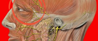
Why x-rays of the head and other organs are harmful?
X-ray is an examination that allows you to see the inside of any human organ. However, this effect is achieved through exposure to waves that are considered radioactive. Therefore, even an ordinary person is not recommended to conduct such an examination more often than once every six months.
X-rays are most dangerous in the early stages of pregnancy. After all, at this time the child’s organs begin to form. And any negative impact can lead to serious abnormalities and even fetal death.
Everyone knows that x-rays are dangerous. However, what explains its negative impact? We propose to talk about this in more detail.
Explanation of the negative effects of x-rays:
- During exposure to x-rays, thin tissues begin to actively divide, being trapped by denser tissues. Thanks to this, a picture appears in which organs and bones are visible.
- However, due to such exposure, DNA strands are broken. Because of this, a large number of free radicals are formed, which are highly chemically active.
- Because of this, some cells either mutate or die.
If for an ordinary person such exposure in small quantities does not pose a threat, then for the fetus, especially in the first trimester of its development, x-rays are very dangerous. Because of it, the most unexpected consequences can arise.
X-ray in early pregnancy: what to expect from it?
The most unfavorable period for X-rays is the first trimester of pregnancy. At this time, the fetus is actively growing due to the continuous division of cells that form structures important for its life, and, in particular, survival. These are the brain and spinal cord, the basic nerve centers, minor damage to which (mutation) can subsequently turn into a pathology incompatible with life.
The table provides so far only hypothetical data on the relationship between the concepts of “X-ray and pregnancy” and the consequences of exposure of the fetus to X-ray radiation above 1 mSv. However, they cannot be considered completely unfounded, since in some cases electromagnetic waves can actually cause fetal malformations.
What are the dangers of x-rays during early pregnancy?
Gestational age | Possible consequences of X-ray exposure on the fetus |
1 Week | Death of the embryo due to the cessation of cell division. |
2 - 3 week | Miscarriage due to non-viability of the embryo, ectopic pregnancy. |
4 – 5 week | Disruption of the development of embryonic brain structures (microcephaly), pathology of the chorion, yolk sac, amnion (temporary embryonic organs), spleen and digestive organs. |
6 – 7 week | The liver, adrenal glands, and thyroid gland suffer. The result is hormonal disorders in the child. The heart is especially vulnerable; abnormal development of muscles, valves and vessel walls is possible. Immune deficiency as a result of exposure to X-rays on the developing thymus gland. |
8 – 9 week | Risk to the ovaries and bronchi. |
10 – 11 week | Incorrect formation of fetal teeth. |
12 – 13 week | Anemia, leukemia, bone marrow dysfunction. |
Thus, the first trimester is the time when it is necessary to avoid any contact with the X-ray beam. But can pregnant women have their teeth taken? Perhaps the only safe procedure for early stages is a dental examination using special equipment with a minimum dose of radiation.
The effect of X-rays on the fetus when irradiating various parts of the body
It should be taken into account that the mechanism of disorders in tissues exposed to radiation is quite complex and, as a rule, depends on the type of radiation, the size of the dose received and the type of body cells.
The fact is that different tissues of the human body are differently sensitive to X-rays. The most sensitive areas are the genitals, lungs, colon, bone marrow, stomach, and lens of the eye. When an X-ray is taken of the upper part of a pregnant woman, namely the chest, neck, mouth or arms, the rays do not directly affect the fetus. And x-ray examination of the abdomen, pelvis, back and kidneys implies direct contact with the baby. Based on the above, we can conclude that X-rays should be taken during an interesting situation only in cases of extreme necessity, when diagnosis cannot be postponed until the birth of the child. In addition, every expectant mother should discuss in advance with her doctor an x-ray during pregnancy, or better yet, refuse to have it performed at all or choose the safest diagnostic methods for pregnant women.
X-ray in the second and third trimesters of pregnancy
The risk is now significantly reduced. All the basic systems of the fetus have already been formed by this time; the only system in relation to which the regime of increased vulnerability remains is the circulatory system. The likelihood of developing anemia exists at all stages of pregnancy. Doctors believe that in the middle of pregnancy, X-rays of the limbs or head do not affect the fetus, since the rays do not specifically affect it.
Is it possible to do dental x-rays for late pregnant women? Yes, if no similar procedures have been performed before and if we are not talking directly about the prenatal period.
X-ray: radiation features and safe dosages
Before you figure out whether pregnant women can take a dental photograph, you need to understand the importance of this procedure. If you do not use this technique, then in severe cases of dental disease the dentist will have to work blindly, which will not guarantee the quality of the treatment performed and does not exclude the occurrence of any complications in the future.a
Therefore, this is an important diagnostic procedure that must be carried out if the doctor has given a referral for this type of examination.
With the help of an x-ray, the doctor will be able to:
- identify dental diseases;
- determine the pathological state of periodontal tissues;
- determine and diagnose the condition of the bone tissues of the oral cavity;
- are there any caries or foci of inflammation;
- see the anatomical structure of the jaws.
IMPORTANT: The dentist himself determines which dental x-ray to take during pregnancy.
It can be assigned:
- outside the mouth;
- inside the mouth;
- survey.
Modern equipment, according to doctors, is not dangerous when examining the teeth of pregnant women. If we are talking about a Soviet-era radiograph, it is not recommended to take dental photographs for pregnant women, especially in the early stages of pregnancy. Since in 5% of cases, irradiation with such devices at 1 rad leads to disturbances in the development of the fetus and retardation of its development.
But these are quite rare cases that arise as a result of a pathological pregnancy. And X-ray radiation can only aggravate and become an impetus for the negative consequences of fetal development. In healthy women, no abnormal phenomena are observed after irradiation .
If a photograph of a tooth during pregnancy is suggested to be taken using a visiograph, then this method is completely safe, and this type of procedure should not be feared. Its radiation can be compared to the amount of radiation if a pregnant woman spent the entire first half of the day in the sun.
The X-ray beam of such equipment cannot penetrate into the deep layers of the skin, especially into the uterus . Therefore, such an x-ray examination is not dangerous for the child’s health. The dose for such an examination is no more than 0.0001 rad, so if necessary, a woman can take several pictures at once without negative consequences in the future.
IMPORTANT: Staying in the sun in summer is several times more dangerous than taking a dental x-ray.
A pregnant woman should also know that despite the safety of the procedure, dental x-rays are not recommended for pregnant women in the first trimester, as this is the most important and responsible period in the formation of the unborn child in the womb.
Modern equipment for dental x-rays
Pregnancy after x-ray
Doctors have an unspoken rule that during pregnancy the total radiation exposure to the child should not exceed 0.3 mSv. This dose of radiation corresponds to a single X-ray of the lungs.
If the specified value is slightly exceeded, the pregnancy outcome can also be positive. However, with repeated studies using X-rays, when the radiation exposure is more than 30 mSv, doctors usually insist on terminating the pregnancy, believing that in these cases no one can guarantee the birth of a physically healthy and mentally functional baby. This happens if a woman does not know about pregnancy, while undergoing any course of treatment that requires mandatory x-ray diagnostics.
At the same time, you should not assume that pregnancy and x-rays are incompatible. If you treat this with understanding and calm, listen to the doctor’s recommendations, and use special protective devices, then the likelihood of a negative impact on the fetus will be minimal.
When pregnant women should not have a dental x-ray
First of all, even before the start of pregnancy, a woman should have all problem teeth treated. If for any reason it is necessary to undergo an examination, you need to know that it is categorically undesirable to take an x-ray in the first trimester. Why shouldn’t pregnant women have dental x-rays done in the early stages?
- A negative impact of radiation on the fetus cannot be ruled out, since it is during this period that the internal organs, nervous, digestive, and cardiovascular systems of the unborn baby begin to develop.
- X-rays can destroy the natural formation processes of the fetus.
- There is a high probability of mutagenic intrauterine development of the fetus.
- There is a danger of miscarriage in the early stages.
- Complications during childbirth are possible.
X-ray in dentistry is a reliable diagnostic method
X-ray in 1st trimester
The most dangerous period for conducting an X-ray examination is the first. It is during the formation and formation of the most important organs - the heart, spine, lungs, organs of vision - that the risk of developing birth defects and pathologies is high. In case of repeated studies, miscarriage or fetal death may occur. If an X-ray was taken on a woman in very early stages (up to 4-5 weeks), the doctor may recommend termination of pregnancy, since the likelihood of having a child with genetic pathologies caused by a violation of the structure of cellular DNA is very high.
important
If an examination is necessary for the expectant mother for health reasons, it is necessary to always use protective equipment (a lead apron on the stomach) and warn the doctor about your situation.