Causes
In most cases, this pathology is associated with a genetic predisposition or gene mutation in the body.
After careful study, scientists have identified a number of chromosomal mutations that provoke rapid growth of bone tissue.
The bones gradually begin to thicken at the bottom. After some time they take on an arched shape.
The occurrence of this disease can be triggered by the following factors:
- excessive physical activity;
- intoxication of the body with chemical and synthetic substances;
- injury to certain parts of the bone mass;
- frequent inflammatory processes;
- radiation exposure;
- oncological processes;
- bone mass osteomyelitis;
- cirrhosis of the liver;
- impaired functioning of the thyroid gland;
- hormonal disbalance;
- leukemia;
- severe vitamin deficiency;
- atrophy;
- exhaustion of the body after a serious illness.
If any changes are detected, it is recommended to seek medical help. Timely treatment will help avoid serious health problems.
Etiology
In the vast majority of cases, bone hyperostosis acts as a hereditary disorder, with the proliferation of bone tissue occurring simultaneously on several bones. Moreover, against this background, signs of damage to other internal organs and other congenital pathologies appear.
However, the following sources can cause this disease:
- a colossal increase in the load on one limb in the absence of the other;
- serious injuries, supplemented by an inflammatory or infectious process;
- acute intoxication with toxic substances - this includes lead, arsenic and bismuth;
- long-term effects of radiation on the body;
- the occurrence of osteomyelitis or osteomalacia;
- formation of oncological tumors;
- previously diagnosed neurofibromatosis;
- echinococcosis;
- the presence of syphilis or cirrhosis of the liver;
- systemic blood diseases, among which are leukemia and lymphogranulomatosis;
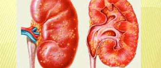
- a wide range of kidney pathologies;
- pathological conditions of an autoimmune nature;
- dysfunction of the endocrine system, namely the pituitary gland and thyroid gland;
- lack of vitamins A and D;
- Paget's disease, provided it occurs at the last stage of sclerosis;
- pathological fractures;
- rheumatic ailments.
In some cases, it is not possible to establish the cause of the development of the pathology - in such cases they speak of idiopathic hyperostosis.
Classification of hyperostosis
In medicine, there are two classifications of pathological changes in bone tissue. These include:
- local;
- generalized.
The first category of disease occurs as a result of the proliferation of bone tissue on one element. This is due to constant physical activity, which contributes to severe deformation. This manifestation is directly related to cancer or chronic types of diseases.
This pathology most often occurs in women with disrupted hormonal levels. There is a lack of calcium in bone tissue. Bones become brittle or even plastic.
The generalized type is an uneven thickening of the bone corset. A striking example of such a manifestation is Caffey-Silverman syndrome. The disease appears in childhood.
During puberty, cortical hyperostosis may occur. The patient has modifications in the shoulder and lumbosacral regions. The body takes on an hourglass shape. Such symptoms indicate the presence of Camurati-Engelmann syndrome.
According to the method of bone growth, the disease is divided into:
- periosteal;
- endosteal.
The periosteal type is caused by a pathological process in the area of the spongy substance. The internal contents are many narrowed lumens that disrupt normal blood flow. In this case, the phalanges of the fingers, shin bones, and the area of the forearms are modified. In most cases, tibial hyperostosis occurs. The pelvic floor takes on an irregular oblong shape.
The endosteal type of disease is accompanied by strong growths in the periosteum area. Internally, the cortical and spongy parts of the bones are affected. The bones become uniformly thickened, which provokes the appearance of curvatures in the area of the feet and hyperostosis of the inner plate of the frontal bone. The skull takes on an irregular oblong shape.
During the study, the formation of a large amount of bone substance is noted, which can provoke compression damage to the brain.
Find out what ozokerite treatment is.
Clinical manifestations
Symptoms and clinical signs of hyperostosis were described by various doctors and received their names. They have taken root in medical practice in the form of syndromes with characteristic manifestations.
Therefore, it is better to consider the clinic from the perspective of a syndromic approach.
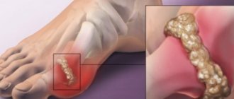
Periostosis ossificans or Marie-Bamberger syndrome
Bone changes are located in multiple foci on both sides in the forearms, legs, metacarpals and metatarsals.
Symptoms:
- fingers look like “drum sticks”;
- nails become flat and wide like “watch glasses”;
- pain in joints and bones;
- increased sweating;
- change in skin color from pallor to redness;
- Possible growth of the nose and the appearance of excess skin on the forehead.
At the same time, patients suffer from relapses of arthritis in the joints of the arms and legs.
Radiographs reveal symmetrical thickening of bone tissue in the diaphyses of long bones and layers in the periosteal areas.
Morgagni-Stuarat-Morel syndrome
Hyperostosis of the frontal bone or frontal is found in menopausal women.
Symptoms:
The photo shows hyperostosis of the frontal bone
- severe headaches;
- obesity;
- manifestation of male sexual characteristics (hair on the face and body);
- insomnia, irritability;
- menstrual irregularities.
Diabetes mellitus and hypertension are often associated.
X-ray is determined: thickening of the frontal bone, proliferation of bone tissue in the area of the sella turcica, damage to the vertebrae.
Blood tests: increased levels of somatotropic and adrenocorticotropic hormones.
Caffey-Silverman syndrome or infaltal hyperostosis
This form is typical for young children. It begins as an infectious disease: loss of appetite, fever, the baby becomes restless.
Typical manifestations:
- limited swelling appears on the arms and legs, face, rarely painful (there are no signs of inflammation);
- When changes appear in the lower jaw area, the face becomes “moon-shaped”.
X-ray reveals compacted bone tissue in the area of the clavicles, long bones, lower jaw, and curvature of the tibia.
Blood tests: accelerated ESR, leukocytosis.
Cortical hyperostosis
The disease is hereditary in nature, the lesion is generalized.
Symptoms:
- disruption of the innervation of the facial muscles in the area of the facial nerve;
- exophthalmos (bulging eyes);
- decreased vision and hearing;
- chin augmentation;
- thickening of the collarbones.
The disease manifests itself in adolescents. X-ray: compaction of bones in the cortical zone.
Camurati-Engelman disease
The disease is congenital.
The diaphysis areas of the humerus, femur and tibia are affected.
Symptoms appear in childhood:
- the child has stiffness and pain in the joints;
- poorly developed muscles;
- a “duck” gait is formed.
Forestier's disease
Forestier hyperostosis causes back pain
Forestier's disease differs from other types by simultaneous damage to the ligamentous apparatus of the joints and the development of immobility.
Most often it affects the thoracic and lumbar spine.
Mostly men over 50 years of age are affected.
It is noted that muscular and overweight people are most susceptible.
Symptoms:
- pain in the spine, often in the morning after sleep, prolonged position without movement, physical activity;
- inability to bend over or turn the body to the side;
- patients feel unwell at the end of the working day.
On a radiograph in frontal and lateral projections, signs of ossification of the vertebral bodies and tendons are determined.
Symptoms
The clinical picture of the disease largely depends on the type and form of manifestation of the pathological process. In rare cases, hyperostosis occurs as a result of external influences. These are mainly injuries, rickets or some viral disease.
If the pathology is caused by a genetic mutation or predisposition, then in this case a rapid growth of layers and a change in the appearance of bone tissue are observed. Such a modification can be detected through diagnostics.
With chromosomal abnormalities, a number of symptoms are identified that indicate the presence of pathology in the body:
- fingers take on an irregular shape. At first glance they resemble drumsticks;
- enlargement of the frontal lobe. It begins to take on an ovoid shape;
- wide nail plates;
- frequent pain in the joints and bones;
- skin changes. It takes on a bluish tint;
- excessive sweating;
- MRI signs;
- arthritis in the area of the phalanges of the fingers.
Local cortical hyperostosis has pronounced symptoms:
- the eyes acquire bulging shapes;
- chin enlargement, it becomes sharper;
- decreased visual function;
- hearing impairment;
- compaction of the clavicular region.
Symptoms of bone hyperstosis
The most common general manifestations of hyperostosis, regardless of location, are:
- excess bone tissue;
- limb deformity;
- bone pain and...
Often, local symptoms may not be as pronounced as the general ones - manifestations of disorders of the general condition of the body depend on the pathology that provoked the development of hyperostosis. The most common ones are:
- autonomic disorders (changes in skin color, increased sweating);
- deterioration in general health, which is manifested by incomprehensible weakness and lethargy
and others.
The clinical picture of hyperostosis that occurs in various pathologies can differ radically. The most common options are described below. Their names may initially mean nothing to patients, but the described signs are common and, when they appear, should prompt the patient to think that he has hyperostosis.
Diagnostics
The disease can be detected using diagnostics. It includes:
- radiography;
- magnetic resonance imaging;
- computed tomography;
- encelophography.
Before starting diagnostic procedures, the patient is recommended to consult with the following specialists:
- endocrinologist;
- gastroenterologist;
- phthisiatrician;
- traumatologist;
- oncologist;
- pediatrician.
These specialists will help identify the root cause of such a deviation. If the disease manifests itself at an early age, the patient undergoes a series of medical tests for possible genetic mutations. The results usually indicate the part of the chromosome that has undergone some kind of change.
Before treatment for hyperostosis of the frontal bone, a history of the disease and a test for genetic mutations in the family line are performed. After this, a thorough examination of the affected and modified sections is performed. X-ray images display the clinical picture of the development of the pathological process.
The MRI procedure indicates the presence of modifications and parts of bone tissue that are susceptible to mutation. Here the focus of pathology is indicated with an accuracy of 1 mm.
Next, the patient must undergo a series of medical tests, such as:
- general urine analysis;
- general blood analysis;
- ultrasonography;
- biopsy.
When all the results are ready, the attending physician will select the appropriate treatment for this pathology.
Marie-Bamberger syndrome
Marie-Bamberger syndrome (systemic ossifying periostosis, hypertrophic osteoarthropathy) is excessive growth of bone tissue, described by the Austrian physician Bamberger and the French neurologist Marie. It manifests itself as multiple, usually symmetrical, hyperostoses that occur in the area of the forearms, legs, metatarsals and metacarpals. Accompanied by a characteristic deformation of the fingers: the phalanges thicken in the form of “drumsticks”, the nails take on the appearance of “watch glasses”. A patient with hyperostosis experiences pain in the bones and joints. Autonomic disorders (redness and pallor of the skin, sweating) and recurrent arthritis of the metacarpophalangeal, elbow, ankle, wrist and knee joints with a blurred clinical picture are also observed. Possible enlargement of the nose and thickening of the skin on the forehead.
Hyperostosis in Marie-Bemberger syndrome develops secondarily, as a reaction of bone tissue to an imbalance of acid-base balance and chronic lack of oxygen. The cause of the syndrome is malignant tumors of the lungs and pleura, chronic inflammatory diseases of the lungs (pneumoconiosis, tuberculosis, chronic pneumonia, chronic obstructive bronchitis, etc.), intestinal and kidney diseases, as well as congenital heart defects. Less commonly observed in liver cirrhosis, lymphogranulomatosis and echinococcosis. In some cases, hyperostosis occurs spontaneously, without connection with any disease.
X-rays of the legs, forearms and other affected segments reveal a symmetrical thickening of the diaphysis due to the formation of smooth, even periosteal layers. At the initial stages, the density of the layers is less than that of the cortical layer. Subsequently, the layers become more dense and merge with the cortical layer. With successful treatment of the underlying disease, the manifestations of Marie-Bemberger syndrome decrease and may even disappear completely. To reduce pain during exacerbation, NSAIDs are used.
Treatment
Treatment of bone hyperostosis directly depends on the cause of its occurrence. If the disease is associated with a complication due to viral microflora, then to eliminate it, a thorough examination is carried out to identify pathogenic pathogens. For each patient, a specific dosage and duration of therapy are selected.
If the disease was caused by a chromosomal mutation, the following treatment is selected to eliminate it:
- corticosteroid hormones (“Prednisolone”). They help prevent the formation of new bone growths in the forehead, collarbones and pelvis;
- drugs to increase the body’s immune defense (“Interferon”, “Immunal”);
- diet;
- change in diet;
- massotherapy;
- physiotherapy.
The duration of treatment largely depends on the severity of the disease and the speed of its progression. Some patients are prescribed lifelong maintenance therapy. The patient is under constant medical supervision.
What is magnetic laser therapy and magnetotherapy?
Frontal hyperostosis - etiology
An increase in bone tissue is possible in any part of the body. Most often diagnosed in the forehead area. Normally, the skull plate has a smooth surface with clear contours. If pathology is detected, the patient is diagnosed with frontal hyperostosis.
Quite often, patients are diagnosed with Morgagni-Stuart-Morel syndrome. Clinical and radiological disease, with characteristic signs of changes in the plates of the frontal bone, as well as the bones of the parietal part of the skull.
The disease develops over the course of
, which indicates the growth of inert tissue along
skull poses a particular threat
in a woman's life. But in fact, hyperostosis at this time reaches its
stages.
The disease is typical for women over 70 years of age. In isolated cases, it can occur after 40.
Prevention and prognosis
You can prevent the occurrence of a pathological process by following the following recommendations:
- complete cessation of bad habits. This is mainly the use of alcoholic beverages, toxic and synthetic substances and smoking;
- change in diet. The menu should contain dishes rich in vitamins and microelements;
- you need to lead an active lifestyle;
- monitor the condition of the body;
- promptly treat viral and colds.
Causes of pathology
The main reason for the appearance is heredity or the body’s response to acute osteomyelitis and damage in the radiation zone. Additional reasons are:
- injuries resulting in the development of inflammatory processes;
- exposure to arsenic, lead;
- oncology;
- syphilis;
- echinococcosis;
- leukemia;
- can develop when the body reaches the stage of oterosclerotization;
- kidney and liver diseases that develop into chronic ones. Also lack of the required amount of vitamin A, D.
Often the disease is diagnosed in athletes or people who have a constant load on one of the bones of the skeleton. For example, if one leg is in constant motion and the other is at rest, then development is guaranteed. Therefore, the load should be uniform and the same for the body as a whole.
Structure and anatomy
The frontal bone is a part of the skull and its base, which consists of four sections:
- Two orbital.
- Arched nasal.
- Frontal scales. Bone lobules located vertically. They are the ones we are interested in.
The frontal scales consist of:
- The outer smooth surface, which has an elevation in the lower part, is the remains of a frontal suture. As a child, he divided the bone into two halves.
- Two temporal ones.
- The inner surface has a concave shape along the midline of the upper part.
It is this internal part, to which the falciform process of the meninges is attached, that will be discussed below. But first, it is important to understand what hyperostosis is in general and how it manifests itself on the inner surface of the frontal bone.