Probably each of us experiences unpleasant sensations bordering on fear upon hearing the diagnosis of a “malignant tumor.” All over the world, scientists are struggling to figure out where it comes from and how to treat it with a 100% guarantee, but alas, the results so far are disappointing. The diagnosis of “adenomatous polyp” does not sound so gloomy, and few people not involved in medicine can explain what it is. Meanwhile, this disease is considered a precancerous condition and is therefore extremely dangerous. Those who are diagnosed with it need to take action immediately to preserve their health, and possibly their lives.
Characteristics of polyps
At their core, polyps in the human body are an area of the mucous membrane that has grown for some reason. That is, they can appear in any organ covered with mucous membrane. According to medical statistics, adenomatous polyp, otherwise called adenoma and which is a benign tumor, causes a lot of trouble. The definition of “benign” means that in some place of the body cells suddenly began to divide uncontrollably, but so far they fully or partially retain the functions of the affected organ or tissue and do not metastasize. It is this important sign that gives a chance to completely cure them. Therefore, an adenomatous polyp is not a death sentence. However, without action, most benign tumors become malignant. Thus, polyps that are only 1 cm in size are highly likely to contain invasive cancer cells, that is, those that have already metastasized. True, the small size of the growth does not provide a 100% guarantee of safety, since there are cases where cancer developed from a single villus of a polyp.
Treatment
This pathology can only be combated surgically. This is the only way to slow down the progression of the disease and prevent the development of cancer.
Surgery involves removing those parts of the large intestine that are damaged by polyps. Healthy areas are preserved.
The type of surgery depends on the number of polypous tumors. If there are few of them and they are grouped in one part, removal is carried out endoscopically.
Sometimes it is necessary to completely remove the large intestine to create a permanent ileostomy. This is done if there are a large number of polyps in this part of the gastrointestinal tract. This treatment method is not suitable for young people. Therefore, doctors try to maintain healthy parts of the intestines. But if there is a high risk of developing a cancerous tumor or if the patient’s condition is serious, it is necessary to carry out just such an intervention.
Drug therapy for polyposis is ineffective. It can only be used to combat symptoms.
After surgery, patients should be regularly examined by a doctor in order to promptly notice relapses of the disease.
Classification
The mucous membranes in humans are multilayered and, depending on the organ they cover, have epithelium of different structure. Adenoma grows on those mucous membranes whose epithelium is represented by a glandular structure, that is, it includes many glands. Based on this, polyps can appear in the stomach, gall bladder, intestines, and organs of the genitourinary system.
In addition to location, there are a number of classification criteria:
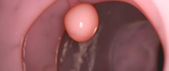
1. By type of base: on legs (stem) or on a wide platform (sessile). It is believed that a sessile adenomatous polyp metastasizes faster. The photo above shows what a large sessile polyp looks like on the wall of the intestine.
2. By size: small, medium, large. As long as adenomas are less than 1 cm, they are least likely to become malignant. Conversely, among adenomas larger than 1 cm, approximately 13% have cancer cells, and those larger than 2 cm have a 51% chance of developing into cancer.
3. In appearance: spherical, oval, mushroom-shaped, dense, soft.
4. By localization: single, nested, multiple. The latter degenerate into cancer approximately 2 times more often.
Morphological characteristics
Adenomatous polyp of the intestine, stomach, and absolutely all organs are structured differently, which greatly affects the prognosis of recovery. They are like this:
1. Ferrous. They consist of many glands and connective tissue rich in blood vessels. They are divided into benign, with signs of atypia (cells lose their shape, their nuclei become denser) and with malingation (atypical cells and glands are immersed in the muscular and submucosal layers of the epidermis, that is, in fact, they are preparing for metastasis).
2. Villous. These polyps are velvety in appearance, similar to cauliflower heads, and often have a rough surface. Their likelihood of malingia is higher than 60%.
3. Glandular-villous.
4. Hyperplastic. Very small, soft, retain the normal structure of the mucous membrane.
5. Juvenile. They do not malign and consist of cystic glands and dense stroma.
6. Fibrous. In the stroma they have many significantly dilated vessels, which is why they resemble inflammatory infiltration.
7. False.
Reasons for appearance
Why polyps begin to grow, there are no exact answers yet. Some scientists believe that they appear in people in old age, others deny this. Statistics say that in young children the chance of detecting adenomas is 28%, in people under 30 years old - 30%, and in elderly people over 70 years old - only 12.8%. The maximum number of detections of adenomas occurs at the age of 40-50 years.
That is, an adenomatous polyp of the rectum or other organ can appear in a person of any age, even in infants (there is a known case of diagnosing a stem polyp in a 2-month-old baby in the stomach). Scientists include the most likely reasons:
— pathologies during embryonic development;
— heredity (children whose parents have experienced adenoma are also about 2 times more likely to get it);
- inflammatory processes in the gastrointestinal tract (gastritis, colitis, dysentery, problems with bowel movements and others);
- unhealthy diet;
- impaired regeneration of the mucous membranes of the stomach and intestines after exposure.
Etiology
The etiology of the development of neoplasms remains unknown, therefore, when establishing the causes, they start from the risk factors that provoke this process.
Typically, adenomatous intestinal polyps appear in predisposed people. This should be understood as cases of benign tumors and cancer, including other localizations in the patient himself and his immediate family.
However, such heredity does not always manifest itself at all. The following risk factors (triggers) are identified:
Adenomatous polyp of the stomach
This organ ranks first in terms of predisposition to the appearance of adenomas. According to one of the classifications, based on morphological characteristics, the following types of gastric polyps are diagnosed:
- tubular;
- papillary;
- mixed (papillotubular).
They are located unevenly in the stomach. Thus, in the upper third of 2241 examined patients, polyps were found in 2.1%, in the middle third these figures reached 17%, and in the lower third there were already 66.8% of pathologies.
The development of malignant tumors in the stomach occurs according to the following simplified scheme: normal epithelium - formation of a polyp - its development into carcinoma - cancer. More often, such a scenario develops over two years, maximum three, but there are isolated cases when people lived with polyposis for about 20 years.
The causes of adenomas in the stomach are common - heredity, developmental pathologies at the embryonic level, inflammatory diseases, mainly gastritis, junk food, alcoholism, chronic gastrointestinal diseases. Also, according to scientists, various neuropsychiatric disorders contribute to the growth of mucous membranes in the stomach.
Diagnostics
An adenomatous polyp in any organ is almost impossible to detect without hardware diagnostics. For the stomach it includes:
- Ultrasound;
- X-ray with thick barium liquid (effective in approximately 4.6% of cases);
- gastroscopy;
— fibrogastroscopy;
- biopsy;
- gastrolaparoscopy.
No less important are laboratory tests of gastric juice, blood and the reaction to occult blood in the contents of the stomach.
The maximum result is obtained from examinations using several methods at once.
Adenomatous polyp of the colon
This disease is in second place of honor after gastric polyposis. According to statistics, polyps in the colon are recorded with the following frequency:
— women – 46%;
- men - 53%.
The dependence of the development of the disease on age is as follows:
— patients from 41 to 60 years old – 56%;
— from 31 to 40 years old – 23%;
– from 14 to 30 years – 10%.
The transformation of polyps into a malignant tumor has some dependence on their number. So, if there are 5 or more of these formations in the rectum, they develop into cancer in 100% of cases.
In the colon, polyps are also unevenly distributed. Thus, 13% of all cases are registered in the ascending part, 13.5% in the transverse colon, 73.5% in the sigmoid part and rectum. The causes of polyps in the intestines are approximately the same as when they occur in the stomach, but doctors give priority to inflammatory diseases. Thus, among 455 examined patients who were found to have polyps, 30% suffered from chronic illnesses (colitis, proctosigmoiditis and others), and 16.4% suffered from dysentery. Poor nutrition also plays a big role. For this reason, colitis is detected in more than 50% of cases.
Varieties
Before starting treatment, the doctor needs to determine the severity of the lesion by classifying the disease. His further tactics will depend on this. The following types of polyps are distinguished:
| By prevalence: | By structure: | By localization and ICD-10 code: |
At the same time, it is important to clarify the nature of multiplicity, since differences in the volume of treatment depend on this. This group includes the following subspecies:
|
|
|
Villous adenomas are most susceptible to malignancy (malignancy).
The main precancerous pathology is considered to be familial adenomatosis (polyposis), in which a large number of constantly progressive neoplasms appear.
The following types of familial polyposis are distinguished:
- Classic option. Installed most often. Symptoms appear from 14-16 years of age with a risk of malignancy by 30-40 years;
- Heavy current. Signs of the disease are already observed in children. More than several hundred polyps are identified endoscopically. Malignancy occurs between 18 and 25 years of age;
- Weakened flow. No more than 100 neoplasms are found throughout the colon, in total located on the right side. The clinical picture manifests itself by the age of 40 with malignancy 10-15 years later.
- Polyposis syndromes (Peutz-Jeghers, Solinger-Ellison, Turk, Allfield, Cronkite-Canada);
Adenomas localized in the initial sections are inaccessible for visual observation, therefore they become malignant more often than others. This is usually due to the fact that pars recta or sigmoid adenomas are easier to detect and remove before cancer develops.
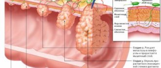
The main danger of a polyp is if its cells begin to actively proliferate. It is this actively dividing part that transforms into adenocarcinoma (first in situ - not extending beyond the lamina propria of the mucous membrane), and then into invasive cancer.
A proliferating adenomatous polyp of the colon is identified after a histological examination of a biopsy taken during endoscopy. However, there is a very fine line between a precancerous condition and your own malignant tumor, so you need to act immediately.
Polyps in the gallbladder
Adenomatous polyp of the gallbladder is a rare disease, occurring in less than 1% of all patients with polyposis. According to statistics, the disease most often affects people after 45 years of age. The gallbladder is a very small organ; in adults it is only up to 14 cm in length and up to 5 cm in width. In structure, it resembles a pouch with thin walls, a wider body, a tapering neck and a very narrow part from which the bile duct originates. The most severe situation is the location of polyps in the cervix or duct. In this case, the exit of bile into the intestines is blocked, and patients develop yellowness of the skin and whites of the eyes. In addition to this sign, there are other signs that a polyp may have grown in the gall bladder:
- aching pain;
- renal colic;
- nausea (especially in the morning);
- bitterness in the mouth;
- indigestion.
The causes of the disease can be inflammation of the gallbladder and its mucous membrane, improper metabolism, poor nutrition, and heredity.
Diagnosis is carried out using ultrasound and ultrasonography. Treatment is mainly surgical, consisting of removal of the gallbladder. Only in some cases can the doctor prescribe medication instead of surgery - Ursosana or Ursofalka.
Symptoms
Adenomyomatous polyposis in most cases does not have pronounced symptoms in the initial stages of development. The presence of outgrowths is detected during endoscopic diagnosis, the reason for which was concomitant diagnoses.
The first symptoms of the problem appear 5 years after the formation of the growth. During this time it grows to 20-30 mm in diameter.
When affected by adenomatous bowel disease, the patient experiences the following symptoms:
- feces become like tar;
- the anus is bleeding, the blood is bright scarlet;
- there is mucus in the stool;
- anal itching;
- the daily number of urges to empty the bowel increases, and the process of defecation itself becomes painful;
- constipation;
- diarrhea;
- intestinal obstruction with giant polyps.
If the adenoma is localized in the stomach, the following symptoms appear:
- bleeding in the stomach;
- burning sensation in the esophagus;
- the skin is dotted with purple-bluish spots;
- nausea, vomit looks like coffee grounds;
- pain in the lower abdomen, in the epigastric region, near the navel;
- bloating;
- frequent urge to have bowel movements;
- bad breath;
- belching;
- flatulence.
The fact that an adenomatous growth has appeared in the uterus is indicated by:
- nagging pain in the lower abdomen that occasionally bothers a woman;
- pain radiates to the lower back and perineum;
- menstrual irregularities in the form of prolonged delays in the absence of pregnancy or in the form of spotting in the middle of the cycle;
- metrorrhagia;
- heavy menstruation, which may cause stomach pain;
- discomfort and pain during intimacy;
- infertility.
A neoplasm that originates in the uterus can spread to the vagina.
The presence of gallbladder adenoma is indicated by:
- yellowness of the skin;
- dull pain in the right hypochondrium;
- colic in the liver;
- bitter taste in the mouth;
- severe skin itching;
- high heart rate;
- high blood pressure;
- aversion to those dishes that you previously liked;
- nausea and vomiting.