| Systemic scleroderma | |
| Clinical manifestations of scleroderma. | |
| ICD-10 | 34. |
| ICD-9 | 710.1710.1 |
| OMIM | 181750 |
| DiseasesDB | 12845 |
| MedlinePlus | 000429 |
| eMedicine | derm/677 ped/2197ped/2197 |
| MeSH | D012595 |
Systemic scleroderma
(Greek σκληρός - “hard” and δέρμα - “skin”) is an autoimmune disease of connective tissue, with characteristic damage to the skin, blood vessels, musculoskeletal system and internal organs (lungs, heart, digestive tract, kidneys), based which is a violation of microcirculation, inflammation and generalized fibrosis.
Etiology
Perhaps the disease has a genetic predisposition. However, reliably provoking factors for its occurrence are external harmful factors such as hypothermia, vibration at work, and previous infections of the nervous system. The development of inflammation of small vessels leads to the growth of collagen and fibrous tissue around them, as well as a specific change in their walls - thickening, loss of elasticity, and possibly even complete closure of the lumen of small vessels. These changes, in turn, lead to disruption of the blood supply to all organs and tissues involved in the pathological process. Insufficient blood supply to tissues leads to their thinning (for example, the mucous membranes of the esophagus and stomach), or, conversely, thickening (the walls of the alveoli in the lungs), disruption of their basic functions (absorption in the gastrointestinal tract, removal of carbon dioxide by the lungs, contraction of muscle fibers).
Forecast
The prognosis for systemic scleroderma is generally unfavorable. The lowest five-year survival rate (30-70%) is associated with the diffuse form. Predictors of an unfavorable prognosis are pulmonary and renal syndromes and the onset of the disease in patients over 45 years of age. The limited form and chronic course of the disease have a more favorable prognosis and better survival, with them it is possible to plan pregnancy and have a successful delivery. Patients with systemic scleroderma are subject to dispensary registration and observation every 3–6 months.
Diagnostics
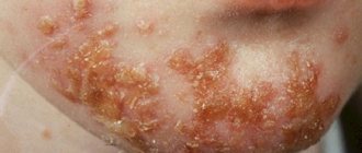
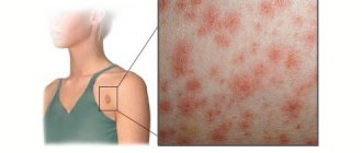
The clinical manifestations of scleroderma are very diverse, since the disease affects almost all organs and tissues.
A characteristic feature is skin damage, which occurs in most patients with scleroderma. Diagnostic symptoms are mask-like appearance of the face (extremely reduced facial expressions, giving the impression of tension in the skin of the face) and changes in the hands (thin and inactive fingers, with large nails and thickening of the terminal phalanges).
The diagnosis of systemic scleroderma is reliable if one “major” or two “minor” criteria are met (American College of Rheumatology).
- "Big" criterion:
- Proximal scleroderma: symmetrical thickening of the skin in the toes, extending proximally to the metacarpophalangeal and metatarsophalangeal joints. Skin changes can be observed on the face, neck, chest, and abdomen.
- "Small" criteria:
- Sclerodactyly: The above skin changes limited to the fingers.
- Digital scars are areas of skin retraction on the distal phalanges of the fingers or loss of substance from the finger pads.
- Bilateral basal pulmonary fibrosis; reticular or linear nodular shadows, most pronounced in the lower parts of the lungs during a standard x-ray examination; There may be “honeycomb lung” type manifestations.
Treatment of scleroderma with folk remedies
When treating scleroderma with traditional medicine, it is possible to reduce the intensity of symptoms and achieve a slower progression of the disease.
It should be remembered that folk remedies should not be used as the only method of treatment. This can lead to a decrease in the therapeutic effect and the development of a number of complications. Only complex treatment using not only folk remedies, but also pharmacological remedies can produce results.
The following traditional medicine recipes are used in the treatment of scleroderma:
- St. John's wort infusion . To prepare it, take mint, St. John's wort, plantain, raspberries and sweet clover, chop them and mix them. After this, three tablespoons of the mixture are poured with five glasses of boiling water, left for 6-8 hours and filtered. The infusion is drunk for two months, 50-100 milliliters per day.
- Meadowsweet decoction. A tablespoon of herb is poured with water (two glasses) and boiled for 15 minutes. The resulting decoction is infused for several hours, after which it is drunk twice a day, 100 milliliters. This treatment helps reduce joint pain.
- Onion . Take an onion, bake it in the oven and chop it finely. One tablespoon of the resulting substance is poured with two tablespoons of kefir and a teaspoon of honey. A compress is made from the resulting composition and applied to areas affected by scleroderma at night, four times a week.
- Infusion of horsetail . Take one tablespoon of a mixture of lungwort, knotweed and horsetail, add water (200 milliliters) and keep in a water bath for 15 minutes. The resulting broth is cooled, filtered and drunk in an amount of 50 milliliters before meals.
- Rhodiola tincture . Used to strengthen the body's immune defense in case of scleroderma. Take it for three months, three times a day, at the rate of two drops per kilogram of weight.
Scleroderma is a fairly rare disease, so there are few traditional methods of treating it. In most cases they are ineffective. They are used exclusively to raise the morale of the patient.
Clinic
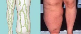
Changes in the blood vessels of the hands lead to the development of Raynaud's syndrome - a sharp vascular spasm with coldness and pain in the fingers. Another specific lesion is articular changes in scleroderma. They manifest themselves as inflammation with a fairly rapid impairment of joint mobility and the formation of so-called contractures, that is, irreversible stiffness due to the growth of fibrous tissue in the joint and loss of elasticity of its capsule.
Of the internal organs with scleroderma, the kidneys, lungs, and heart are most often affected. Changes in them lead to a decrease in all functions of the organ. For example, with kidney damage, this is manifested by an increase in renal failure. As a result, general intoxication of the body develops in combination with loss of protein and ions.
Most of the changes in scleroderma, such as muscle and bone pain, can resemble symptoms of rheumatism or rheumatoid arthritis. Differential diagnosis with these diseases is carried out on the basis of x-ray examination and immune tests.
Scleroderma ICD 10
Scleroderma belongs to the group of diffuse connective tissue diseases and is characterized by progressive fibrosis of internal organs, skin, decreased immunity and vascular pathologies.
Scleroderma code according to ICD 10 is M.30. - M.36. Most often, the disease occurs in women of childbearing age.
In recent years, due to the improvement of diagnostic methods for scleroderma, it is often possible to begin early treatment of the disease, which improves the prognosis and increases the life expectancy of patients. The disease has virtually no effect on the reproductive function of women (it is significantly reduced only in the presence of renal complications).
Symptoms (signs)
Clinical picture
• Raynaud's syndrome occurs in 90% of patients with diffuse and in 99% of patients with limited SS. Renal scleroderma crisis is considered as a visceral equivalent of Raynaud's syndrome.
• Skin involvement is detected in 95% of patients. The early (edematous) phase of damage to the endothelium of small vessels, following an increase in the permeability of the vascular wall, passes into the next (indurative phase), characterized by increased collagen synthesis. Over time, atrophy of the skin and its appendages occurs (atrophic phase) with a characteristic adhesion of the skin to the underlying tissues •• Bilateral swelling of the hands, then thickening of the skin of the fingers, face, forearms, development of induration and atrophy. In the diffuse form - rapid spread to the chest and abdomen; in the limited form, changes affect only the fingers and face •• Telangiectasia in 30% of cases of diffuse and 80% of limited SS •• Subcutaneous calcification (in 5% of cases of diffuse and 45% of limited forms) is localized in places often subject to trauma (fingertips, elbows, knees) - Tibierge-Weissenbach syndrome •• Hyperpigmentation alternating with areas of depigmentation - “salt and pepper” skin color changes. The skin becomes tense, shiny, fused with the underlying tissues •• Trophic disorders: ulcerations, ulcers, alopecia.
• Joint damage •• More than 50% of patients with SS complain of swelling, stiffness and pain in the joints of the fingers, wrist and knee joints (80% of cases of diffuse and 90% of limited forms) •• In the early stages of the disease, rheumatoid-like arthritis may develop, but typical for SS, a fibrosing joint process with severe limitations of mobility is considered •• Tenosynovitis with carpal tunnel syndrome and ligament friction noise during movement •• Flexion contractures •• Osteolysis of the nail phalanges.
• Muscle damage •• Diffuse muscle atrophy •• Non-inflammatory muscle fibrosis •• Inflammatory myopathy, clinically similar to dormant myositis (a variant of the cross syndrome).
• Damage to the gastrointestinal tract •• Esophagus: esophagitis (dysphagia, diffuse dilation of the upper parts, narrowing in the lower third, weakened peristalsis) or metaplastic changes in the mucous membrane of the distal part of the esophagus (Berrett's metaplasia). Esophageal motility disorders and reflux occur as a result of replacement of SMCs of the lower third of the esophagus with collagen; transversely - the striated muscles of the upper third of the esophagus are usually not damaged. Strictures of the esophagus develop (a consequence of constant reflux), and ulcers form in the area of the esophageal-gastric junction •• Stomach and duodenum: atony and dilatation. Severe fibrosis leads to impaired iron absorption •• Small intestine: dilatation, weakened peristalsis, malabsorption syndrome, small intestinal bacterial contamination syndrome •• Large intestine: diverticulosis, constipation.
• Liver damage. Primary biliary cirrhosis is observed in CREST syndrome.
• Lung damage •• Basal and then diffuse pulmonary fibrosis. Interstitial lung lesions occur due to fibrosis of peribronchial and perialveolar tissues. Patients complain of progressive shortness of breath during exercise •• Dry pleurisy •• Pulmonary hypertension in the absence of interstitial fibrosis.
• Heart damage •• Left ventricular failure •• Myocardial fibrosis - rare •• Myocarditis - rarely, mainly in patients with symptoms of dermatomyositis •• Acute pericarditis (usually as echocardiography is a finding) •• Endocarditis with the formation of a defect (extremely rare) •• Angina pectoris appears due to fibrosis of the coronary arteries •• Heart failure develops rarely, edema syndrome is not observed.
• Kidney damage •• AKI (scleroderma renal crisis) develops as a result of fibrosis of the interlobular arteries in combination with some vasoconstrictor stimulus (increased diuresis, blood loss, surgery) •• Malignant arterial hypertension •• Proteinuria in 30% of patients with SS. Changes in urinary sediment are minimal •• Rapidly progressive renal failure •• Hyperreninemia in 90% of cases •• Microangiopathic hemolytic anemia and thrombocytopenia.
• Damage to the nervous system •• Polyneuritic syndrome is associated with Raynaud's syndrome or develops independently •• Neuropathies of the cranial nerves are rarely noted (mainly the V pair of cranial nerves).
• Sjogren's syndrome (in 20% of cases).
• Damage to the thyroid gland •• Hashimoto's thyroiditis •• Fibrous atrophy.
Causes
The origin of the disease is unknown. Therefore, the likelihood of developing the disease in most cases is high due to the fact that it is not possible to prevent it in advance using preventive measures. But using medications can significantly improve the patient’s quality of life and eliminate unpleasant symptoms.
Conventionally, factors contributing to the development of the disease include:
- infections (scarlet fever, tuberculosis, diphtheria);
- autoimmune disorders (caused by malfunctions of the immune system).
Malfunctions of the immune system can be caused by vaccinations or allergic reactions previously suffered by the patient. The development of scleroderma can occur due to severe stress, hypothermia, and disruption of the endocrine system.