| Acute and chronic apical periodontitis | |
| Periodontal process under the root | |
| ICD-10 | 04.404.4, 04.504.5 |
| DiseasesDB | 29362 |
| MedlinePlus | 001059 |
Periodontitis
is an inflammation of the periodontium, characterized by a violation of the integrity of the ligaments that hold the tooth in the alveolus, the cortical plate of the bone surrounding the tooth and the resorption of bone tissue from small sizes to the formation of large cysts.
Classification
Based on etiology, the following types of periodontitis are distinguished:
- Infectious.
- Traumatic.
- Medication.
- Iatrogenic.
Classification of periodontitis
This classification is accepted in Russia, foreign classification is different.
- Acute periodontitis (periodontitis acuta): acute serous,
- acute purulent.
- Chronic periodontitis: fibrous,
- granulating,
- granulomatous.
- Exacerbation of chronic periodontitis.
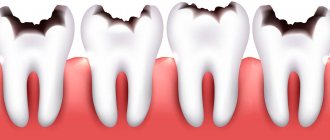
Periodontitis - photos of symptoms and treatment
Schematic example for the treatment of periodontitis
Example of chronic periodontitis
Example of marginal periodontitis
It is known that treatment of periodontitis with antibiotics is most often a very effective means of combating this disease; these drugs help most patients.
Read about various remedies against tooth sensitivity here.
About inflammation of the submandibular salivary gland, see here.
Etiology
- Infectious periodontitis
is mainly a complication of caries. Both primary (when the process is a consequence of untreated caries, and then pulpitis or periodontal disease), and secondary (when the process has an iatrogenic cause). Based on the method of bacterial penetration, periodontitis is divided into intradental and extradental (intradental and extradental). The latter includes periodontitis, which develops as a result of the transition of the inflammatory process from surrounding tissues (osteomyelitis, sinusitis).
- Traumatic periodontitis
occurs as a result of both a significant, single impact (a blow from a fall or hitting the face with hard, heavy objects), or as a result of a minor but chronic injury (an oversized filling, biting off a wire or thread in the absence of nearby teeth). In case of injury, the process is usually acute.
- Drug-induced periodontitis
most often develops with improper treatment of pulpitis, when potent drugs enter the periodontium (for example, paste containing arsenic, formalin, phenol) or irritating materials (phosphate cement, pins). Periodontitis that occurs as a result of allergic reactions that can cause a local immunological reaction is also classified as medicinal.
The main cause of the development of periodontitis in children is infection, when microorganisms, their toxins, and biogenic amines coming from the inflamed necrotic pulp spread into the periodontium.
The likelihood of developing periodontitis increases in people who smoke[1]. According to researchers, cigarette smoke and its components contribute to the formation of biofilm, which includes various pathogenic microorganisms, such as Staphylococcus aureus
,
Streptococcus mutans
,
Klebsiella pneumonia
and
Pseudomonas aeruginosa
.
Types of disease
Depending on the route of infection, there are:
- intradental periodontitis – the infection enters through the apical canal of the problematic tooth,
- extradental - the disease occurs due to the spread of an infectious process from another area of suppuration (for example, an adjacent tooth),
- retrograde – infection penetrates through the gum, from its edge.
Acute apical periodontitis
Acute periodontitis is characterized by the presence of sharp localized pain of a constant nature. Initially, with acute periodontitis, a mild aching pain is noted, which is localized and corresponds to the area of the affected tooth.
Later, the pain becomes more intense, tearing and pulsating, sometimes radiating, which indicates a transition to purulent inflammation. The acute apical process lasts from 2-3 days to 2 weeks. Conventionally, it is possible to identify 2 stages or phases of the course of acute periodontal inflammation:
- First stage
. The periodontal intoxication phase occurs at the very beginning of inflammation. It is characterized by the occurrence of prolonged, continuous pain of an aching nature. Sometimes this is accompanied by increased sensitivity when biting on a sore tooth. There are no visible changes in the tissues surrounding the tooth; with vertical percussion, increased sensitivity of the periodontium is noted.
- Second stage
. The phase of the pronounced exudative process is characterized by continuous pain. There is pain when biting on a tooth; Even a slight touch of the tongue to a sore tooth causes pain. There is a feeling of the diseased tooth moving out of the dental arch (symptom of an overgrown tooth). Percussion of the tooth is sharply painful. Irradiation of pain is noted. The appearance of exudate and inflammatory acidosis contribute to the swelling and melting of collagen fibers of the periodontium, which affects the fixation of the tooth, it becomes mobile. The spread of serous and serous-purulent infiltrate is accompanied by the appearance of soft tissue edema and the reaction of regional lymph nodes.
The general condition of the patients suffers: malaise, headache, body temperature (due to dental pain) rises to 37-38 °C, leukocytosis, and increased ESR are observed.
Radiologically, in acute periodontitis, no changes in the periodontium are noted.
| This section of the article has not been written. According to the plan of one or more Wikipedia contributors, a special section should be located in this place. You can help by writing this section. |
Chronic apical periodontitis
- Chronic fibrous periodontitis.
Diagnosis of this form is difficult, since patients do not complain and also because, for example, chronic gangrenous pulpitis can give a similar clinical picture.
Objectively, with chronic fibrous periodontitis, changes in the color of the tooth are noted, the crown of the tooth may be intact, there is a deep carious cavity, probing is painless. Percussion of the tooth is often painless, there are no reactions to cold or heat. Necrotic pulp with a gangrenous odor is often found in the tooth cavity.
In the clinic, the diagnosis of chronic fibrous periodontitis is made on the basis of an x-ray, which shows deformation of the periodontal fissure in the form of its expansion at the root apex, which is usually not accompanied by resorption of the bone wall of the alveoli, as well as cement of the tooth root.
Fibrous periodontitis can occur as a result of acute periodontal inflammation and as a result of healing other forms of chronic periodontitis, pulpitis, or occurs as a result of overload with the loss of a large number of teeth or traumatic articulation.
- Chronic granulating periodontitis.
It often manifests itself in the form of unpleasant, sometimes mild pain (a feeling of heaviness, fullness, awkwardness); There may be slight pain when biting on a sore tooth; these sensations occur periodically and are often accompanied by the appearance of a fistula with purulent discharge and the ejection of granulation tissue, which disappears after some time.
Hyperemia of the gums of the diseased tooth is determined; when pressing on this area of the gum with the blunt end of the instrument, a depression appears, which does not disappear immediately after removal of the instrument (symptom of vasoparesis). When palpating the gums, the patient experiences discomfort or pain. Percussion of an untreated tooth causes increased sensitivity and sometimes pain.
Enlargement and tenderness of regional lymph nodes are often observed.
X-ray examination of chronic granulating periodontitis reveals a focus of bone loss in the area of the root apex with unclear contours or an uneven line, destruction of cement and dentin in the area of the tooth apex.
- Chronic granulomatous periodontitis
is often asymptomatic; patients less often complain of discomfort and slight pain when biting.
The anamnesis contains indications of past periodontal trauma or pain associated with the development of pulpitis. When granuloma is localized in the area of the buccal roots of the upper molars and premolars, patients often indicate protrusion of the bone according to the projection of the apexes of the roots.
Objectively, the causative tooth may not have a carious cavity, the color of the crown is often changed, the presence of a carious cavity with decay of the pulp in the canals is noted, and finally, the tooth may be treated, but with poorly filled canals. Percussion of the tooth is often painless; upon palpation of the gum from the vestibular surface, a painful bulge may be noted, corresponding to the projection of the granuloma.
An X-ray examination reveals a picture of a clearly defined rarefaction of rounded bone tissue. Sometimes you can see destruction of tooth tissue in the apex and hypercementosis in the lateral parts of the root.
A favorable outcome of granulomatous periodontitis with timely and correct treatment is the transition to the fibrous form. In the absence of treatment or incomplete filling of the root canal, the granuloma turns into a cystogranuloma or root cyst of the tooth.
- Aggravated chronic periodontitis.
More often, granulating and granulomatous periodontitis exacerbates, less often fibrous periodontitis. Since exacerbation occurs in the presence of destructive changes in the periodontium, pain when biting on a tooth is not as severe as in acute purulent periodontitis. As for the remaining symptoms (constant pain, collateral swelling of soft tissues, reaction of the lymph nodes), they can increase in the same sequence as in acute purulent periodontitis.
Objectively, the presence of a deep carious cavity is noted (the tooth may be untreated or filled), absence of pain on probing, sharp pain during percussion, both vertical and horizontal, to a lesser extent. The tooth may be discolored and movable. Upon examination, swelling, hyperemia of the mucous membrane and often the skin are determined; over the area of the causative tooth there is a smoothness of the transitional fold; palpation of this area is painful. There is no reaction of dental tissues to temperature stimuli.
Exacerbation of chronic fibrous periodontitis is radiographically accompanied by a decrease in the clarity of the boundaries of rarefaction of bone tissue, the appearance of new foci of rarefaction and osteoporosis, according to the inflammatory focus. The X-ray picture of granulomatous periodontitis in the acute stage is characterized by loss of clarity of the boundaries of rarefaction of bone tissue in the apical part of the tooth, blurred periodontal line in the lateral sections of the periodontium and clearing of the bone marrow spaces along the periphery of the granuloma. Aggravated chronic granulating periodontitis is radiographically characterized by more pronounced eroded contours of the rarefaction focus against the background of a general blurred pattern.
The electrometric reaction on the part of the periodontium in all forms of periodontitis is over 100 microns or completely absent. Therapeutic measures for periodontitis go beyond the treatment of only the causative tooth and consist of the active release of the body from the infectious focus, thereby preventing sensitization of the body, preventing the development of inflammatory processes in the maxillofacial area and diseases of the internal organs.
| This section of the article has not been written. According to the plan of one or more Wikipedia contributors, a special section should be located in this place. You can help by writing this section. |
Pathogenesis
Microorganisms penetrate into the periodontium most often through the root canal during pulpitis. When infection penetrates, inflammation occurs. Periodontitis is also possible in other conditions: trauma, cracked tooth root, long-term exposure to arsenic in the dental cavity, sepsis. The periodontal gap is filled with interstitial fluid and, along with the ligamentous apparatus of the tooth, plays the role of a shock absorber under chewing loads. The periodontium and its constituent elements are rich in receptors that respond to pressure, which increases with periodontitis, so inflammation causes severe pain. With inflammation, exudation occurs (sweating of fluid). Swelling and exudation are responsible for the main, but not the only sign of the disease - pain. If there is an outflow of this fluid through the root canal of the tooth, the pain is less pronounced and conditions are created for the development of chronic periodontitis. Otherwise, acute periodontitis develops - first serous and then purulent.
In the diagnosis of chronic inflammatory process in the periodontium, X-ray data are of decisive importance. In this case, the x-ray picture of periapical changes in each of the roots of a multi-rooted tooth may be different. X-ray is not observed in acute periodontitis. With chronic fibrosis, narrowing or, more often, expansion of the periodontium. In chronic granulomatous, the picture is of a clearly defined loss of bone tissue with a rounded shape. Sometimes you can see destruction of tooth tissue in the apex and hypercementosis in the lateral parts of the root. Chronic granulating - a focus of rarefaction with corroded contours, destruction of cement and dentin in the area of the tooth apex. Exacerbation of chronic periodontitis is determined by the form of inflammation preceding the exacerbation, the duration and severity of the inflammatory process [2] [ unauthorized source?
][3][
unreputable source?
].
Possible complications
- a consequence of acute periodontitis can be the development of periostitis (flux), abscess, phlegmon, osteomyelitis;
- gradual destruction of bone tissue during granulomatous periodontitis can lead to spontaneous tooth loss;
- granulomas transform into cysts, which are able to grow into the maxillary sinus, causing sinusitis;
- when a cyst forms, a chronic fistula can form, opening either into the oral cavity or onto the surface of the skin of the perimaxillary region;
- a persistent focus of infection in chronic periodontitis can cause septic complications from other organs and systems (for example, septic endocarditis).
Link to cardiovascular disease
According to many epidemiological studies, there is an association between periodontal disease and markers of certain cardiovascular diseases. [4][5] Scientists have not yet been able to establish the nature of this connection, despite many attempts to create an explanatory theory.
Thus, in a recent multicenter, prospective, placebo-controlled study, STABILITY4, it was shown that the loss of a large number of teeth was associated with a high risk of stroke, death from cardiovascular and other causes.[6] Patients who were completely missing teeth had the highest risk of these diseases, the highest levels of inflammatory markers (hs-CRP, IL-6 and Lp-PLA2), and the highest levels of cardiovascular risk markers (GDF15, troponin T , atrial natriuretic peptide).[7]
The national study FINRISK-1997, conducted at the University of Helsinki (Finland), included 8,446 Finnish patients aged 25 to 75 years, also showed that defective dentition is associated with future development of cardiovascular complications, diabetes and high mortality.[ 8] The deterioration in prognostic indicators reached 140% in people with more than 5 teeth missing, compared with patients with a complete dentition.
Neither this nor previous studies show exactly what reasons link the poor condition of teeth and the cardiovascular system.
Treatment
Treatment is a step-by-step procedure with a dentist, ending with filling the tooth and its roots. First, to eliminate inflammation, it is necessary to surgically drain the exudate (purulent or serous). Physiotherapy, warm rinses with heated mineral water, sulfa drugs, and broad-spectrum antibiotics are prescribed. If the treatment is ineffective, and also if the tooth cannot withstand the tightness, it must be removed.
Treatment of chronic periodontitis includes three main stages:
- mechanical preparation (expansion, cleaning),
- antiseptic treatment (disinfection),
- canal filling.
Mechanical treatment is carried out with the aim of completely removing the decayed root pulp and the layer of infected dentin from the canal walls. Disinfection of the canal is often completed with the use of intracanal ultrasonic physiotherapy.
Then procedures are carried out to combat inflammation in the jaw and stimulate reparative processes in the bone. Absorbable anti-inflammatory and antibacterial pastes are placed into the root of the tooth. Physiotherapeutic procedures are used.
After stopping the inflammation in the periodontium, the canals are very carefully and thoroughly filled. In 85% of cases, complex treatment of periodontitis is effective and a cure occurs.
If, after the combined therapeutic effect, it is not possible to eliminate the granulomas, they resort to apical resection of the tooth root, followed by fixation of the tooth in the jaw alveolus. Sometimes all the measures taken are ineffective, in which case the affected tooth has to be removed. After stopping the inflammatory process in the bone, the issue of prosthetics or tooth implantation is decided.
Diagnostics
Detection of inflammatory processes in periodontal tissues is carried out using basic and additional diagnostic methods, however, the determination and subsequent treatment of granulating periodontitis in chronic form causes certain difficulties due to the absence of pronounced clinical symptoms.
Differential diagnosis of chronic apical periodontitis is carried out with the following processes:
- average caries;
- chronic gangrenous pulpitis;
- acute apical periodontitis at the stage of relief of inflammatory phenomena.
In addition, before treating granulomatous or granulating periodontitis, the dentist, based on an x-ray examination, must identify the distinctive features of one form from another.
Prevention
It is impossible to completely eliminate the likelihood of developing caries and its complications such as pulpitis and apical periodontitis, however, if you follow simple tips, it is easy to reduce the frequency of visiting the dentist with tooth pain.
- Clean your mouth daily using a toothbrush and toothpaste. In addition, it is recommended to use floss (dental floss), which allows you to remove food particles and microbial plaque from contact surfaces and other hard-to-reach places.
- For hygienic care, dentists advise choosing toothpastes enriched with fluoride compounds. However, you should pay attention to the dosage of this substance: for adults, the preventive concentration of fluoride is 1.5 thousand ppm, and children who have not yet reached 12 years of age require a dose not exceeding 1 thousand ppm.
- Reducing carbohydrate foods in the daily diet, especially those containing sucrose.
- Timely cleaning of tartar.
- When a carious process develops, it is necessary to contact a dentist for treatment so that the disease does not spread deep into the tooth.
- Regular visits to the dentist for diagnostic examinations.
Maintaining dental health at a high level will help avoid the occurrence of apical periodontitis.
How to prevent the disease
Preventive measures to prevent the formation of apical periodontitis include:
- Visit the dental office once a year. If the condition of the teeth is unsatisfactory, there are fillings or other inflammatory diseases, then at least 2 times a year.
- Oral hygiene You should brush your teeth 2 times a day, rinse after meals, and use dental floss to remove food particles from hard-to-reach places. It is advisable to replace a simple toothbrush with a mechanical one, because... it makes more movements per minute, which contributes to high-quality cleaning of teeth from plaque.
- Healthy eating. Eliminate foods containing large amounts of sugar from your diet. Eat fruits, vegetables, herbs, boiled meat, fish - rich in vitamins and other beneficial elements necessary for the body to function normally.
- Strengthening protective functions. If the immune reaction is weak, the doctor prescribes immunostimulants and vitamin complexes.
- Rejection of bad habits. Smoking, drugs and alcohol thin tooth enamel and affect the formation of tartar.
- Treatment of identified problems with teeth and gums. Lack of or incorrectly administered therapy leads to a chronic course of the disease or other complications.
An incorrectly selected treatment method or a long-term lack of medical care affects the disease: the acute period changes to a chronic one.
Complications of the disease include: periostitis, abscess, osteomyelitis, phlegmon and sepsis, the formation of cysts and granulomas.
In advanced cases that are not amenable to restorative therapy, the affected tooth is removed.
Notes
- KY Zee.
Smoking and periodontal disease // Australian Dental Journal. - 2009. - T. 54 (September). — P. 44-50. - doi:10.1111/j.1834-7819.2009.01142.x. - Acute periodontitis
- Pathogenesis of apical periodontitis
- Lockhart PB, Bolger AF, Papapanou PN, et al. Periodontal disease and atherosclerotic vascular disease: does the evidence support an independent association?: a scientific statement from the American Heart Association. Circulation 2012;125(20):2520-44.
- Dietrich T, Sharma P, Walter C, Weston P, Beck J. The epidemiological evidence behind the association between periodontitis and incident atherosclerotic cardiovascular disease. J Clin Periodontol 2013;40 Suppl 14:S70-84.
- Held C. et al. Characterization of cardiovascular endpoints, impact of event adjudication and effects of darapladib in the STABILITY trial //EUROPEAN HEART JOURNAL. - GREAT CLARENDON ST, OXFORD OX2 6DP, ENGLAND : OXFORD UNIV PRESS, 2020. - T. 36. - P. 1104-1104.
- “New” Periodontal Disease and Cardiovascular Disease (English) (inaccessible link) (10/21/2015). Retrieved December 12, 2020. Archived October 26, 2020.
- Missing teeth predict cardiovascular events (English) (06/05/2015). Retrieved April 29, 2020.