Enterobiasis is a disease that develops due to the penetration of pinworms (roundworms) into the body. According to statistics, pathology is most often diagnosed in children, less often in adults. Parasites develop only in the human body, so it is impossible to become infected through animals. Enterobiasis is considered a “dirty hands” disease. Therefore, failure to comply with personal hygiene rules is the main reason for the development of the disease. To identify a pathological process in the body, a special study should be carried out. But how to do this, and where to get tested for enterobiasis, you need to figure out. So sooner or later this information will be useful to everyone.
Enterobiasis, what kind of analysis is this?
This procedure includes laboratory tests that will help confirm or refute the suspicion of the presence of parasites in the body. Analysis for enterobiasis and worm eggs is important, since it is on the basis of the data obtained that the doctor will be able to correctly establish the diagnosis and prescribe adequate treatment.
After fertilization in the intestines, the worms come out through the anus. This happens so that the females can lay eggs in the folds of the skin. Therefore, to detect their presence in the body, an enterobiasis test is used, which includes a scraping, smear, and stool examination.
The therapeutic procedure is carried out based on clinical indications. As a preventive measure, the analysis is carried out once a year. The most accurate research result can be achieved by collecting material three times at intervals of 7-10 days.
This procedure is prescribed for children from 1 year of age. In order to thoroughly understand what kind of analysis enterobiasis is, you need to familiarize yourself with the conditions for its conduct.
How is the analysis carried out?
For better access to the perianal fold, the patient spreads his buttocks with his hands. The laboratory assistant collects the material with a cotton swab or adhesive tape, depending on the method of subsequent research.
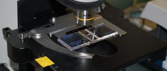
- Scraping from perianal folds. The material is collected with a cotton swab soaked in glycerin. It is passed along the perianal area, capturing the border of the anal mucosa. A cotton swab is dipped into the solution, then taken to the laboratory, where it is centrifuged and microscopically examined for the presence of pinworm eggs.
- Graham's method. This method involves using adhesive tape about 10 centimeters long. Take one end with tweezers and apply it to the skin in the perianal area, creating tight contact between the adhesive tape and the skin. Then peel off and transfer to a cover glass, without air bubbles. Microscopy is performed in the laboratory.
- Imprint method using glass eye rods according to Rabinovich. The sticks, dipped into the glue solution with the wide end, are dried, maintaining their sticky properties for several days. To avoid injury to the skin, the sharp edge of the edge is removed. The stick is applied to the perianal fold from different sides, then placed in a test tube and taken to the laboratory, where microscopic examination is carried out. In this way, the eggs are preserved for a long time and lie freely on the adhesive base.
It is recommended to carry out the analysis 3 - 5 times with an interval of up to 7 - 10 days to increase the accuracy of diagnosis.
When is the test scheduled?
Infection of the body with pinworms does not go away without leaving a trace. With an increase in the number of parasites, severe dysbiosis develops, intestinal dysfunction and excessive flatulence. The person becomes irritable as sleep is disturbed.
In addition, other characteristic signs of the disease appear:
- itching in the anal area at night;
- periodic abdominal pain for no apparent reason;
- nausea after eating;
- vomit;
- gnashing of teeth in sleep;
- causeless cough;
- persistent cold;
- frequent allergic reactions;
- redness of the perineum;
- bowel dysfunction;
- increased appetite or its complete absence;
- weight loss with a normal diet;
- drowsiness;
- fast fatiguability.
The presence of at least several of the signs listed is a reason to be tested for enterobiasis.
A certificate of the procedure performed is also required for:
- admission to kindergarten, school;
- pregnancy;
- visiting the pool;
- registration of a medical record;
- inpatient treatment.
Therefore, every person should know where to get tested for enterobiasis and how to do it correctly.
Scraping for enterobiasis: when to take it
Symptoms of enterobiasis should be a good reason to consult a doctor for advice. This pathology is very common in our country, so anyone can become infected with it. Infection can occur if the following factors are present:
- consumption of insufficiently thermally processed meat and fish products;
- neglect of personal hygiene rules;
- presence of bad habits: biting nails, licking fingers. Most often, this is how babies become infected;
- sharing household items with an infected person, as well as close physical contact (handshakes, hugs);
- eating poorly washed vegetables and fruits, especially in the summer, when there is a risk that insects have landed on the food;
- drinking untreated water from natural sources.
Weak immunity promotes infection. It is believed that if a child has symptoms of pathology, parents also need to get tested, since this disease is transmitted through household contact. Weak immunity contributes to pinworm infection
The disease has an incubation period that lasts from 12 days to two weeks. It is after this period of time after infection that the first signs of the disease begin to appear:
- there is itching and discomfort in the anal area after bowel movement;
- discomfort and pain appear in the gastrointestinal tract;
- gas formation increases;
- food is poorly digested, which causes constipation and diarrhea;
- the color of the skin of the genital and excretory organs changes;
- rashes on the skin;
- inflammatory processes, infections of the genitourinary system (thrush, urethritis);
- Children can often observe an increased interest in the genital area and anus.
How to prepare for the procedure?
To get the most reliable results, you need to prepare in advance for the procedure. The main rule is to avoid taking anthelmintic drugs. If this is not done, the presence of parasites in the body cannot be established.
When collecting stool for testing for enterobiasis, adults and children should:
- exclude fatty foods from the diet in advance;
- carry out the act of bowel movement independently (without using laxatives).
When prescribing a smear or scraping, you must adhere to the following recommendations:
- the procedure should be carried out in the morning;
- You should not swim before taking the test.
In order for the child to behave calmly, parents must explain in advance what kind of test for enterobiasis it is and how painless it is.
How long to wait for the result of scraping for enterobiasis?
You can find out the results of enterobiasis scraping within 24 hours. The material is submitted several times to accurately confirm the result. The test for fecal enterobiasis is done in the morning, so you can find out the result on the same day. It is necessary to correctly collect the test for enterobiasis several times over 10 days. How long to wait for the result depends on which diagnostic method was used.
If you scrape once, it may show a false result, so to confirm the diagnosis you need to collect tests twice. You can do a smear correctly at home. It is better to have your child tested in a clinic.
Smear in adults
Collection of material for analysis can be carried out directly in the laboratory or independently at home. It is necessary to understand that the biomaterial retains its qualities for no more than 2 hours at a temperature of +4-+8 degrees, so it must be delivered to a medical facility as quickly as possible.
The procedure is carried out in the following sequence.
- Purchase a special container with a cotton swab from the pharmacy for storing biomaterial.
- Wear sterile gloves.
- Immediately before the procedure, dip the tip of a cotton swab in glycerin.
- Spread your buttocks by bending forward.
- Run a cotton swab along the folds around the anus, pressing lightly on it.
- Place the collected material in a container and close it tightly.
A smear is taken in the same order in the laboratory. Before the procedure, the healthcare worker must wear a mask and sterile gloves. After this, ask the patient to lie on the couch on his side or while standing, bend forward and spread his buttocks. Then use a cotton swab dipped in saline or glycerin to run along the folds near the anus. Place the material in a special container.
After collection, all collected containers are placed in a special box at the required temperature, where they await their turn for examination. In this mode they can be stored for 8 hours.
Based on this, we can draw a conclusion about how an enterobiasis test is taken in the form of a smear at home and in the laboratory.
Preventive measures
Compliance with basic rules of personal and household hygiene is the basis for the prevention of this unpleasant disease. Let us note the main ones:
- From an early age, a child must be taught to wash his hands with soap before eating, after visiting the toilet, and after returning from a walk;
- wash your intimate parts and change your underwear twice a day;
- bed linen must be changed once a week and washed with boiled water;
- It’s probably unnecessary to talk about keeping the apartment clean and removing dust every day;
- In some cases, when the disease is recurring in the family, it is necessary to get rid of pets.
Perianal scraping
This procedure for collecting material is somewhat different from taking a smear, so it’s worth understanding how an enterobiasis test is taken in this case, and whether it can be done independently.
Perianal scraping in the laboratory is carried out as follows.
- The healthcare worker should wear sterile gloves and prepare the slide.
- Peel off the adhesive tape from the device.
- Apply it with the sticky side to the folds near the anus.
- Return the tape to the glass and smooth it out.
After this, the biomaterial is transferred for research.
If necessary, this procedure can be carried out independently. To do this, it is recommended to use wide tape 2 by 5 cm and degreased glass. To obtain the biomaterial, you need to apply the tape with the sticky side to the anus, and then transfer it to the glass, smoothing the surface.
It is important to transfer this material to the laboratory as quickly as possible so that it does not lose its relevance. There is only 2 hours for this, so it is best to collect samples immediately before leaving for the laboratory.
What is a scraping for enterobiasis?
Fecal analysis (scraping) for enterobiasis - detection of pinworm eggs (helminths, the clinical manifestations of which are itching in the anal area and intestinal disorders). Enterobiasis is a disease caused by small parasitic worms - pinworms. Pinworms do not exceed 1 cm in length. Usually one end of this worm is pointed (hence the name), and the other is rounded. The color of parasites can vary: from whitish or yellow to dark or even black.
Live pinworms can crawl or squirm. Pinworms are nocturnal: it is at night that females enter the rectum and onto the skin around the anus, causing discomfort and itching, lay eggs in the folds of the skin and die.
Infection occurs through dirty hands or contaminated objects. A child, scratching or touching the skin in the perineal area, contaminates his hands with worm eggs. Infection can also occur when hands are contaminated from bedding or underwear, where eggs fall from the body of a sick person. The role of flies and cockroaches in the transfer of pinworm eggs has been proven. A child, scratching or touching the skin in the perineal area, contaminates his hands with worm eggs
Repeated infections through the anus are possible, when individuals that have matured to the larval stage move from the skin to the intestines. Enterobiasis is very common and is considered the most common infection in the World. In some children's groups, up to 100% of children are infected with pinworms.
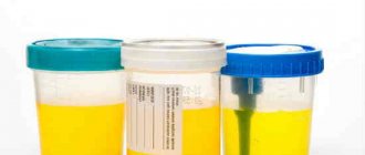
How is stool collected for analysis?
In order for the analysis for enterobiasis to be valid in this case, it is necessary to reduce the likelihood of errors when collecting biomaterial to a minimum.
If a helminthic infestation is suspected, experts recommend carrying out the procedure several times, since there is a possibility of conducting the study during the “silent period.” This means that the female parasite is localized in the intestine, waiting for the eggs to mature. If the analysis was carried out precisely at this time, then detecting signs of the presence of worms in the body is very problematic.
Most doctors are inclined to believe that stool examination is the least informative test for enterobiasis. According to statistics, it is possible to detect worms using this method only in 30% of cases. This is due to the biological characteristics of parasites. Since they do not lay eggs in the intestinal lumen, but crawl out to the perianal folds to do this.
But collecting stool when analyzing for enterobiasis makes it possible to identify the presence of other types of parasites (roundworms, tapeworms).
Before collecting biomaterial at home, it is necessary to exclude the use of:
- cleansing enemas;
- antibiotics;
- rectal suppositories;
- laxatives.
To know how to take an enterobiasis test correctly, we suggest that you familiarize yourself with the basic recommendations for the procedure.
- It is necessary to prepare a container for storing samples in advance. An opaque container with a tight lid is best for this. It can be purchased at a pharmacy. It comes with a special spoon, which makes collecting the material much easier. There is no need to sterilize the container, just wash it well and dry it.
- The volume of the test sample should be approximately 2 teaspoons, otherwise it will not be enough to obtain reliable results.
- It is important that the stool intended for analysis does not contain additional impurities in the form of urine and vaginal discharge. Therefore, it is recommended to collect material after urination. And to prevent menstrual flow from getting in, you should use a tampon.
- Samples for research must be taken from the top, bottom and middle of the stool. This is necessary to increase the awareness of the study. If obvious signs of the presence of parasites were found, then it is necessary to place them in a prepared container and transfer them to the laboratory along with the biomaterial.
- After collecting stool, it is recommended to transport it to the laboratory as quickly as possible. But if this is not possible, then the container with the material should be stored in the refrigerator at a temperature of 0-+4 degrees. In this mode, it retains its information content for 24 hours. In case of temperature changes during storage, it is necessary to collect samples again.
- It is best to use morning stool for analysis. Before submitting for testing, you must write your name on the container, as well as the date and time of sample collection.
- When submitting stool for enterobiasis, you must prepare for the fact that this procedure must be carried out 3 or more times. Only in this case can maximum reliability of the study be achieved.
If feces have already been collected, but there is no way to transfer it to the laboratory and preserve it properly, then it is recommended to preserve the material. To do this, you need to use a 50% glycerin solution. In this case, it should be mixed with distilled water at the rate of 50 ml of glycerin 90 ml of water. The biomaterial can be stored in the resulting preservative for 2-3 weeks, provided that the volume of the working solution is 3 times larger than feces.
Preparation
In order for the results to be as reliable as possible, several conditions must be met.
It is recommended three days before testing stool for worm eggs:
- Avoid taking medications that have antibacterial and anthelmintic effects.
- Do not use medications that increase peristalsis. Laxatives should also be avoided.
- Remove foods that have a pronounced coloring effect from your usual diet.
If, shortly before the planned stool test for worm eggs, any instrumental examination of the large intestine was carried out, you must make sure that 14 days have passed after it.
The collection of biomaterial must also be carried out in accordance with certain requirements. It is necessary to ensure in advance that there is a container in which the stool sample will be placed. Any small jar made of glass or plastic with a tight-fitting lid can be suitable for this purpose. It must be thoroughly washed and dried. The best option is a sterile disposable container, which can be purchased at any pharmacy. In addition, it simplifies the process of collecting biomaterial due to the included spoon.
It is recommended to take stool samples from several areas of stool, both externally and internally. The minimum amount of material is 2 teaspoons, but not more than 1/3 of the volume of a standard disposable container. To avoid getting a distorted result when collecting stool from a small child, it is important to ensure that no urine gets into the sample.
Material for analysis of worm eggs can be stored for several hours. It is allowed to collect it in the evening and place it on the bottom shelf of the refrigerator door overnight, and deliver it to the laboratory in the morning. But the best option is when a person brings morning feces for examination. Fulfillment of this condition is a guarantee of obtaining the most reliable result.
To test for enterobiasis, you do not need to collect biomaterial. This type of analysis is considered simpler - the scraping process takes a few seconds. In addition, it does not require special preparation. The only condition is that before taking samples for enterobiasis, it is prohibited to carry out hygiene measures in the morning. This is due to the fact that female pinworms lay eggs at night. If you wash them off with water after waking up, even if infected, the result will be negative.
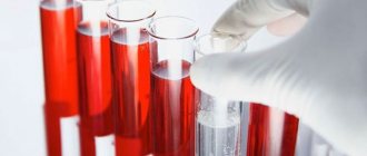
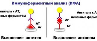
Blood analysis
With this type of research, the results are considered the most informative, since they can confirm or refute with maximum accuracy the fact of infection of the body with parasites. In medicine, this diagnostic method is called enzyme immunoassay for enterobiasis. What kind of research this is needs to be figured out.
When a foreign invasion enters the human body, the immune system tries to defend itself against this invasion. At the same time, it produces specific antibodies for the fight, and the type of immunoglobulins directly depends on the strain of parasites. Therefore, when collecting venous blood for research, it is possible to determine with maximum accuracy the type of invasion that is present in the body.
The amount of antibodies produced also indicates how long the pathological process lasts. This is determined by the type of immunoglobulins, since as the disease progresses they differ from those produced at the beginning of infection.
Based on this, it becomes clear: if antibodies are present in the blood, then infection has occurred, and vice versa. There are cases when the analysis shows a small amount of immunoglobulins in the blood. In medicine, this is considered a borderline condition, that is, it is impossible to confirm or deny the fact of infection. In this case, the doctor prescribes a repeat test after a few days, depending on the characteristics of the patient’s immune system.
Venous blood sampling for analysis for enterobiasis is done in a laboratory setting. The procedure is carried out in the morning on an empty stomach. No special preparation is required before the procedure.
This study is used much less frequently than previous methods. It is prescribed for severe and long-term disease. In this case, an analysis for enterobiasis helps to accurately determine the type of parasite, and is also necessary for making a final diagnosis. Only based on the data received, the doctor prescribes the necessary course of treatment.
Treatment of enterobiasis
The first key to successful non-drug treatment of pinworms is strict adherence to personal and household hygiene requirements. The first condition is to strictly keep your hands clean, wash them with soap as often as possible, and never bite your nails or put your hands in your mouth. Next, change your panties and bed linen daily! Iron only with a hot iron. Third, take a shower at least once during the day, and in the morning and evening, be sure to thoroughly wash the perineal area with soap.
Using an enema with soda will help fight itching, and if you don’t hesitate to put a tampon treated with Vaseline into the anus at night, you can achieve a three times more dramatic reduction in invasiveness than when taking medications.
Treatment of enterobiasis in adults and children is carried out with the drugs Pirantel, Mebendazole, Vermox, some of which are quite toxic and should not be given to expectant mothers and preschool children.
Side effects from the action of drugs caused by intoxication of the body include abdominal pain, nausea, and in some cases, headache. The decay products of worms are effectively removed from the body through cleansing enemas or taking activated charcoal.
How to test for enterobiasis in children?
In children, testing for invasion is much more common. This is due not only to the high level of infection at this age, but also to the need to obtain a medical certificate for all preschool and school institutions. Therefore, parents must know how to test their child for enterobiasis and what is necessary for this.
The collection of biomaterial from children is carried out using a scraping or smear in a special treatment room on the direction of a pediatrician. A pediatric test for enterobiasis is a completely painless procedure, and therefore parents must first explain to the child that there is nothing wrong with this.
The collection of biomaterial occurs in the morning. There are two ways to take it.
- Smear. The nurse puts the baby on the couch and takes off his underwear. She dips a cotton swab in saline solution and, spreading her buttocks, runs it along the folds near the anal outlet.
- Scraping The child is also placed on the couch. For collection, adhesive tape is used, which is applied to the anal folds for a few seconds. After this, the tape is glued to the glass and sent for testing.
Sometimes samples for testing should be collected at home. Therefore, parents should know how to take a test for enterobiasis from a child, and how quickly it needs to be delivered to the laboratory.
The main condition for the correct implementation of the procedure is increased attention to disinfection, since otherwise the biomaterial will be damaged.
Main stages of analysis.
- Prepare a container, a cotton swab or glass with adhesive tape in advance.
- Wash your hands thoroughly with soap and dry.
- Wear sterile gloves.
- Put a diaper on the bed.
- Lay the baby on his side and remove his underwear.
- Gently spread the buttocks and wipe the anal folds with a cotton swab dipped in glycerin or stick with adhesive tape.
- Place the stick in the container or stick the tape to the glass without touching the sticky side with your hands.
- Close the container tightly.
After samples for research have been collected, they must be taken to the laboratory. The resulting biomaterial retains its information content for 2 hours, so it is not recommended to hesitate.
Tests are taken in the morning. To obtain the most accurate results, it is recommended to test for enterobiasis three times every few days.
What is enterobiasis?
Enterobiasis is an infectious disease that occurs when the human intestine is damaged by parasites - pinworms. The main symptom of the disease is severe itching in the anal area. The disease is also characterized by allergization of the body.
Enterobiasis is considered one of the most common parasitic diseases in Russia. Children of primary school and preschool age are more susceptible to it, however, adults can also get enterobiasis.
The pinworm, the causative agent of the disease, is a milky-white round worm with a pointed end of the body, which explains its name. The parasite has a suction cup on its body, thanks to which it is held in the intestines of the host. The size of an adult parasite reaches one centimeter. The main danger to humans are female pinworms, as they lay eggs. Male parasites are excreted from the intestines in feces after fertilization.
Features of infection
The disease is contracted by ingesting mature parasite eggs. This can happen when eating dirty vegetables and fruits, contaminated water, not following personal hygiene rules, or inhaling dust that contains worm eggs.
Important! Enterobiasis is referred to as “diseases of dirty hands.” To protect a child from infection, parents should teach him the rules of personal hygiene from early childhood. After visiting the street, public places, the toilet, or playing with pets, you must wash your hands with soap.
When ingested by the human body, under the influence of digestive enzymes, the shells of the eggs are destroyed and larvae emerge from them. Moving through the gastrointestinal tract, the larvae go through several molts and turn into adults.
After the fertilization process, the males die, and the females remain parasitizing in the intestines. Here they feed on intestinal contents. After fertilization, the process of egg maturation occurs in the uterus of the female pinworm. But in order to lay eggs, the helminth must enter an oxygen environment - exit the host’s body. This is explained by the fact that the vagina of a female pinworm has a muscular sphincter, which is in a closed state in the oxygen-free environment of the human intestine.
We advise you to find out what foods your pancreas loves.
Read: what are the causes of sheep feces in humans.
Once in an environment containing oxygen, the pinworm's reproductive tract relaxes and eggs emerge. Pinworms often crawl out of the rectum at night, since the anal muscles are relaxed during night sleep. After the parasite crawls out of the host's body, it dies.
Within a few hours, the eggs laid in the anus mature. The crawling of helminths and the release of a special sticky substance during egg laying causes severe itching. By scratching the skin around the anus, a person transfers worm eggs to his hands. After this, they can get on household items, bedding, food and directly into the mouth, after which re-infection occurs.