Ultrasound of the prostate: what is it?
Transabdominal ultrasound of the prostate is a modern non-invasive method for diagnosing diseases of the pelvic and abdominal organs. In medicine, transabdominal ultrasound of the prostate gland is one of the most accurate methods for identifying prostate pathologies in men.
Using a special sensor, the doctor can assess the size of the prostate, look at its contours and consistency, identify neoplasms, and identify the presence of an inflammatory process.
The procedure is carried out quickly, painlessly, and does not cause any discomfort. In addition, ultrasound exposure through the abdominal wall is considered absolutely safe for the body, in contrast, for example, to the influence of radiation during an x-ray.
Indications
Why is a transabdominal ultrasound of the prostate performed?
The main indications for transabdominal ultrasound of the prostate gland in men may be:
- feeling of pain and difficulty urinating;
- urinary incontinence;
- frequent urge to urinate, often accompanied by discomfort, pain and a feeling of incomplete emptying of the bladder;
- pain in the back (sacral region), perineum and lower abdomen;
- accelerated ejaculation during sexual intercourse;
- decreased potency;
- discomfort during bowel movements;
- burning in the perineum and urethra.
Indications for ultrasound anatomy of the prostate gland may also be an increase in the size of the gland (which can be noticed by the doctor during an examination by palpation), poor results of laboratory tests, and also as a preparation for surgical interventions.
What can it reveal?
What does a prostate ultrasound show?
Carrying out such a diagnosis helps the doctor recognize the following male diseases:
- Prostate adenoma.
- Acute and chronic prostatitis.
- Prostate cancer. This is especially important, because according to statistics, the risk of cancer increases with age, and this disease can be completely asymptomatic for a long time.
- Gland cysts, often developing against the background of prostatitis. Ultrasound examination of the prostate allows not only to detect the presence and location of a cyst, but also to estimate its size.
Preparation
Preparing for a prostate ultrasound in men is not difficult:
- The patient will need to come to the test with a full bladder. Before doing an ultrasound of the prostate (1-1.5 hours before), you need to drink 0.5-1 liter of cool boiled water.
- You are not allowed to urinate before the procedure.
- You shouldn't drink too much liquid either. The patient should feel a slight urge to avoid discomfort before and during the procedure.
- For a prostate ultrasound, it is recommended to take napkins, a clean towel and socks with you.
Technique
How to do an ultrasound of the prostate gland?
The procedure is carried out quickly and usually does not take more than 10-15 minutes:
During the procedure, the patient does not experience any discomfort, unlike transabdominal examination, which is carried out through the abdominal wall, transrectal ultrasound of the prostate involves the insertion of a special sensor into the rectum .
Although many patients try to avoid this procedure, it is considered the most reliable and accurate. The prostate gland is in contact with its wall to the rectum, so the doctor can examine the organs most carefully.
The way of preparing for this study also differs. Typically, patients are asked to eat a light diet the night before their appointment and to have a cleansing enema in the morning. If the patient uses rectal suppositories or ointments, they are canceled on the eve of the prostate ultrasound procedure.
How to do a prostate ultrasound - photo:
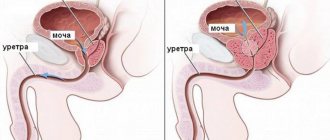
Transabdominal ultrasound of the prostate gland - photo:
Ultrasound of pelvic organs / Gynecological
Take care of the woman!
For this you need so little...
Why do you need to look after your health? The answer is simple: so that you don’t have to monitor the course of the disease. Our body is unique: it tries to the last to keep us “in line,” inventing incredible tricks to compensate and smooth out the onset of the disease.
This is especially true for the female body: nature has given it more endurance and resistance for the sole purpose of ensuring that human life on our planet does not stop. But modern women often try to join “men’s games” instead/together for this purpose. What result? The statistics of gynecological diseases are not reassuring.
But any disease can be prevented if you promptly - systematically - monitor your health, ask your body, which faithfully serves us: what is going on inside? Gynecological ultrasound examination in this case has no equal - it provides detailed information about condition of the female reproductive system.
What can you learn from an ultrasound of the female reproductive system?
Using this method, the doctor can examine in detail the entire uterine cavity and the endometrium, the layer lining the uterine cavity. The ovaries, bladder and pelvic cavities will receive special attention. It will be possible to find out the functional state of these organs. In addition, tumors can be detected using ultrasound at the earliest stages, when treatment is as gentle as possible.
Preparation for gynecological ultrasound.
The procedure itself can be of three types:
- transabdominal ultrasound, when the doctor looks at the organs through the anterior abdominal wall;
- transvaginal gynecological ultrasound, when a sensor is inserted into the vagina,
- transrectal, when the sensor is inserted through the rectum.
A transvaginal examination provides the clearest picture; in addition, it does not require special preparation. During a transabdominal examination, the key to image clarity is a full bladder, so an hour before the procedure you will have to drink a liter and a half of water and not urinate. Before transrectal examination, a cleansing enema is necessary.
Why once every six months?
It is with this frequency that your body should be examined using ultrasound: if any changes have occurred during this time, they are usually insignificant and require minimal medical correction. Moreover, this rule is no less important for a teenage girl - a future mother, for whom such a procedure will help maintain her health and then conceive a baby without any problems. This means that human life on earth will continue - and isn’t that what women exist for?
Indicators of norm and pathology
During the ultrasound, the necessary indicators are recorded:
- The doctor assesses the size of the gland. Normally, they should be up to 2.5-3 cm in length and 4 cm in width. A significant excess of these indicators (especially in men after 35-40 years) indicates pathologies and deviations from the norm.
- Using a special formula, the doctor determines the volume of the prostate. Normally, it should not exceed 24-26 cm.
- Then the mass is calculated. The normal value is 17-18 grams.
- In addition to the basic parameters, the doctor also determines the appearance and shape of the prostate gland. It should be symmetrical, resembling the shape of a chestnut fruit. The contours are clear and clearly visible.
- Deviations can also be indicated by an increase or decrease in the echogenicity of the organ, as well as signs of blockage of the ducts.
These parameters may vary slightly among patients. Everyone's body is different, so this may not indicate any pathology.
When is a transabdominal examination prescribed?
This procedure should only be prescribed by the attending physician: gynecologist, proctologist, urologist. Under no circumstances should you self-diagnose. Indications for such an examination may include a variety of diseases of organs located in the abdominal cavity. For women, these are the appendages and uterus, and for men, these are the prostate gland.
When is this research method prescribed for women?
This study is prescribed for women:
- To determine the parameters of the uterus and appendages. Helps determine the presence of any pathologies in the reproductive system.
- If there are all the signs of an ectopic pregnancy.
- If there is suspicion of adhesions and tumor.
- To verify the presence of pregnancy in the early stages.
- To monitor fetal development throughout pregnancy.
- Determine if the pregnancy is developing or if it has frozen.
- Confirms the presence of serious diseases in the fetus in the last stages of pregnancy.
- Helps determine the size and location of a cyst or fibroid.
- To diagnose a malignant tumor.
- Helps detect endometriosis, polyps and other pathological diseases of the appendages.
- Helps in identifying endometrial cancer.
- In case of failure of the ovaries.
- For polycystic and multifollicular ovaries.
- For problems with conception.
Read also: The testicle goes into the groin area in men
When is this research method prescribed for men?
This study is prescribed for men:
- For pain in the groin.
- For discomfort in the perineum and scrotum.
- With painful and frequent urination.
- With difficulty passing urine.
- If you have the urge to go to the toilet, but cannot void.
- If there is a constant feeling of a full bubble.
- For suspicious and unnatural discharge from the urethra.
- If you have problems conceiving.
- For chronic diseases of the reproductive system.
- If you have problems with potency.
- If the man is already over forty years old.
- For sexually transmitted infectious diseases.
- When blood clots appear in the urine.
How often should the procedure be performed?
To prevent diseases of the male genital area, it is necessary to undergo annual diagnostics. This is especially true for men over 35-40 years of age.
Transabdominal ultrasound is considered one of the simplest, most accurate and accessible procedures that allows identifying prostate diseases even at the earliest stages of development. Don't forget about preventative visits to your doctor. Taking preventive measures is much easier than dealing with an existing problem. Be healthy!
And you can find out about harmful and beneficial products for the male organ here.
Indications for use
Transabdominal ultrasound of the prostate is performed when the following symptoms appear:
- frequent urination, especially at night;
- discomfort when urinating (pain, burning along the channel);
- nagging pain in the perineum or lower abdomen;
- rectal lumps;
- erectile disfunction;
- infertility.
Transabdominal ultrasound of the prostate gland is prescribed by urologists and sex therapists as a preventive examination to exclude inflammatory processes and additionally visualize the pelvic organs. Prostate diseases and disorders of the organ’s structure are the root cause of male infertility. Ultrasound is performed as an auxiliary method for pathologies of the scrotum, penis, bladder, varicose veins of the small pelvis, and varicocele. The study is prescribed for gland cysts, acute or chronic prostatitis, prostate adenoma or the appearance of stones in it. The study is carried out if there are contraindications to the transrectal method (TRUS), for example, with:
- anal sphincter fissures;
- rectal polyps;
- proctitis and paraproctitis, fistulas;
- hemorrhoids in the acute stage.
Patients are not embarrassed during this examination and may not delay their visit to the doctor. This is the initial diagnostic method, prescribed after tests. Transabdominal ultrasound is suitable only for men of asthenic physique.
How to prepare
You can prepare for the examination within a few hours. In order for the ultrasound sensor to better visualize the organ through the abdomen, it is necessary to refrain from eating food three to four hours before the start. It is better to carry out the procedure after the morning toilet. If it is scheduled for the afternoon, it is recommended to limit yourself to a light breakfast. In case of difficulty with bowel movements, a cleansing enema is performed in the evening or on the eve of the test.
Before performing a transabdominal ultrasound of the prostate, it is necessary to avoid increased flatulence, that is, gas formation. It is not recommended to consume cabbage, sweets, or carbonated drinks on the appointed day. If you are prone to flatulence, before the procedure you need to take a tablet of Espumisan or Domperidone to improve intestinal motility.
One and a half to two hours before the test, you need to drink 1–1.5 liters of water. If the urge to urinate is strong before the ultrasound, it is necessary to empty the bladder, but not completely. If the volume of residual urine is less than 100 ml, the prostate cannot be visualized on the device screen.
How is a transabdominal ultrasound performed?
The patient lies on his back on the couch. The doctor lubricates the sensor with a special gel. It allows you to eliminate the air gap between the surface of the sensor and the abdomen. This will make the research result more accurate. The sensor is applied to the man's lower abdomen. The study lasts 7-10 minutes. During the procedure, the specialist examines not only the prostate, but also the bladder.
The sensor is moved across the abdomen with light, smooth, sliding movements, without lifting it from the surface of the body. An image appears on the ultrasound machine screen. The doctor reads the size of the gland, its structure, identifying possible changes. The device performs calculations. The patient is given the results of the examination in writing and in the form of a photographic image. The conclusion indicates the parameters of the organ under study and the identified pathologies.
How is the examination carried out?
After preparing for the procedure, the patient comes to the doctor’s office and takes the correct position lying on his back. This provides transabdominal access to the prostate. The doctor coats the ultrasound probe with gel and places it firmly on the patient’s lower abdomen. The examination is done within seven to ten minutes, while a specialist examines the bladder.
With light and smooth movements of the sensor across the abdomen, the doctor finds the projection of the organ. Then the size of the organ is recorded on the screen of the ultrasound machine, and its structure is examined for obvious changes. The calculations are made by computer. The patient receives the examination result in hand with all the parameters and a printed picture.
How they do it
A transabdominal prostate ultrasound is performed very simply, without causing any discomfort or pain in the patient. The examination is done in the supine position. To ensure good contact of the sensor with the skin of the abdomen, a special gel is used, which improves the passage of ultrasonic waves. The pelvic organs are usually examined in two planes - transverse and longitudinal, which allows you to examine the prostate and bladder from different angles.
During the examination, emphasis is placed on the volume of residual urine in the bladder, since this indicator is of great importance for diagnosing prostate pathologies. To determine the exact amount of urine remaining, the patient empties the bladder, after which the doctor continues the examination. The norm is the absence of fluid in the bladder, let's say the remaining urine is 50 ml; exceeding the norm indicates problems with the genitourinary system. The duration of a transabdominal ultrasound is 15–30 minutes.
Decoding the results
To decipher the results, you need to know the norms for the size and shape of the prostate gland. The organ has smooth and clear edges on ultrasound. If the scan is transverse, then the thickness and width of the prostate are determined. The norm for length for patients from 18 to 50 years is from 2.5 to 4.5 centimeters, and width - from 2.8 to 4.3 cm. With age, the organ can moderately hyperplasia, so the attending physician will take into account the age category of the man when calculating the size – the upper limit of normal may increase slightly.
It is difficult to determine the volume on a transabdominal ultrasound of the organ due to the presence of only two indicators, without the thickness of the prostate. An approximate calculation is based on the fact that the organ is spherical in shape, therefore, according to the formula, the volume should not exceed 28 cubic centimeters for healthy men who are sexually active. With these indicators, you can contact a urologist for further examination or a preliminary diagnosis.
Advantages and disadvantages
Transabdominal prostate examination has a number of advantages:
- the research is carried out quickly;
- no special preparation of the patient is required;
- no contraindications to the procedure;
- relatively low cost of the procedure.
Another examination method is transrectal ultrasound of the organ. It is carried out by inserting a sensor through the anal sphincter into the intestine. Transabdominal ultrasound examination of the prostate gland does not require placing a transducer directly into the body. But it also has disadvantages:
- it is impossible to determine the initial changes in the structure of the organ;
- Difficult visualization of the prostate in patients with abdominal obesity.
With the help of transabdominal examination, only the size of the organ, the symmetry of the lobules, and localization in the pelvis are determined. The remaining parameters are difficult or impossible to determine. Therefore, transabdominal ultrasound is often prescribed as a primary procedure before other types of diagnostics.
What is the price
Ultrasound of the prostate gland is performed transabdominally in every region of Russia. The examination apparatus is standard and does not require special attachments. The main thing is proper preparation of the patient and knowledge of the doctor. The cost varies depending on the region, the novelty of the device itself, and the presence of additional Doppler studies of blood flow in the organ. If Dopplerography is added to transabdominal ultrasound, the final cost increases by 100–300 rubles.
Source egopik.ru
A transabdominal prostate ultrasound is performed to diagnose prostate diseases. In general, ultrasound is one of the accurate methods with which you can check the condition of internal organs and understand whether treatment therapy is effective. Most often, this method of examination is carried out for the purpose of preventing and identifying human diseases. Ultrasound is also performed externally if there are contraindications to other ultrasound methods.
Preparation for the procedure
Carrying out a routine examination according to the rules requires preparation, which will reduce the level of intestinal gases and improve image quality:
- 3 days before a transabdominal ultrasound, a man is recommended to exclude from his diet foods that increase the level of gases in the intestines. Food should be light, heat-treated and without coarse plant fibers.
- Taking medications is suspended as directed by the doctor, especially if the procedure is carried out in 2 stages with a study of the function of urinary outflow and blood circulation.
- It is advisable to avoid alcohol intake, since its negative effects can distort the function of the blood vessels that supply the prostate.
- On the eve of an ultrasound of the prostate gland using the transabdominal method, dinner should be no later than 18 hours, and a cleansing enema is done if there is a tendency to constipation. In normal cases, it is enough to limit yourself to taking a mild laxative.
- You must bring water with you to the examination; you must not smoke, eat or take any medications until the diagnosis is completed.
In emergency cases, examination of the prostate gland is carried out without preparation.
Reasons for performing ultrasound
The doctor issues a referral for a transabdominal ultrasound of the prostate gland in the following cases:
- infertility;
- discomfort during urination;
- discharge from the genital organ;
- constant feeling of fullness of the bladder;
- pain in the perineum, lower back or lower abdomen;
- compactions in the gland, which can be identified by rectal palpation of the organ;
- burning;
- overestimation of PSA in the blood;
- decreased potency.
Usually the doctor gives a referral for a transabdominal ultrasound after receiving a urine or blood test.
The doctor may also send the patient for testing if abnormalities are detected in the sperm analysis.
There are no contraindications for this diagnostic method. However, if a person is obese, it will be difficult to see his internal organs. Thanks to this diagnostic method, the presence of the following diseases can be detected:
- acute and chronic prostatitis;
- BPH;
- prostate cyst;
- malignant formation;
- the presence of abscesses and purulent formations;
- hypertrophy;
- scarring.
One of the advantages of this ultrasound method is that this method is the most affordable, and many men undergo this examination in a free clinic, having received a referral from a urologist. Also, if you urgently need to obtain data on the condition of the prostate, you can perform a transabdominal ultrasound without special preparation.
When is a pelvic ultrasound necessary?
Ultrasound examination is prescribed to identify diseases and pathologies of organs and structures of the genitourinary system.
This analysis is considered one of the most informative, safe and painless. For an ultrasound of the pelvic organs, preparation is not difficult. Therefore, it is recommended for use in examining patients of any age and gender. The doctor refers the patient for this test when there is a suspicion of the following diseases:
- The appearance of neoplasms (tumors, adenomas, fibroids)
- Formation of cysts and polyps
- Pathological change in the structure of organs
- Presence of stones (stones, sand) in the bladder
- Cystitis
- Inflammation
- Infertility
- Salpingo-oophoritis
- Endometriosis
- Problems in the development of genital organs in children
- Fetal development disorders during pregnancy
- Prostatitis
Such ailments may be accompanied by the following symptoms:
- Pain in the lower abdomen and groin
- Bloody, mucous, or purulent discharge in the urine
- In women – bleeding not during menstruation or after menopause, cycle disorders
- Problems with urination (pain, urge too often, inability to empty the bladder)
- Pain in the lumbar region
If you experience such sensations and discomfort, you should immediately consult a qualified doctor.
Ultrasound is also prescribed to women in such cases as:
- Establishing the presence of pregnancy and its timing
- Monitoring the condition of organs:
- Before and after an abortion
- Before and after surgery
Both men and women are recommended to undergo a preventive examination of the genitourinary system every six months or at least once a year.
What is the essence of the method
In general, ultrasound of the prostate gland can be performed using two methods: transrectally and transabdominally. The first type of procedure confuses many people, since the device’s sensor is placed in the man’s anus, for which he is completely unprepared. But this method is more accurate for identifying various human diseases.
The external or transabdominal method of ultrasound is more accessible to people, as it is performed even in free clinics. The advantage of this method is its painlessness and lack of discomfort, which cannot be said about another method of conducting research.
The procedure is very simple. The patient should sit on the couch, on his back. The doctor spreads a special gel on the sensor for better research results. This substance is necessary to prevent air from passing between the sensor and the abdomen. As a rule, the prostate is examined in the transverse and longitudinal plane; the doctor can also specify other planes. Usually, along with an ultrasound of the prostate, the doctor examines the bladder. During the procedure, ultrasound waves impact the prostate from different angles.
As a rule, after the examination, the patient may already know the results, since the doctor sees them on the monitor during the ultrasound. In some cases, the uzologist does not announce the results of the study, but issues them along with photographs of the examined organ. In this case, the urologist himself must decipher the received data for the patient. The duration of such a procedure does not exceed half an hour; generally, 15 minutes are enough to examine the internal organs.
In some cases, the doctor may conduct an examination after emptying the bladder to determine residual urine. This indicator is also one of the important ones for diagnosing prostate diseases.
In order to accurately determine the remaining body fluid, you need to empty your bladder naturally. If you wait too long to go to the toilet, the examination results may be false.
The only unpleasant moment when carrying out this method is discomfort on the body after applying the cold gel. It is recommended to wipe it off after the procedure, as marks may remain on clothing.
Description of the procedure
Most often, a pelvic examination begins with a transabdominal ultrasound, after which the doctor asks the patient to empty a full bladder and performs either a transrectal or transvaginal ultrasound scan.
During examination through the anterior abdominal wall, patients lie on a couch. When pressing on the abdomen with an ultrasonic sensor, you may experience discomfort and a strong desire to go to the toilet, since the bladder is full. Other unpleasant sensations usually do not occur.
Transrectal and transvaginal ultrasound examination of the pelvic organs are more uncomfortable procedures, since patients have to expose intimate parts of the body.
During transrectal ultrasound, men lie with their legs slightly bent at the hip and knee joints, with their backs to the doctor. A rectal ultrasound sensor with a disposable condom on and a special gel applied is inserted into the rectum to a shallow depth. Discomfort may occur both during insertion of the sensor and during its movements in the rectum. If pain occurs during the study, you must immediately inform your doctor.
During a transvaginal examination, women lie on their backs with their knees bent and legs slightly apart. A condom is also placed on the vaginal sensor and a conductive gel is applied, which simultaneously acts as a lubricant. The doctor inserts the prepared sensor into the vagina to a depth of 3-4 cm. This procedure feels less unpleasant than an examination on a gynecological chair. Pain and severe physical discomfort occur only in patients who have serious pathological changes in the internal genital organs.
Zubkova Olga Sergeevna, medical observer, epidemiologist
16, total, today
( 52 votes, average: 4.69 out of 5)
Ultrasound of the thyroid gland - indications, norms, preparation
Examinations to determine ovulation
Related Posts
Normal examination parameters
Only after receiving the results of the study can a conclusion be made about the condition of the prostate gland. The shape of the prostate on ultrasound should be triangular or oval. The contours of the organ should be smooth and the lumen of the ducts should be free.
It is known that the growth of the indicators of this organ ends by the age of 25. As a rule, the volume of the prostate in an adult man should not exceed 28 cubic centimeters, and its dimensions should be within 4.1 * 4.2 * 2.3 cm. If these figures are too high, then the doctor must make a diagnosis and prescribe treatment for the patient .
Normally, the amount of residual fluid in the bladder should not be more than one tenth of the total amount of emptying. If its volume is more than 40 ml, then this condition indicates a disease. In order to diagnose the condition of the prostate gland, it is necessary to conduct such studies several times, comparing the results.
It will be difficult for a person who does not have a medical education to decipher the results of the study. If no pathologies are found, the ultrasound should result in the following conclusions:
- density is normal;
- no changes in blood vessels;
- homogeneous tissue structure;
- gland parameters are within normal limits.
If the patient has some kind of prostate disease, then the test results may include the following values:
- a strong change in size, up to 7 centimeters, indicates prostate adenoma;
- with enlarged lymph nodes and unclear boundaries of the organ, neoplasms are observed;
- with inflammation or prostatitis, increased or decreased echogenicity is observed.
Also, in the presence of stones and cysts, this can be seen on the results of an ultrasound of the prostate.
Disadvantages and advantages of the method
As mentioned above, most often the doctor prescribes the transrectal ultrasound method, since the external type has disadvantages. After the procedure, you can only obtain the following internal organ data:
- form;
- size;
- symmetry;
- gland volume;
- location.
This method is based on scanning internal organs, but due to low sensitivity it is difficult to detect the disease at an early stage of the disease. The advantages of this method include the following features:
- such an examination allows you to see all the necessary parameters for diagnosing prostate diseases;
- this method is one of the available ones and its price is not so high compared to other diagnostic methods;
- Thanks to such an examination, you can check the condition of many of the man’s internal organs;
- the procedure does not harm the person; it does not require making incisions or punctures.
Another advantage of this procedure is its duration. Also one of the positive aspects is the minimal preparation for the study.
In addition, this procedure is safe for humans, since it does not expose them to radiation. To conduct the study, frequencies from 3.5 to 7.5 MHz are used, which is enough to identify pathologies.
How to prepare
External ultrasound requires special preparation to ensure that the examination data is accurate. The main methods of preparation for the procedure are the following:
- 2 hours before the test, drink at least a liter of liquid (it is best to drink non-carbonated mineral water);
- three days before the procedure, do not eat foods that increase gas formation;
- Do not urinate for 2 hours before the ultrasound;
- 10 days before the study, avoid taking aspirin and NSAIDs.
It is important to take exactly one liter of fluid before the procedure, as insufficient volume will complicate ultrasound examination and visualization. And if you drink a lot of liquid, on the contrary, the patient will want to urinate during the procedure.
This diagnostic technique is used to identify many diseases. This type of procedure will be less expensive than other prostate ultrasound methods. The cost of the procedure in different clinics is excellent, but generally it does not exceed a thousand rubles, when a rectal examination will cost more.
Source proprostatu.ru
Preparing for a transabdominal ultrasound
No special complicated preparation is needed for a pelvic ultrasound. But some nuances still need to be taken into account.
Before the procedure, drink several glasses of water to keep your bladder full. This is necessary for better visualization of organs, because ultrasound passes well through liquids, but poorly through gases. A full bladder pushes the intestines out of the pelvis, which also makes the task easier.
A few days before the ultrasound, it is necessary to exclude gas-forming foods from the diet . These include:
- Legumes.
- Dairy products.
- Fresh vegetables.
- Fried food.
If you have increased gas formation, regardless of nutrition, then take activated charcoal or drugs that reduce gas formation.
Do not perform a transabdominal ultrasound and an intestinal examination using contrast agents on the same day. This may affect the information content of the pelvic examination. As a last resort, alternate the procedures correctly so that they do not interfere with each other.
For women, this examination is done on days 7-10 of the menstrual cycle.
Conducting transabdominal ultrasound (video)
Contraindications and dangers
This ultrasound examination has no contraindications. If the preparation is strictly followed, the results will be informative and accurate in any case.
Ultrasound does not cause harm; the patient is not ionized or irradiated during the procedure. At the same time, he does not feel pain, since the procedure does not violate the integrity of the skin.
Ultrasound does not aggravate any diseases and does not affect the body in any way. It works on the same principle as natural ultrasound, which some animals can generate.