There are many types of skin cancers. One of these is Bowen's disease, which was described by a dermatologist from America and named after him.
The pathology is squamous cell carcinoma, which is located in one place and tends to grow to the periphery. Foci of the disease can reach several centimeters in size. Carcinomas are painless and may develop plaques or scaly surfaces.
Reasons for appearance
The exact reasons why Bowen's disease occurs have not yet been established. However, it is known for certain that the process of cell degeneration is affected by exposure to sunlight. Old age is also a risk factor. Most often, the disease is diagnosed in people who take drugs that suppress the immune system, such as immunosuppressants, cytostatics and glucocorticoids.
The likelihood of developing pathology increases in patients who are infected with the human papillomavirus, especially type 16. In addition, other risk factors include prolonged exposure to arsenic. Hydrocarbons and mustard gas also play a role in the development of Bowen's disease (in men more often than in women).
Adverse external influences on superficial skin cells disrupt metabolic processes, which accelerates their death. The new cells that are formed change at the genetic level, which ultimately causes disruption of their functions and structure. Initially, the middle, spinous layer of the epidermis is affected, its cells begin to change and divide abnormally.
Until the tumor penetrates the membrane that separates the middle layer of the skin and the epidermis, it is designated as a carcinoma confined to a single location within the epithelium. Metastasis in this case is excluded, although the formation is considered malignant.
Symptoms of intraepidermal cancer
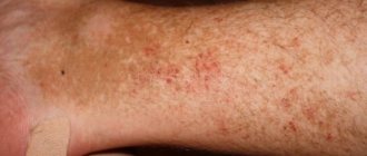
The most typical sites of infection are the skin of the face, extremities and perianal area. Painless brown papules the size of a pea form on the skin, which subsequently merge with each other and form an extensive lesion. There are eczema-like, hyperkeratotic and warty forms of the disease.
After many years, squamous cell invasive skin cancer forms at the site of the dermatosis. A characteristic sign of transition to cancer is the appearance of a dense node, which soon ulcerates. An accurate diagnosis is established by histological examination of the affected skin. Hypertrophy of the integumentary epithelium is detected; atypical cells with pathological nuclear division are found in the germinal layer of the epidermis (Malpighian layer).
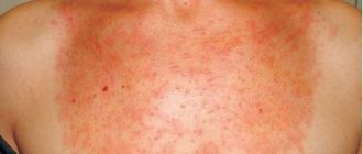
The disease manifests itself by the appearance of single (in some cases, multiple) lesions of pink-red or red-brown color with a poorly defined border, which gradually transform into plaques, slightly rising above the level of unchanged skin.
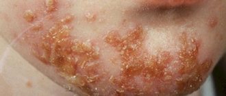
Plaques are characterized by slow peripheral growth, round or oval shape; less often, skin defects have polymorphic outlines. The surface of the inflammatory focus is uneven, granular, covered with yellowish crusts and scales, which, when removed, reveal a wet, shiny, bloodless wound.
Visually, inflammatory foci are distinguished by variegation: zones of hyperpigmentation are adjacent to areas of clinically unchanged skin.
As it progresses (with long-term existence of the pathology), superficial ulceration with the formation of partially cicatricial erosions, an increase in the area of the lesion, or the merging of several foci into one extensive lesion are noted.
On the oral mucosa, Bowen's disease usually manifests itself in the form of single, flat, slightly sunken lesions, sometimes with areas of increased keratinization (but more often papillomatous). Lesions in the eyelid area, most often the upper eyelid, are also predominantly papillomatous in nature. The disease, localized on the mucous membranes, proceeds much more intensely, with early malignancy.
As a rule, the disease is not accompanied by subjective disorders; occasionally patients complain of itching or burning.
Usually, in the absence of appropriate treatment, the disease progresses steadily, although cases of lifelong progression without progression have been described.
In order to start treatment in a timely manner, you need to know what symptoms this disease has. Thanks to this, it is possible to stop the development of the disease in the early stages and prevent the appearance of cancer, as well as reduce the number of possible complications.
If you suddenly find that irritation or spots of unknown etiology have appeared on your skin, you should visit a dermatologist who will prescribe a more thorough examination.
Bowen's disease manifests itself in several stages:
- First, a spot appears with blurred edges. Over time, it not only grows, but also turns into a flat plaque. The affected surface constantly itches, and specific scales form here. When they are removed, the surface of the plaque is slightly moist, but does not bleed at all.
- Over time, the plaque grows and absorbs more and more healthy skin. Gradually the edges begin to rise and growth intensifies.
- If the disease is not stopped in the early stages of development, Bowen's disease develops into a new form of cancer. In this case, there is a sharp appearance of ulcers of various sizes on the entire surface of the plaque.
- A neoplasm on the mucous membranes initially leads to the appearance of red, smooth lumps, which later turn into bleeding ulcers.
Symptoms of Bowen's disease
Photos of the external manifestations of the disease are presented later in the article. The main symptom of the disease is reddish-brown spots on the skin, growing from the center to the periphery. The spots have clear boundaries and raised ring-shaped edges. In some cases, the lesions appear as flaky patches of skin. The formations are flat, with raised edges, oval or round in shape with regular outlines. Sometimes these skin lesions can be itchy, but most often they are painless. In the future, ichor or pus may begin to be released from them, and crusts may also form. Small growths stand out on the granular and uneven surface of the disease focus.
Bowen's disease in women may appear as a wart-like growth with cracked skin or hyperpigmentation. Most often there is one focus of the disease, but in 15% of patients there is multiple localization.
Etiology, pathogenesis, clinical picture[ | ]
The disease occurs in older people, mainly women. It usually occurs on the torso, upper limbs and perineum. The occurrence of Bowen's disease is associated with skin injuries, exposure to ultraviolet radiation, and contact with substances containing arsenic (for example, drugs or substances from the chemical industry.
When the disease occurs, solitary or in a third of cases multiple lesions appear on the skin: at first it is a small red spot or plaque of irregular or round shape with peeling. Such plaques are formed when reddish papules and nodes merge. White or yellow scales on the plaque are easily removed, revealing a red, weeping surface underneath.
The plaque is clearly demarcated from the surrounding tissues, gradually grows and eventually rises above the skin level. The plaque has a motley appearance due to the appearance of zones of atrophy, hyperkeratosis, and growths. In some cases, Bowen's disease causes several gradually merging lesions. Gradually the process spreads to the entire layer of the epidermis. Over time, the growing lesions transform into squamous cell carcinoma, which allows us to talk about Bowen's disease as a precancer.
When diagnosing, Bowen's disease should be distinguished from psoriasis, actinic keratosis and basal cell carcinoma.
When the disease progresses
As the disease progresses, ulcers and erosions form, which gradually heal and scar, increasing in size and affecting an increasingly larger surface of the skin.
Most often, the symptoms of Bowen's disease appear on areas of the skin that are open, but sometimes the pathology is localized on the palms, soles and genitals. It also happens that the disease is localized in the oral cavity, and in this case this clearly refers to a precancerous condition, since there is a high probability of malignancy. Lips and gums may also be affected.
Symptoms of the disease and types of pathology
Bowen's disease is most often localized on the upper extremities, genitals and body, but in fact the disease can be located on any part of the skin and mucous membranes. The mucous membranes most often affected are the soft palate and tongue. The tumor is never localized to the female genital organs. This localization occurs much more often in men.
Initially, a red spot appears on the skin, small in size and with uneven edges. After some time, the spot begins to increase in size and transforms into a scaly plaque. The plaque has a yellowish-red color and uneven edges. In some cases, a heterogeneous color of the plaque is noticed; there are lighter and darker areas on it.
As the tumor progresses, its structure changes: signs of atrophy, thickening of the epidermis, and warty growths appear. The tumor on the skin is slightly raised, but does not have the ridge shape characteristic of skin cancer. The disease lasts for a very long time, years and decades, with slow growth and possible transition to squamous cell skin cancer.
There are several forms of Bowen's disease:
- pigment form;
- anular;
- verrucous;
- nail bed disease.
The main sign of Bowen's disease is the appearance of a neoplasm (foci of dyskeratosis) on the surface of the skin - single or several at once.
Over time, a wart may form on the affected area.
When the neoplasm is located on the surface of the mucosa, the plaques may have a papillomatous velvety surface.
Although the growth is more common on exposed skin, it can affect the palms and soles of the feet. If Bowen's disease is diagnosed, located in the oral cavity, it is considered an obligate cancer with a high degree of malignancy - in most cases it degenerates into cancer. Externally, this neoplasm looks like an unevenly colored spot, which can be located in the oral cavity and nasopharynx (root of the tongue, inside of the cheeks, soft palate, palatine arches, behind the molars, on the lips).
DETAILS: Skin rashes during pregnancy
Sometimes when a tumor appears, the patient may experience some soreness or itching. With further development, the focus of the neoplasm may deepen, and white areas similar to leukoplakia may appear on its surface.
Bowen's disease is divided into the following forms:
- anular (ring-shaped) – the most common, accompanied by the appearance of rounded lesions with raised borders in the shape of a ring;
- warty (verrucous) - manifested by the appearance of small growths on the surface, similar to warts;
- pigmented (spotty) – accompanying the accumulation of melanin in cells and subsequent darkening of the skin;
- Bowen's disease of the nail bed, leading to destruction of the nail plate.
One form that is in many ways similar to Bowen's disease is Queyra's erythroplasia. It is characterized by the appearance of reddish, rough or smooth spots on the skin of the inner flesh and head of the penis, which may be accompanied by peeling, pain, ulceration, and itching. During urination or ejaculation, blood may be released from the urethra.
If Bowen's disease is diagnosed in the genital area in women, it can occur in the form of vulvar intraepithelial neoplasia. Most often it is associated with infection with type 16 papillomavirus. Red, rough spots appear on the surface of the skin of the perineum, and a burning or itching gradually occurs.
Diagnostics
If a doctor suspects a patient has Bowen's disease, it is necessary to determine the presence of external signs of the disease, as well as carefully collect anamnesis. It is necessary to carry out differential diagnosis, since the pathology has symptoms similar to many dermatological diseases. Sometimes patients do not immediately notice the problem, since the spots formed on the skin do not cause discomfort. For this reason, a careful examination of the patient is considered an important stage of diagnosis.
In addition, a piece of the affected tissue is taken for a biopsy. This study will exclude other diagnostic options and confirm Bowen's disease in women and men (the photo shows what the pathology may look like). Without a biopsy, it is impossible to accurately determine the danger of the lesion and choose treatment methods.
Consequences and complications of Bowen's disease
The main threatening consequence of Bowen's disease is progressive malignancy into squamous cell carcinoma with invasion into the surrounding soft tissue and metastasis.
In addition, the following complications are possible:
- infection of the inflammatory focus;
- ulceration of plaques;
- sepsis.
Data from studies of the frequency of association of Bowen's disease with malignant neoplasms of internal organs are ambiguous: the authors indicating the presence of a connection found cancer of internal organs in 57% of those who died from Bowen's disease. However, in additional studies, a significant increase in the incidence of internal organ cancer was observed only in patients with lesions on closed areas of the skin (33%), while in Bowen's disease on open areas of the skin, the incidence of visceral cancer was only 5%.
The main terrible consequence of Bowen's disease is considered to be malignancy into squamous cell carcinoma and its germination into the surrounding soft tissue with subsequent metastasis.
Other complications may include:
- inflammation infection;
- ulceration of plaques;
- sepsis.
What attention is paid to
When examining damaged tissue, the specialist pays attention to the following signs:
- Acanthosis with the presence of elongated and thickened outgrowths.
- Horny surface of the skin.
- Paraketosis of focal type.
- Disorderly position of spinous cells.
- Large brightly colored nuclei and atypia.
- Cellular vacuolization.
- Mitotic figures.
As the disease progresses to the cancer stage, the following changes occur:
- Destruction of the basal protective membrane.
- A sharp change in the shape of cells with acantholysis plunging deep into the dermis.
Treatment of Bowen's disease
A standard treatment regimen for the pathology has not yet been found. Therapy is selected individually, depending on the location of the disease, the patient’s age, the number and size of lesions, the person’s health status and other indicators. Quite often, therapeutic methods are combined.
Patients diagnosed with Bowen's disease are offered various treatment options:
- Cryotherapy.
- Chemotherapy.
- Photodynamic therapy.
- Electrical destruction.
- Surgical intervention.
It is quite difficult to predict which method will help a particular patient, although all of the above procedures are effective in themselves. For this reason, each patient receives an individual therapy program.
In some cases, the specialist opts for a wait-and-see approach. This happens if the patient is elderly or the disease is localized on the extremities, in places that can be protected from sun exposure. The patient is recommended to regularly visit a specialist, and at the first symptoms of progression of the formation, surgical intervention is performed.
Treatment methods
The choice of treatment method depends on the form of Bowen's disease, its location and size. Surgical treatment of the disease is most often practiced. If the size of the lesion exceeds 5 cm in diameter, the tumor is removed surgically. The surgical method includes not only traditional excision of the tumor with nearby tissue, but also such removal methods as:
- laser destruction;
- cryodestruction;
- radio wave therapy.
Surgical treatment methods are not used in case of localization of Bowen's disease on the mucous membranes. To treat diseases on the mucous membranes, close-focus fluoroscopy or photodynamic therapy are used.
Treatment is carried out by dermatologists. Apply 30-50% prospidin ointment in the form of daily applications under compress paper for three weeks, cryodestruction and surgical treatment. The most effective surgical treatment is excision of the affected skin within healthy tissue, followed by radiation therapy. The prognosis is favorable provided radical surgical treatment is performed before the appearance of metastases.
View specialized medical institutions
Very often, Bowen's disease is confused with eczema, psoriasis, lichen planus and solar keratosis, squamous cell epithelioma and basal cell carcinoma. To make an accurate diagnosis, a thorough examination of the patient is carried out based on the clinical picture: the appearance of the plaque, location, size and growth rate. To confirm the diagnosis, a histological examination may be prescribed.
Treatment options for Bowen's cancer will depend greatly on the patient's age and the size and location of the tumor. The most common method is excision of lesions with a surgical knife or electric knife. In particularly severe cases, radiation therapy may be used. In the early stages (if the size of the lesion is less than 2 cm), applications of 5% fluorouracil ointment are common, which is also prescribed in cases where Bowen's disease is diagnosed in the oral cavity. If a lesion measuring 2 cm is detected, cryodestruction may be prescribed.
Treatment of Bowen's disease is surgical with excision within healthy tissue with mandatory histological examination. Cryodestruction and radiation therapy are also used.
Treatment of the disease depends on the area and severity of inflammatory changes.
For small skin defects (up to 2 cm), ointments and applications with cytostatics and the use of dermatotropic antipsoriatic drugs are indicated.
If the size of the inflammation is more than 2 cm, it is recommended to remove it using surgery, electrocoagulation, cryodestruction, close-focus radiotherapy, laser therapy.
The treatment of this disease is influenced by many different factors: from the general condition and medical history of the patient, to the characteristics and localization of the tumor.
First of all, you need to know which doctor you should contact not only for diagnosis, but also for subsequent treatment. If symptoms appear, you should contact an oncodermatologist who can rule out dermatitis and other skin diseases. He will take and prescribe all the necessary tests. After receiving the results, treatment is prescribed, depending on the severity of the disease.
So, in the initial stages, when the disease only manifests itself, good results are shown by exposing the affected area to liquid nitrogen. In this case, special medications are also widely used, which should strengthen the immune system and suppress the pathogenic environment. The course of treatment is carried out for two months under the supervision of a specialist.
Repeated tests should be taken periodically and monitored for the development of the disease. If the growth of pathogenic and mutated cells continues, then the course of treatment should be repeated or switch to a more radical method.
On mucous membranes, lesions should be treated with extreme caution. Most often, radiotherapy is used here. This treatment method is quite lengthy and should be carried out under the supervision of specialists for several weeks. After a break it is recommended to repeat it.
If there are many lesions that have affected the skin or they are too large, then experts recommend using a radical laser removal method. This method is quite traumatic and requires removing the affected area with healthy skin around the perimeter.
The surgical method is considered one of the most effective, since it removes all the affected cells and those that could be in contact with them. This prevents relapse of the disease, but requires long-term rehabilitation. If a very large area has been affected, cosmetic surgery may be required in the future to eliminate the scar after the procedure.
A variety of medications, creams, ointments and sprays are widely used in the treatment of Bowen's disease. With a complex effect, treatment usually takes place much faster. The most popular in this case is glyciphone ointment 30%. Other products are also widely used, which should be used only as prescribed by a doctor and the accompanying instructions:
- Efudix cream;
- fluorouracil ointment;
- Immunosupportive drugs are indispensable.
DETAILS: Papular dermatitis - symptoms and types
All drugs and medicines are prescribed by a doctor only after a detailed examination and depend on the chosen treatment method and the severity of the disease.
Today, traditional methods of treating Boeun's disease are especially popular, but they should not be abused. It is best to turn to a specialist for help, and use traditional methods of treatment only as a supportive form.
So, hemlock infusion is considered very popular, which is used to wipe the affected areas of the skin. To do this, mix a tablespoon of herb with a glass of lime water. There are many other traditional medicine recipes, but they should be used only after consultation with a specialist.
Treatment methods are selected individually, taking into account the patient’s age, location of the tumor, size and number of lesions, and other factors. A combination of several methods is often used.
Treatment methods used in the treatment of the disease:
- chemotherapy;
- photodynamic therapy;
- cryotherapy;
- electrical destruction;
- curettage;
- surgical intervention;
- sometimes a wait-and-see approach is used.
The surgical method is quite often used in treatment; this method is very effective, since tumor cells do not penetrate deep into the skin and do not metastasize. The tumors are removed and sutures are applied; a scar is later formed in their place. The operation itself is performed under local anesthesia. To obtain the best cosmetic effects, micrographic Mos surgery is used.
Curettage is another method of surgical intervention that involves curettage and subsequent electrocoagulation. With this method, the affected tissue is first scraped out with a curette, and then the lesion is cauterized. This type of treatment is performed with pain relief. Most often it is necessary to conduct more than one session. After cauterization, a scar may remain.
Chemotherapy is applied externally to kill tumor cells. To do this, use ointment with fluorouracil, which is applied to the affected area twice a day for a week, then take a break and repeat again. Thus, 4-6 courses are carried out (usually this treatment is used in combination with other treatment methods). Imiquimod is also used to treat Queir's erythroplasia and large lesions on the legs.
Cryotherapy (cryodestruction), used for treatment, is considered a minimally invasive method, as it has little effect on unaffected tissue. Cryotherapy uses liquid nitrogen or argon, which is directly applied to the affected area. When the affected tissue freezes, a crust forms on top, and after a few weeks it should fall off. This method is used at the initial stage.
Phototherapy is used for large foci of neoplasm. To carry out treatment, a light-sensitive substance is applied to the lesions and then exposed to a certain type of light irradiation. Abnormal cells are destroyed. As a rule, phototherapy requires several sessions.
Due to the fact that there is a danger of the disease degenerating into cancer, the use of folk remedies is not considered effective. Whenever using traditional methods of treatment, you must coordinate their use with your doctor and not use it instead of the main treatment.
Description of therapeutic techniques
As mentioned above, there are a number of treatments for Bowen's disease. Some of them are listed below.
- Chemotherapy drugs are used to destroy tumor cells. These include Imiquimod and 5-Fluorouracil. For a week, ointments with the same active ingredients are applied twice a day to the affected areas of the skin, then a break is taken for several days, and the course of treatment is repeated. In this way, up to 6 courses of treatment are carried out.
- Surgical intervention. This is the most effective treatment method, since malignant tumor cells do not metastasize and are located on the surface of the skin, without penetrating too deeply. The operation is performed with local anesthesia.
- Curettage. It consists of curettage followed by electrocoagulation. The affected tissue is scraped off with a special instrument, and then the source of the disease is cauterized. In some cases, several sessions are necessary.
- Cryotherapy. This is a minimally invasive method of therapy, which consists of a slight effect on healthy tissue surrounding the lesion. After treatment, the tissues freeze and form a crust, which disappears after a few weeks. The method is suitable for single formations at the initial stage.
- Phototherapy. Used for large affected areas of the skin. The procedure consists of applying a special cream, which tends to accumulate in the affected cells, and subsequently exposing them to light irradiation. Cells that have absorbed the photosensitizer from the cream die. The cream is applied 4-6 hours before the irradiation procedure. Several sessions are required for recovery.
- Radiation therapy. Previously, this method was widely used, but today preference is given to safer methods. After radiation therapy, poorly healing skin lesions form. As a rule, this technique is used if surgical intervention is not possible.
- Laser therapy. This method is not widespread. However, it has shown some positive results. A number of studies are needed to evaluate the effectiveness of this method.
We looked at the treatment of Bowen's disease. We will consider the forecast in the next section.
How is Bowen's disease treated today?
In medical practice, there are the following methods of treating this disease:
- Freezing with liquid nitrogen:
In the postoperative period, redness, swelling and blisters are observed in the frozen area. For extensive defects, this procedure is carried out in several stages.
- Curettage:
The essence of the manipulation is local scraping of the epidermis under local anesthesia.
- Radical excision:
The abnormal area of skin is surgically removed. After the operation, the wound is sutured, which may subsequently result in the formation of a scar.
- “5-fluorouracil” cream:
This drug is able to control and normalize the process of cell division, that is, the growth of formation. The action of the cream is aimed at destroying mutated elements.
- “Imiquimod” cream:
This remedy was originally developed for the treatment of genital gangloma. Recent studies have established the effectiveness of the cream in the fight against Bowen's disease. A side effect of this is inflammation of the dermal layer.
- Photodynamic therapy:
The patient is treated with a chemical on the affected area of the body, after which the growth is irradiated with light waves.
- Radiation therapy:
Highly active radiological radiation leads to the destruction of the mutation and restoration of the skin.
- Chemotherapy at the local level:
Such treatment is rarely carried out. It consists in the local introduction of cytostatic substances into the pathological focus.
Prognosis for this disease
With this disease, especially if the therapy is carried out on time, a favorable prognosis is given. When the source of the disease is removed, the patient can be considered completely healthy. The risk of a skin lesion degenerating into an invasive form of cancer reaches 3-5%.
The probability of such degeneration increases to 10% when localized in the genital area or Queyra's erythroplasia. Some scientists, however, believe that transformation into cancer occurs in 80% of cases. These discrepancies indicate differences in climatic conditions, intensity of exposure to sunlight and detection of the disease in different countries.
When the diagnosis is accurately established, the best option is to remove the tumor using medication or surgery. This will protect the patient and reduce the risk of cancer.
How to determine the beginning of transformation?
There are a number of signs by which you can determine the beginning of the transformation of Bowen's disease into cancer:
- Bleeding.
- Formation of a lump in the affected area.
- Ulceration.
- Enlarged lymph nodes.
- Skin thickening.
- Change in color of affected skin.
The appearance of the above symptoms indicates the need to urgently consult a doctor before metastasis occurs and cancer cells begin to spread throughout the body.