Liver lipomatosis is a pathology accompanied by the formation of nodes in the structure of the gastrointestinal tract. These formations are benign, consist of fat cells, and can disrupt the functionality of adjacent tissues and organs. In addition, there is a risk of the tumor transforming into malignant.
In the early stages, the disease has subtle symptoms, making diagnosis difficult. However, as the pathology develops, specific symptoms of liver disease arise: nausea, pain in the right hypochondrium.
Lipomatosis of the liver and pancreas simultaneously occurs very often.
Basic information about this pathology
Hepatic lipoma is a benign formation consisting of adipose and connective tissue, as well as processes of nerve cells. The consistency of the formation is soft, has mobility, and does not cause pain.
Liver lipomatosis develops due to excessive accumulation of adipose tissue in the body, which gradually begins to replace healthy hepatocytes. Against the background of the pathological process, the liver begins to enlarge, its activity is disrupted, as well as the synthesis of hepatic iron enzymes, as a result of which the organ loses its ability to cleanse the blood of toxic substances.
Most often, lipomas are diagnosed in women 25-50 years old. They appear as a result of disruption of the process of formation of fat cells, expansion of the lumen of the ducts of the sebaceous glands.
Signs of lipomatosis of the liver and pancreas depend on the form of formation.
Liver lipoma: what is liver lipomatosis and what are the treatment methods
Often a cyst poses a serious threat to human health and life. One of the threatening types of fat is liver lipoma. The presence of this pathology indicates organ dysfunction and general intoxication.
Lipoma is a benign formation caused by the growth of connective tissue. The disease develops gradually, especially in areas with a small layer of fat. The site of the tumor can be any part of the body or any internal organ.
Genesis of the disease
The cyst is formed from fat cells, nerve fibers and muscle connective tissue. In terms of density, the tumor is often soft, painless and mobile. The average age of patients is 25-50 years.
Liver lipoma most often occurs in women due to the structure and formation of adipose tissue and the way it is distributed. Tumor growth is associated with impaired adipocyte production and an increase in free space in the sebaceous glands.
Cysts vary in type of consistency:
- Lipofibromu - soft structure, labile, contains fat cells;
- Fibrolipomu - dense, inert, consisting of the connective tissue of the organ;
- Myolipomu - a rigid and immobile cyst formed by fibers;
- Angiolypomu is a dense consistency with its own blood supply system.
The nature of the neoplasm is mainly of exogenous origin. Under certain conditions it can become cancerous.
Stages of liver lipomatosis
8 1
| Degree of tumor development | Appearance of lipoma |
| The light form covers 1/3 of the liver. The cyst is medium in size and has virtually no symptoms. | |
| 2 | It affects half the organ, which affects the general condition of the patient. Heartburn, flatulence, belching and a feeling of heaviness in the epigastric region may occur. |
| 3rd | An area involving more than 50% of the liver. It is characterized by a serious condition and disruption of the digestive tract, destabilization of blood sugar levels. The organ is not able to produce somatostatin, glucagon and insulin. The patient's condition deteriorates significantly. There is an increased risk that the tumor will develop into a malignant one. |
The form of formation differs from lipomas:
- diffuse - small fats localized throughout the liver and practically not affecting the functioning of the glands;
- insular (connective) - large lesions of one or more types that block the ducts of the endocrine gland and cause symptoms of gastrointestinal dysfunction.
There is also a nodular and mixed form of lipoma. The latter is characterized by a large amount of fat deposits in the organ and latent flow.
Causes
The factors that cause excessive fat accumulation in the liver are not fully established. Among the most likely causes of cyst development are:
- Pathological changes in cellular metabolism in the body;
- Genetic predisposition in family history;
- Consistency of the body with toxins and waste;
- Decomposition of lipid metabolism (metabolism) with an increase in the level of lipophilic alcohol in the blood;
- Presence of diabetes mellitus;
- Hormonal imbalance caused by physiological processes in the body;
- Decreased activity of the pituitary gland and thyroid gland;
- Pathologies of the pancreas and liver;
- Obesity;
- Alcohol addiction.
The tactics for treating tumors depends on the source of the cyst formation, the stage of development and the age of the patient. Adults are more likely to suffer from fatty liver than children.
Lipoma symptoms
At the first stage, the disease is asymptomatic. Only when the tumor has reached a large size does it show the characteristic signs of a lipoma. Fatty liver manifests itself in the following symptoms:
- Diseases of tactile sensations in the peritoneum and right hypochondrium;
- Nausea;
- loss of appetite or appetite;
- Conditions of general loss of performance;
- Increased fatigue and depression;
- Increased nausea reflex, indigestion, flatulence, etc.
- Dark pigment spots on the skin;
- Liver loss, palpation.
In addition to the typical signs of oily skin, hyperplasia of the spleen and liver is observed, which becomes the plasma form of the disease. Fatty liver does not go away on its own and requires immediate medical treatment. The tumor is discovered accidentally or during the treatment of complaints in the epigastric region.
Complications
Missing or untimely treatment of fats can have serious consequences for the health and life of the patient. Possible complications of trigger lipoma are:
- dysfunction of the entire HIT system;
- loss of filtration properties of the liver;
- sealing of the cyst and its transition to a malignant form (liposarcoma);
- compression of surrounding tissues, leading to necrosis;
- severe pain in the epigastric region of the peritoneum and abdomen;
- inflammation of the cyst and development of associated pathologies
A large tumor can rupture with subsequent damage to the integrity of nerve endings and tissue vessels.
Diagnostics
To determine lipoma and differentiate it from other diseases, examination methods are used:
- ULTRASOUND. Allows you to determine the size and structure of the cyst;
- X-ray. Prepared using a contrasting medium;
- MRI. Detailed neoplasm showing the exact clinical picture;
- CT. Helps determine the degree of density and content of lipoma;
- laboratory tests, including general tests of urine, blood, biochemistry, sexually transmitted diseases, hepatitis and HIV;
- pinpoint aspiration biopsy;
- cytological examination;
- oncological research.
With the help of complex diagnostics, it is possible to assess the extent of damage to an organ lipoma and make a prognosis for its further development. The course of treatment is determined only by the examination results and the final diagnosis.
Treatment methods
Removal of liver lipoma is exclusively surgical. If the tumor is small, the condition and genesis of the cyst are controlled without surgery. The radio wave or strip method is used to extract the tumor.
In the first case, the operation is performed without contact, which eliminates blood loss and pain. The degree of complications and injuries in this case is minimized. When cutting fat, a special radio wave scalpel is used, which emits waves of different lengths and frequencies. Using this method helps protect healthy tissue from damage.
The second method of treating liver lipoma is performed under general anesthesia, by cutting the skin and resection of fat from the organ cavity. Depending on the situation and clinical indicators, endoscopic techniques may be used.
- The rehabilitation period after surgical treatment is short and lasts 1-2 weeks.
- To prevent relapse of the disease, it is recommended to create favorable conditions for preserving the body and carry out a number of restorative procedures.
Prevention
Preventative measures to prevent liver fat from forming or reoccurring:
- Periodic cleansing of the body from accumulated toxins and waste;
- Treatment of diseases and disorders of the digestive tract;
- Maintaining a diet with low-fat foods;
- Exercise through sports, fitness, aerobics, etc.
- Moderate food consumption and avoidance of bad habits;
- Availability of regular medical examinations.
Maintaining a solid body can significantly reduce the risk of liver lipoma and its consequences.
© 2020 — 2020, MedPechen.ru. All rights reserved.
Forms of the disease
The disease can have several variations:
- Nodular lipomatosis. It is characterized by the formation of one or more lipomas located in the subcutaneous fatty tissue (hypodermis).
- Diffuse lipomatosis. This form is characterized by a severe course and the appearance of a larger number of formations.
- Mixed. The pathology is characterized by a slow course, accompanied by the appearance of a large number of lipomas on various organs.
The reason for the formation of lipomas[ | ]
| This section is missing references to information sources. Information must be verifiable, otherwise it may be questioned and deleted. You may edit this article to include links to authoritative sources. This mark was set on July 14, 2020 . |
There is an opinion that an important role in the development of lipoma is played by metabolic changes caused by a deficiency of regulatory enzyme proteins, but it is necessary to understand why enzyme deficiency, or in other words, fermentopathy, occurs. And the reason lies in the doubling of the long arm of chromosome 12 12q13-15, where the loci of the gene responsible for the synthesis of the TAG lipase protein are located, the only function of which is the breakdown of fats into energy and water. That is, for the first time it has been established that lipomas grow not due to excessive deposition of fats in adipocytes, but due to blocking of fat breakdown pathways.
Familial lipomatosis is inherited in an autosomal dominant manner and manifests itself at a young age.
Stages of the disease
Taking into account the severity of the pathological process, liver lipomatosis is divided into the following stages:
- First phase. Fatty formations affect up to 1/3 of the liver. The pathology is characterized by a blurred clinical picture, liver functionality is slightly impaired.
- In the second phase, about half of the hepatocytes are replaced by adipose tissue, the patient notes symptoms such as a feeling of heaviness in the right hypochondrium, belching, and bloating.
- At the third stage, more than 50% of tissues are damaged. The patient notes the appearance of digestive disorders, the blood sugar level rises sharply. The likelihood of wen degeneration into malignant formations will increase.
Why does lipomatosis of the liver and pancreas occur?
Causes and symptoms of lipoma of the liver and pancreas
With liver lipoma, most patients indicate the following symptoms:
- pain in the right side;
- loss of appetite;
- change in general health for the worse;
- prostration;
- difficulty concentrating;
- weakness;
- pigmentation of the armpits and neck;
- digestive problems;
- constipation
When palpating the area where the organ is located, you can feel how the liver protrudes somewhat from under the ribs, as it increases in volume. However, it is worth considering the fact that other diseases can give similar symptoms. Therefore, to make an accurate diagnosis, you should undergo the necessary examinations and tests.
As for pancreatic lipoma, the causes of its occurrence have not yet been clarified. But it was possible to establish a connection between the appearance of wen and the following factors:
- the patient has previously suffered acute pancreatitis or has a chronic form of it;
- hereditary predisposition to organ lipomatosis;
- low levels of thyroidin (hormone) or high blood sugar in the patient;
- weight gain due to obesity;
- regular abuse of alcoholic beverages.
However, all these factors are not necessary for the development of this disease. There are cases of diagnosing pancreatic lipomatosis in a patient who does not have any of the above pathologies. Conversely, a person suffering from obesity and/or alcoholism has a normally functioning gland.
Symptoms of the presence of a neoplasm on the pancreas are frequent nausea and vomiting, bloating and gas, malfunction of the digestive system, and pain on the right side of the abdominal cavity.
Reasons for the development of pathology
Currently, the exact causes of wen on the liver are not clear. However, experts identify a number of factors contributing to the development of pathology:
- Passive lifestyle.
- Having excess weight.
- Regular consumption of large doses of alcoholic beverages.
- Pathologies of the pancreas and liver that have a chronic course.
- Functional disorders of the pituitary gland and thyroid gland.
- Hormonal imbalances.
- Diabetes.
- Impaired lipid metabolism, elevated cholesterol levels.
- High levels of slagging in the body. As a result of oversaturation with toxins and waste, the functionality of the gastrointestinal tract is disrupted, which significantly increases the risk of lipoma formation in the liver and pancreas.
- Hereditary predisposition. The risk of benign tumors is higher in patients whose close relatives suffered from liver lipomatosis. This is due to a congenital pathology of the twelfth chromosome, which is responsible for lipid metabolism.
The treatment regimen for a tumor depends on the cause of its development. Liver lipomatosis often develops in patients who abuse alcohol. Ethyl alcohol can disrupt the structure of hepatocytes and, as a result, negatively affect the functionality of the organ.
Causes of the disease
There are factors that cause the disease. So, the main causes of lipomatosis:
- Genetic predisposition.
- Weak metabolism.
- Hormonal disorders that occur due to improper functioning of the thyroid gland or pituitary gland.
- Transitional age.
- In case of disruption of body functions (musculoskeletal system, blood circulation, breathing, digestion) with limited motor activity, decreased strength of muscle contraction.
- Drinking alcohol and smoking.
- Chronic diseases.
- Intoxication of the body.
Symptoms of lipomatosis
Diffuse changes in the liver such as lipomatosis, as well as in the pancreas, are diagnosed more often in patients over 30 years of age. At the initial stages of development, the pathology is characterized by latency, and the first clinical signs appear as the wen spreads to neighboring tissues and junctions of nerve cells.
Most often, formations are detected by chance during the diagnosis of other diseases. Over time, the lipoma grows and manifests itself with the following symptoms:
- Pain when palpating the right hypochondrium.
- Loss of appetite, decreased appetite, loss of body weight.
- Increased fatigue, stress, irritability due to hormonal imbalances.
- The appearance of mini-lipomas under the skin.
- Digestive disorders: stool disturbance, bloating, vomiting.
- The appearance of dark spots in the armpits and on the neck.
- Enlarged liver, which can be detected by palpation.
If symptoms of diffuse liver lipomatosis appear, it is important to pay attention to the problem and consult a doctor. The disease does not go away on its own, and only competent therapy will prevent further growth of the tumor and its degeneration into a malignant formation.
Liver lipoma: diagnosis by CT and ultrasound, treatment and nutrition
Liver lipidoma or fat is a dense growth in the liver tissue. Although initially benign, fat has a tendency to become malignant. Liver lipoma is diagnosed in women and men over 45 years of age. At risk are persons with excess weight, diseases of the hepatobiliary system, and alcoholism.
What is a wen in the liver?
- With constant exposure to negative factors, fat increases in size, disrupts the function of hepatocytes (liver cells), and leads to disruption of the digestive and filtration functions of the organ.
- The basis of the pathological process is a violation of the regeneration of lipocytes and adipocytes - the main fat cells.
- Liver lipomas are classified by type of filling :
- Lipofibroma is a mobile neoplasm of soft consistency, consisting of an abundance of adipose tissue and a small amount of connective tissue;
- Fibrolipoma is a pathological tumor consisting of an abundance of connective tissue with a small amount of fat;
- Myolipoma is a tumor whose structure contains muscle fibers and adipose tissue;
- Angiolipoma is a neoplasm with an abundance of vascular filling, characterized by intensive growth due to constant nutrition.
Liver lipoma itself does not pose a direct threat to the life and health of patients, but with growth, injury and other unfavorable factors, the risk of developing a malignant tumor and liver failure increases.
Causes
- Lack of nutritional discipline, unhealthy diet;
- alcoholism;
- overweight, obesity, visceral obesity and fatty liver;
- Constant increase in cholesterol levels;
- Diabetes mellitus of any kind;
- Hormonal disorders;
- Metabolic disorder;
- Kidney failure;
- Injury;
- Hepatitis, cirrhosis;
- Acute toxic factor.
Congenital forms of fatty liver lipomas are known, which are caused by genetic predisposition and disruption of the integrity of the 12th chromosome. The presence of fats on the liver is possible if the mother is alcoholic during the pregnancy of the fetus. As a rule, the formation of focal neoplasia is simultaneously influenced by several negative factors.
Symptoms
Clinical manifestations are expressed in the following symptoms :
- dull, drawing pain in right leg;
- positive pain reaction upon palpation of the right leg;
- weight loss, appetite;
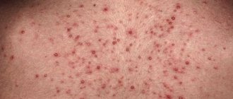
- the appearance of a rash in the body, small fatty nodules on the skin;
- indigestion;
- constant feeling of fatigue.
As organ failure increases, headaches, jaundice, roughness and roughness of the skin are added.
All these symptoms can also occur with other diseases of the internal organs, malignant tumors, so it is so important to respond to signs of stress in a timely manner and consult a doctor.
Diagnostics
A number of laboratory tests are mandatory :
- general and detailed blood tests (especially important indicators for creatinine, urea, bilirubin, alkaline phosphatase, AST, ALAT);
- urine test (general, sterility);
- feces for physical and chemical indicators.
The exclusion of syphilis, AIDS, and viral hepatitis is mandatory. The instrumental methods used include computed tomography and ultrasound.
Liver lipoma on ultrasound
Ultrasound examination of the liver and organs of the hepatobiliary system is an accessible and safe method for assessing the condition of organs, their structure and anatomy.
Ultrasonic fats are thin hypoechoic inclusions in capsules located in the thickness of adipose tissue. If necessary, use Doppler to visualize blood flow and vascular components.
Liver lipoma on CT scan
Computed tomography is a modern diagnostic method based on the ability of tissue to absorb ionizing radiation of varying intensities. In other words, a CT scan is an x-ray image equipped with modern computer programs.
CT helps to determine the size, contours, and structural changes in the liver tissue to answer the question about benign neoplasms and tumors.
If fatty lumps are located deep in the retroperitoneum, a contrast medium is used. With the help of a CT scan, doctors can clearly determine the type of new fat formation.
Note! If the diagnosis is doubtful, they order an MRI and biopsy. The last test is usually performed during surgery. The resulting material is sent for histological examination.
Treatment methods
Oncologists note all neoplasms as a possible precancerous condition and recommend removing changes in the focal tissue, regardless of size and filling.
Surgeons, on the other hand, disagree and use waiting tactics in the absence of positive dynamics of growth, distribution, and the formation of new centers.
Drug therapy
Conservative treatment is aimed at restoring liver tissue and eliminating unpleasant symptoms.
A typical drug therapy regimen includes the following medications: :
- hepatoprotectors (phosphogliv, Berlition, Heptral);
- antioxidants (essence, lipin, glutargin);
- vitamins;
- antiviral drugs (Acyclovir, Ribavirin);
- anti-inflammatory and antibacterial agents for active inflammation;
- choleretic agents (chophytol);
- amino acids.
medications are prescribed as symptomatic treatment and are temporary. In severe cases, therapy is carried out in a hospital setting.
Surgery
Surgical treatment is a promising direction in the treatment of fatty liver tumors. Only radical surgery can help get rid of tumors and liposomes forever. There are two main methods of treatment.
Radio wave method
A minimally invasive procedure in which fat is removed using a special radio wave scalpel. The tumor is affected by radio waves of varying intensities.
You may be interested in: Why do nasal polyps grow - the main causes of polyposis, diagnosis
Advantage of the method :
- Painless,
- High efficiency,
- Target and accuracy.
High-frequency energy provokes a surge of energy inside the cells, evaporates the contents of the capsules, and eliminates neoplasms.
The method is used for individual fats not exceeding 1.5-2 cm. After surgery, the risk of infectious complications is lower, and recovery takes less than a week.
Abdominal surgery
During the manipulation, the liver is removed from the bed, examined, and the fat is dissected along with the capsule in healthy tissue. After the wound, the liver is tightly sutured. The resulting tumor is sent for histological examination.
After each type of operation, dietary correction is necessary; provoking factors must be excluded.
Unfortunately, the risk of relapse reaches 75% with the same lifestyle and bad habits.
Nutrition for liver lipoma
Any treatment for various liver diseases is ineffective without a therapeutic diet. Doctors recommend following diet No. 5 and its variations depending on the current condition.
After the operation, he is strictly limited to salted, smoked, fatty meat, fish and carbohydrate dishes. Complete exclusion of alcoholic beverages, sweet lemonade, and sweets.
Basic nutrition :
- weak lean broth with vegetables, chicken breast;
- fresh fruits and vegetables;
- sour milk and dairy products;
- porridge of buckwheat, rolled oats, oatmeal, rice. Broths made from medicinal plants, rose hips, horses, and compotes are suitable for drinking. All drinks should be consumed without sugar. Food should be fractionated, food should only be stewed or stewed.
In the case of chronic liver failure, the diet lasts a lifetime.Why uterine bleeding occurs during postmenopause, which we explained in a separate article. If it is true that aspirin is used against colon cancer, you can find out here. We immediately warn that uncontrolled use of the drug is unacceptable in this situation.
Complications and prognosis
The main danger of lipomatous nodes on the liver is the risk of developing a malignant tumor and the development of organ failure, but this is only possible under the influence of certain factors:
- injuries,
- exposure to toxins and alcohol.
chronic intoxication,
Adding complications demonstrated :
- sharp stabbing pain,
- vomit,
- nausea,
- excessive weight loss.
Doctor of Medical Sciences, Professor A. Astvatsatryan gives additional information about liver tumors:
The prognosis for lipomatous liver disease largely depends on the compensatory resources of the organ, the general clinical history of the patient and the patient’s desire to change his lifestyle.
Treatment is always complex and is aimed not only at removing the tumor, but also at the possible mechanism of provocation.
Timely diagnosis and adequate treatment ensure a normal standard of living and reduce the risk of developing cancer and liver failure.
A peritoneal lipoma of the anterior abdominal wall has been detected, what to do? Read this article from us.
You can make an appointment directly through our resource.
Be healthy and happy!
Diagnosis of this disease
It is very difficult to detect lipomatosis in the early stages of development, since the disease is hidden. In addition, the symptoms of the pathology are similar to those of other diseases.
To diagnose lipomatosis, the following diagnostic methods are used:
- Ultrasonography. Allows you to identify formations, evaluate their location and size. Using this diagnostic method, lipomatosis can be detected at the earliest stages. This study has no contraindications and is absolutely safe.
- CT scan. It is a more informative diagnostic method, allowing the specialist to obtain the most complete picture of the disease.
- Computed tomography using contrast agent. This technique allows for a deeper study of the structure of the lipoma.
- Biopsy. A tumor tissue sample obtained by biopsy is examined for cytology. This research method is the most accurate.
In addition, the patient is prescribed laboratory tests of blood and urine samples, and a biochemical blood test. The latest analysis allows you to detect diseases such as hepatitis, syphilis, HIV, which are a contraindication to surgery. That is, the treatment method directly depends on the results of blood biochemistry.
How to treat liver lipomatosis?
Description of the disease
Liver lipoma consists of adipose tissue. It rarely produces muscle fibers. The condition is classified according to the number of neoplasias:
- single;
- multiple (liver lipomatosis).
Usually the organ contains adipose tissue, but in small quantities. When lipomatosis develops, its amount exceeds the norm. The doctor talks about obesity in the organ. Often this disease can develop in people who follow a strict diet or are vegetarians.
Depending on the degree of liver depletion, the neoplasm is divided into 2 categories:
- mobile, when pressure is applied and the arm is removed, the lipoma moves and returns to its place;
- stationary, i.e. strongly attached to the parenchyma of the organ, this condition is rare and interferes with the functioning of hepatocytes.
The size of the lipoma can vary. It can vary from a few millimeters to several centimeters. The formation is located on the parenchyma, but does not interfere with neighboring organs and tissues.
Education is good quality. It grows to a certain size, then the growth stops. Hepatocytes retain their structure. Lipomania spreads to neighboring organs and cannot enter the bloodstream.
Causes
Until now, it was assumed that the only cause of liver lipoma was obesity. However, so far doctors have found that this disease often manifests itself in people whose weight is within or below normal weight.
There are several reasons for the formation of adipose tissue:
- heredity, i.e. tendency to form fatty infiltrates in parents and children;
- hormonal disorders, such as changes in the concentration of a substance secreted by the thyroid gland;
- metabolic disorders leading to the deposition of adipose tissue in organs and tissues;
- diabetes mellitus, in which not only glucose, but also lipids actively circulate in the blood;
- disorders of the gastrointestinal tract, in which not all substances are excreted from the body, but remain in the body and are redistributed to various tissues;
- chronic inflammatory liver diseases;
- alcohol abuse, since ethyl alcohol toxically disrupts the functions of many organs and tissues;
- mechanical damage or postoperative complications in the liver;
- chronic inflammatory diseases of the pancreas;
- radiation exposure leading to structural changes in many cells and metabolism.
The doctor must determine the root cause of the disease. If a liver lipoma is simply removed surgically, it can re-form if the cause is not addressed.
Symptoms
If the disease is in the early stages, there are no clinical symptoms. This is the most dangerous for the patient, since he does not visit the doctor. Adipose tissue gradually increases and can impair hepatocyte function. When they reach large sizes, the following clinical symptoms appear:
- dyspeptic symptoms (nausea, vomiting);
- weight loss;
- general signs of discomfort (lethargy, fatigue, weakness, drowsiness);
- protrusion of a neoplasm visible on the skin in the area of the right rib;
- soreness in the area of the right rib;
- darkening of the skin in the neck and armpits;
- unlike inflammatory diseases leading to hepatocytes, the formation of lipomas is not accompanied by yellowing of the skin and sclera of the eye;
- the liver grows moderately, protruding slightly under the right ribs, palpation causes pain.
If the disease is neglected, then in addition to hepatomegaly (increase in the size of the liver), splenomegaly develops (increase in the size of the spleen). Cells from these organs can be released into the bloodstream along with fatty tissue. This form of the disease is called plasma.
Symptoms of lipomatosis and liver lipoma are similar to those of other diseases. In order to distinguish them from each other, it is necessary to diagnose the condition of the internal tissues of a person. Only then can the doctor make a reliable diagnosis and prescribe treatment.
Diagnostics
The patient's condition is diagnosed in several stages.
- Take a medical history. This is data obtained from the words of the patient or his close relatives. Based on these data, the doctor can make a diagnosis and plan further examinations.
- General inspection. The doctor assesses the condition of the skin, mucous membranes, and sclera. In this case, it is difficult to suspect liver pathology. The doctor performs palpation (probing) and percussion (tapping) of the abdominal cavity. If the lipoma is large, it can be easily detected by probing. To confirm this suspicion, the doctor will prescribe additional examinations.
- laboratory tests. The mandatory list includes a general blood and urine test, a biochemical blood test, and a program. An increased level of white blood cells and SOE indicates inflammation of the internal structures of the body, which can develop in the later stages of liver lipoma. Fatty inclusions in the blood can be detected. In addition, doctors prescribe a lipid profile to determine the amount of fat. If there is no hepatitis, then there are no changes in blood biochemistry and co-program.
- X-ray. This test can only detect deposits in the liver, but cannot determine their structure and size.
- Ultrasound. Using this study, you can determine the structure and size of fatty inclusions. Assess the condition of blood vessels and parenchyma.
- Biopsy and subsequent histology. Remove a small piece of parenchyma and lipoma. The doctor will evaluate the composition of the cells and show the presence of normal hepatocytes and fatty inclusions. In this case there is no malignant tumor.
- MRT, CT. Based on the results of the study, the doctor sees the layers of the liver on the screen. Evaluates the quality of nerve fibers and blood vessels. You can see how deep the tumors are.
- MRT and CT are not always prescribed. In most cases, ultrasound is sufficient. After the doctor has made an accurate diagnosis, he prescribes treatment. It is recommended to use diagnostic methods during and after treatment. This allows you to estimate the rate of removal of pathological fatty inclusions. If a method does not work, it is replaced by another.
Treatment
To prevent the development of fatty tumors, it is recommended to carry out complex therapy consisting of several methods:
- Diet. Fatty, spicy, salty, smoked, fried foods are completely removed from the diet. A person can eat cereals, low-fat soups, boiled meat, fish, dairy products, vegetables, and fruits. You need to eat 5-6 times a day. If food is divided into small portions, it facilitates the digestion process, which is often affected by metabolic disorders.
- I take vitamins. For the proper functioning of all organs and systems, the human body must be provided with vitamins, amino acids, minerals, and trace elements. For the initial and intermediate stages of the disease they can be used in the form of tablets. If the human body is severely depleted and the digestion process is disrupted, drug treatment can be prescribed using infusion.
- Hepatoprotectors. These are substances that eliminate the possibility of damage to hepatocytes. This is done using phospholipid complexes embedded in their membrane. It becomes strong and impermeable to toxic substances.
- antioxidants. These are drugs that help eliminate cell destruction when exposed to free radicals and oxygen complexes.
- Preparations for milk thistle spots, prescribed in the form of oils, tablets. This herbal remedy protects hepatocytes from damage and stimulates the process of regeneration of new cells.
- Eliminates the root cause. This includes comprehensive treatment of obesity, metabolic disorders, and hormonal changes. It is recommended to stop drinking alcohol completely. If he has become a drug addict, he must go to a drug treatment clinic.
- Enzymes are prescribed for digestive disorders.
You may be interested in: Diet for gastric polyps - the basics of therapeutic nutrition
If the fatty formation is large enough, the doctor recommends a surgical method. Laparoscopy can be used for this, i.e. lipoma removal with a small incision in the abdominal cavity. After surgery, a person needs little time to recover. The fat in the liver is completely removed along with the capsule.
If surgery is performed, but the cause is not eliminated, lipomas and lipomatosis appear again. Self-medication is excluded. This can lead to irreparable complications and worsening of the patient's condition.
Complications
If a person has a liver lipoma, treatment is not carried out, this can lead to irreparable complications for the body. The formation gradually increases in size and causes the following pathologies:
- damage and necrosis of healthy hepatocytes;
- vasoconstriction, leading to interruption of blood flow and parenchymatrophy;
- compression of nerve endings, causing acute pain;
- vascular damage, minor and major hemorrhages;
- hepatitis, which can cause hepatitis;
- cellular malignant tumor (liposarcoma), spread of metastases to neighboring tissues and organs;
- hernia formation.
To avoid causing the above complications, it is recommended to start treatment immediately after diagnosis.
Forecast
The sooner the patient sees a doctor, the better the result. If treatment is carried out on time, it gives a positive result. Complications include bleeding and compression of nerve tissue, which can lead to death.
Prevention
This is recommended for the prevention of lipoma and liver lipomatosis:
- proper nutrition with all foods containing nutrients, vitamins, amino acids and minerals;
- timely treatment of all systemic diseases;
- periodic examination by a therapist who conducts all laboratory tests to identify various conditions and serious diseases;
- Persons with metabolic or hormonal disorders are advised to visit a doctor at least once every 6 months.
Liver lipoma is a disease that must be treated promptly to prevent complications. It is recommended not to carry out self-therapy, as this may lead to a worsening of the patient's condition. If liver lipids are removed in time, it guarantees a favorable outcome.
Causes of liver lipidoma and ways to control neoplasms Link to main publication
Pathology therapy
If suspicious symptoms are detected, the patient should consult a physician. The doctor will prescribe the necessary examination and, if necessary, recommend consultation with specialists.
The causes and treatment of lipomatosis are interrelated.
Therapy for the disease must be comprehensive. The patient is recommended to undergo surgery, use medications and lifestyle changes. With the help of surgery, the formation is removed, and medications can stop the further development of the pathology. The doctor must make a decision about taking medications based on information about the stage, shape, nature, and location of the tumor.
With the help of medications, you can prevent the occurrence of a new lipoma, protect hepatocytes from damage, restore their functionality, and support the liver. During therapy, the use of hepatoprotective drugs, antiviral agents, immunomodulators, vitamins, and antibacterial medications is recommended. Antioxidants, multivitamin complexes, amino acids, and herbal extracts can be used as supportive treatment.
Diagnostics
Diagnosing multiple lipomas is not difficult. Usually, a routine examination at an appointment is enough for the doctor to do this. The doctor clarifies the patient’s complaints, specifies the duration of the disease, and family history. It also turns out that there are concomitant pathologies and allergies to medications (this is taken into account if surgery is required). Then the doctor examines and palpates the location of the formations. Usually this is enough to make the correct diagnosis. In doubtful cases, an ultrasound may be performed.
Choice of drugs for lipomatosis
When choosing medications, the doctor takes into account the suspected cause of lipomatosis (diffuse changes in the liver). For example, if the disease is caused by alcohol damage, it is indicated to prescribe lipoic acid, which can accelerate the cleansing of ethanol from tissues and neutralize its effects. If lipomatosis is at the third stage of development, it is indicated to take amino acids. In order to restore liver structures, it is recommended to use vitamins, essential phospholipids, antioxidants and medications based on milk thistle extract.
It should be noted that tumor cells can remain in the patient’s liver throughout his life. However, if they do not cause discomfort, surgery may not be performed.
Wen treatment
Lipoma therapy involves surgery. Traditional and folk medicine in this case will not be effective and will not cure pathological changes in the liver. After a thorough diagnosis, surgery is performed. It is performed in hospital under general anesthesia. If the tumor is small, the radio wave method is used. With its help, it is possible not to injure other organs of the gastrointestinal tract. Basically, the scalpel does not come into contact with the skin and does not destroy fat cells. If the changed adipose tissue is large, then direct surgery is used. The tissue is cut and the tumor is removed, then sutured. Rehabilitation after such an operation lasts 2-3 months. At the same time, you need to adhere to proper nutrition and regularly come for examinations.
Surgical treatment of this disease
You can get rid of lipomatosis of the liver and pancreas only through surgery. If necessary, the surgeon also removes adjacent tissues that are affected.
The radio wave method can be used to remove small tumors. This method is safer, since the patient does not lose blood during the procedure, and the likelihood of injury or complications is minimal. The formation is removed using a radio wave scalpel.
If the tumor is large, its removal is only possible through standard surgery. During the intervention, local or general anesthesia can be used. The type of anesthesia is determined taking into account the patient’s age and the severity of the disease.
Treatment
Treatment of lipomatosis is carried out in three directions:
- surgical removal;
- pharmacological treatment;
- lifestyle correction.
The method is chosen individually, taking into account the type of disease, size and prevalence of lipomas.
The main method of treating lipomatosis is surgical excision of subcutaneous formations, although it does not exclude relapses. Superficially located small lipomas are removed using a minimally invasive method - through a small puncture using a laser or cryodestruction. This type of surgery is performed on an outpatient basis under local anesthesia.
READ ALSO: What to do if a man gets pimples on his scalp?
Symptomatic therapy is aimed at reducing pain and combating disorders of the gastrointestinal tract. For lipomatosis caused by hypofunction of the endocrine glands, hormone replacement therapy is prescribed.
The structure of a lipoma is identical to the structure of mature adipose tissue: it has a lobular structure, each lobule consists of specialized cells with high cellular activity - adipocytes.
Following a therapeutic diet aimed at normalizing fat metabolism will help slow down the progression of the pathological process. A split diet, a diet low in high-calorie foods, especially fatty and sweet foods, and moderate but regular physical activity are recommended.
Nutrition rules for this pathology
The most important point of complex therapy is following a diet and giving up harmful addictions. With proper nutrition, the load on the damaged organ is reduced, the body is saturated with useful substances.
During therapy for hepatic lipomatosis, the patient's diet should be rich in the following foods and dishes:
- Rice, oatmeal, buckwheat, cooked in water.
- Dairy products, low fat milk.
- Fish, lean meat, steamed.
- Fresh fruits, vegetables.
- Soups based on vegetable broth and without frying.
It is important to refrain from eating foods rich in fats and carbohydrates. The patient is also advised to avoid spicy, fatty foods, coffee, and chocolate.
An essential condition for successful treatment of lipomatosis is abstinence from alcoholic beverages. Harmful addictions (drug addiction, tobacco smoking, alcohol) will only accelerate the occurrence of complications.
Possible consequences of the disease
Most often, the consequences arising from lipomatosis are minimal. However, atypical lipomatosis increases the likelihood of developing such dangerous pathologies:
- If the disease has a protracted course, the wen can degenerate into liposarcoma (malignant tumors). Quite often this occurs as a result of injury, bruise, or damage. If you contact a doctor late, a malignant tumor can develop metastases (secondary tumor foci).
- Large fatty formations can compress neighboring tissues, disrupting nerve-fiber conduction and blood circulation. Such a violation threatens the occurrence of paroxysmal pain, paralysis, and paresis.
- Under the influence of certain factors, an inflammation process occurs, against which the risk of developing hernias and exacerbation of pathologies that have a chronic course increases. In this case, the patient needs more radical and long-term therapy.
Possible complications
Despite the fact that lipoma is a benign formation, there is a risk of its development into oncology. In addition, as its size increases, it can affect surrounding tissues and organs. This leads to necrosis, liposarcoma, or rupture of one of the organs of the gastrointestinal tract. Signs of the pathological condition are:
- pain when palpating the abdominal area;
- compaction in the liver area;
- nausea and gag reflex after eating;
- stitching pain under the ribs;
- rapid loss of body weight.
Return to contents