Main symptoms:
- Ocular hemorrhages
- Blurred vision
- Veil before my eyes
- Appearance of dark spots in the field of vision
- Decreased vision
- Difficulty reading small text
Diabetic retinopathy is a disease characterized by damage to the vessels of the retina and impaired visual perception of the cornea. The pathology develops against the background of insulin-dependent and non-insulin-dependent forms of diabetes mellitus. As a result of its progression, visual function is significantly reduced, up to complete loss of vision (without the absence of timely treatment).
Online consultation on the disease “Diabetic retinopathy”.
Ask a question to the specialists for free: Endocrinologist.
- Etiology
- Mechanism
- Stages
- Symptoms
- Diagnostics
- Complications
- Treatment
- Prevention
To save vision in the presence of diabetic retinopathy, invasive interventions are used. Burning the retina with a laser is highly effective, as is complete removal of the vitreous (this technique is used in difficult situations).
Etiology
The main reason for the progression of diabetic retinopathy is the presence of long-term diabetes mellitus. Medical statistics are such that the pathology is diagnosed in 15 out of 100 people with diabetes mellitus that has lasted for two years. If the duration of the disease is 15 years, then retinopathy is diagnosed in 50% of patients.
Factors that increase the risk of diabetic retinopathy:
- obesity;
- renal failure;
- arterial hypertension;
- vascular atherosclerosis.
Factors that are “triggering” for the progression of the disease:
- adolescence;
- smoking;
- pregnancy (only if treatment for diabetes mellitus was started during the period of pregnancy);
- genetic predisposition.
Causes
The pathology develops against the background of long-term diabetes mellitus. The following factors are important in the development of complications:
- glucose level;
- duration of the disease;
- heredity;
- presence of concomitant pathology;
- pregnancy.
When diabetes lasts less than two years, initial signs of retinopathy are diagnosed in 15% of patients. With a disease that lasts more than 20 years, changes in the fundus of the eye occur in every person.
The risk of retinopathy increases over time
Development mechanism
Retinopathy is a lesion of the retina, located on the fundus of the eye and providing a person with vision. The retina is nourished by small vessels that approach it along its entire length. The pathogenesis of diabetic retinopathy is a change in the condition of the vessels that supply the retina.
In the presence of risk factors, these vessels are damaged and they stop supplying blood to the retina. The process leads to its swelling and gradual destruction until complete loss of vision.
Retinopathy is characterized by changes in the structure of blood vessels
Mechanism of disease progression
Diabetic retinopathy
Diabetic retinopathy begins to gradually develop due to the increased concentration of sugar in the vessels of the retina. As a result, the vascular wall is damaged. Most often, small-caliber vessels that provide nutrition to the retina are attacked. Hyperglycemia leads to:
- when the walls of blood vessels are damaged, a violation of blood microcirculation is observed;
- a certain amount of blood that comes out of damaged vessels permeates nearby tissues;
- due to the fact that the retina ceases to be fully “nourished”, new blood vessels are formed, which are also destroyed (proliferative diabetic retinopathy);
- blocked vessels grow with connective tissue.
Microangiopathy in diabetes
In patients with diabetes, damage to medium vessels (macroangiopathy) and small capillaries (microangiopathy) is possible. Considering the widespread prevalence of metabolic conditions and insulin metabolism disorders (up to 15% of the world's population), the risk of an increase in dangerous vascular pathology is extremely high. One of the first doctors to notice diabetic retinopathy is an ophthalmologist.
The fundus is an ideal place to identify primary capillary disorders. The retina is provided with a large number of small vessels, from changes in which the doctor will quickly and reliably see the initial manifestations of the disease. The pathogenesis of diabetic retinopathy is characterized by the following factors:
- changes in the structure and thickening of the microvascular bed;
- proliferation of capillary endothelial cells;
- slowing blood flow in the retina;
- accumulation of protein substances inside capillaries;
- local expansion of microvessels;
- retinal ischemia;
- formation of new capillaries and veins.
Diabetic angioretinopathy progresses much faster with combined metabolic syndrome, when, in addition to diabetes, the patient suffers from arterial hypertension and obesity.
Stages
Non-proliferative. This stage is the initial stage in the progression of diabetic retinopathy. During the non-proliferative stage, small-caliber blood vessels (local) begin to dilate in the retina. This process is medically called microaneurysm. As a result, hemorrhages occur in the surrounding tissues. The retina is also impregnated with plasma. As a result, it swells along larger vessels.
At the non-proliferative stage, treatment is simpler. If it is started in a timely manner, there is a high probability of complete preservation of visual function. Treatment of the non-proliferative stage is prescribed only by a qualified ophthalmologist, together with an endocrinologist.
Preproliferative stage (progressive). If it develops, damage to the veins that carry blood from the retina is observed. Limited areas of pathological expansion, loops, and duplications appear in them. During the preproliferative stage, hemorrhages in tissues increase more and more.
Proliferative diabetic retinopathy. Its characteristic manifestation is the ingrowth of new blood vessels into various parts of the retina. They can be localized in the area of the visual spot. Many areas of the vitreous body are saturated with plasma that comes out of damaged blood vessels. Due to the fact that the newly formed vessels have fragile walls, microaneurysms appear in them again, provoking new hemorrhages, which can cause detachment of the retina itself. Proliferative diabetic retinopathy often causes vision loss.
Stages of diabetic retinopathy
Pathogenesis
The pathogenesis of diabetic retinopathy is accompanied by serious disorders in the retinal vessels.
In diabetic retinopathy, the structure of the capillaries is more affected. During the pathological process, changes in the properties of blood vessels and capillaries, loss of pericytes are observed, the structure of the basement membrane becomes thinner, and damage and proliferation of endothelial cells also occurs. During disorders of a hematological nature, a process of deformation and intensification of the formation of the “coin columns” sign is noted. Additionally, platelet flexibility and aggregation are reduced, which lead to a decrease in the oxygen transport process.
In response to these negative symptoms, the body begins to grow new capillaries; this is required to restore blood flow to the eye tissues. This process is called proliferation. At this time, destruction of small vessels occurs, which causes fluid and blood to leak into the retinal area. As a result, a process of swelling of the nerve endings occurs, followed by their swelling, and ultimately a spot with a yellow tint is formed.
It would seem that what’s so bad about this is that the vessels collapsed, and new ones formed in their place. But, in fact, new vessels have an imperfect and defective structure; they cannot fully supply the eyes with blood, oxygen and useful elements. The formed vessels have fragile walls, which is why frequent hemorrhages on the retina occur, as well as clots and fibrous tissue, scars and cicatrices on the retina.
Symptoms
The biggest danger of diabetic retinopathy is that it can occur without a single symptom for a long period of time. At the first stage of diabetic retinopathy, the decrease in visual function is so slight that the patient does not notice it at all. As the pathology progresses, the following symptoms appear:
- gradual decrease in visual function;
- objects that are located in close proximity to a person begin to appear blurry to him (a characteristic symptom);
- Difficulties arise when reading small text.
The proliferative stage is supplemented by the following symptoms:
- the decline in visual function progresses;
- dark spots or a veil appear before the eyes. This symptom indicates the presence of intraocular hemorrhages. But it is worth noting that they can disappear on their own.
If, in the event of such symptoms, comprehensive diagnosis and treatment has not been carried out, then complete loss of vision may occur without the possibility of recovery in the future. The danger also lies in the fact that the symptoms of this pathology are very scarce and may indicate the progression of any other eye pathologies. Therefore, people who have previously been diagnosed with diabetes should be regularly examined not only by an endocrinologist, but also by an ophthalmologist.
Preventive actions
Prevention of diabetic retinopathy in both eyes consists of maintaining an optimal balance of sugars in the body, following a diet and optimizing carbohydrate metabolism, normalizing blood pressure and correcting lipid metabolism. These measures will help reduce the risk of ophthalmic pathologies and reduce the risk of accelerating complications if they exist.
Maintaining proper nutrition and systematic physical activity can improve the health of people with diabetes. It is equally important to undergo a timely examination by an ophthalmologist.
Timely preventive measures can prevent eye damage in people with a pathological condition called diabetes mellitus. This is due to the fact that in the last stages of the progression of the anomaly, therapy is not as effective as it should be. And since it is not possible to determine retinopathy in the early stages without a systematic visit to the ophthalmologist, patients feel pathological processes when irreversible consequences occur.
Diagnostics
The most informative methods for diagnosing the disease are:
- ophthalmoscopy;
- fluorescein angiography;
- slit lamp examination.
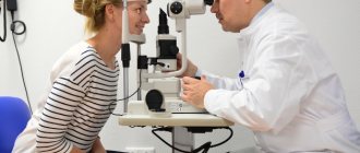
A slit lamp examination using a special lens allows the doctor to accurately diagnose the presence of pathology even at an early stage of development (detection of retinal edema).
With the help of ophthalmoscopy, it is possible to detect the presence of microaneurysms at the initial stage of the pathology. At the second stage, during the examination, the doctor sees small whitish foci, stripes, deformation of the veins of the fundus and already formed foci of infarction in the fundus. During the proliferative stage, ophthalmoscopy can reveal newly formed vessels and clarify their location.
Additional examination techniques:
- visometry;
- perimetry;
- intraocular pressure tonometry;
- electrooculography;
- Ultrasound of the eye;
- electroretinography;
- gonioscopy;
- diaphanoscopy.
Hemophthalmos
Classification of retinopathy
There are three stages of development of this late complication of diabetes:
- non-proliferative or background - stage I
This is the very first stage of retinopathy, which is typically characterized by increased permeability of small vessels of the retina, which is otherwise called microvascular angiopathy.
At this stage, with a long-term course of the disease, it is quite natural that the process of disease progression occurs in the complete absence of any visual impairment. The patient does not notice any obvious symptoms and signs, but the ophthalmologist can observe a completely typical picture: pathological changes in the retina in the form of microaneurysms, hemorrhages, retinal edema, and exudative lesions.
When examining the fundus, small dots or small spots of yellow or white color, often with unclear boundaries, may be noticeable. A slight swelling of the retina may be noticeable in its macular zone (in the central part) or in any area of the large eye vessels.
- preproliferative - stage II
This is the second, one might say, transitional stage of retinopathy.
In this case, the vascular chain of the eye is more distorted and tortuous. In some places, the vessels can be greatly dilated (they seem to swell, swell) while the rest of the vessel remains within acceptable limits. The vessel begins to resemble a thread consisting of small “knots”. These are the so-called loops.
- proliferative – stage III
After preproliferative retinopathy, the third stage begins, when the vessels distorted in this way cease to properly perform their functions.
Blood passes through such crooked tunnels with great difficulty, creating an even greater load on the damaged eye vessels, and this directly indicates that the tissues that depend on them begin to experience hunger. Blood saturated with oxygen and nutrients cannot “break through” to the cells.
This is how retinal nonperfusion is diagnosed (in fact, this is a violation of the blood supply).
If some part of the retina begins to starve, a protective mechanism is activated, resulting in the release of special vasoproliferative substances. They are able to start the process of regeneration, formation, and growth of new blood vessels. This process is called neovascularization.
But the vessel, which has already ceased to perform its function, begins to heal together with the adjacent tissue, which does not receive proper nutrition. This is called fibrosis (overgrowth of connective tissue with scarring).
This is how proliferation starts - the growth of tissue due to cell division.
This process is extremely chaotic, as it is implemented in order to quickly eliminate localization in those places where the blood supply was disrupted and cells began to die.
In this way, the body tries to save the organ from complete “death.” But this can only aggravate the situation and completely deprive a person of vision, since tissue growth triggers the progression of retinopathy, with frequent hemorrhages (vessels become incompetent and cannot replace previously existing ones) and vitreous detachment.
This happens because the newly formed vessels begin to grow along the posterior wall of the vitreous and seem to “displace” it.
Despite such a terrible picture, patients’ vision may deteriorate slightly, since the processes described above proceed extremely slowly (it all depends on the state of health and metabolism of the person). For some people it lasts for 3 or 4 months, for others it lasts for several years.
In this case, a person often consults a doctor as a result of a fall or deterioration of vision in one eye, but in fact, retinopathy progresses in both eyes, the process just progresses a little faster in one of them.
Even if the body quickly patches up the gap, it will not complete its mission. Therefore, fibrosis does not stop on its own, and the patient’s visual acuity (his sensations) do not reflect the seriousness of the situation. The degree of fibrosis determines the feasibility of surgical intervention when deciding whether to refer the patient for laser photocoagulation of the retina.
- diabetic macular edema
This cannot be called a separate degree, but rather an additional factor in proliferative (much more common) or non-proliferative diabetic retinopathy (less common).
This complication leads to blindness, but in the early stages of its manifestation, vision may not deteriorate. Often, at first the patient begins to have difficulty distinguishing small objects or it becomes extremely difficult for him to read, and only after that his vision deteriorates so much that even objects close to him become blurry and indistinct.
Treatment
Treatment of diabetic retinopathy directly depends on what stage of the pathology was diagnosed in a person.
- at stage 1, therapy is usually not prescribed. The doctor prescribes the patient to continue to take glucose-lowering medications, as well as to regularly come for examinations to an ophthalmologist in order to prevent further progression of the pathology;
- the second stage is more dangerous. The patient's condition must be constantly monitored, as this stage can quickly progress to the proliferation stage. The doctor needs to constantly monitor the concentration of glucose in the blood. Therapy includes drugs that help eliminate metabolic disorders. The introduction of special agents directly into the vitreous body is also indicated.
- in the proliferation stage, the only correct method of treatment is the use of an argon laser.
Methods for eliminating the disease:
- diet therapy. The patient's diet is limited to simple carbohydrates and animal fats. At the same time, it must include oatmeal, vegetables, and fruits;
- the endocrinologist develops a specific individual treatment regimen with glucose-lowering drugs;
- vitamin therapy;
- to reduce retinal edema, glucocorticoid hormones are injected directly into the vitreous;
- laser coagulation;
- cold exposure – cryoretinopexy;
- partial extraction of the vitreous;
- removal of the vitreous.


