What is a cataract?
Cataract is a pathological process characterized by clouding of the lens of the eye, which leads to partial or complete loss of vision. The main symptom of cataracts is decreased visual acuity, and depending on the location of the pathological process, myopia or farsightedness may develop.
The most common causes of cataracts are injuries, the presence of certain diseases (for example, glaucoma, diabetes), radiation and other unfavorable factors, for example, age - about 90% of all cases of the disease are observed in the elderly.
The disease cataract got its name from the ancient Greek “καταρράκτης”, which translated means “waterfall, splashes of a waterfall.”
To learn more about this disease, consider the following point.
Development of cataracts
The lens is a transparent, round, elastic body that is located inside the eyeball, opposite the pupil, between the iris and the vitreous body. The lens is formed from elongated epithelial cells, and its substance is made from a mixture of proteins (crystallin). Nutrition occurs through the vitreous body and aqueous humor.
In normal condition and health, the lens is absolutely transparent.
The function of the lens is to transmit and refract light towards the retina. In fact, it is a natural lens.
Cataract is a pathological process in which, due to certain factors, the lens begins to become cloudy, i.e. lose its transparency. In this case, the retina of the eye does not receive enough of the necessary light, and accordingly cannot sufficiently evaluate what the eye sees.
The retina of the eye is the converter of the image visible to the eye and its transformation into a nerve impulse, which then enters the brain, where the residual formation of what we see occurs.
Other common diseases associated with the lens are myopia, farsightedness, and astigmatism.
Cataract is a partial or complete clouding of the lens of the eye, located inside the eyeball between the iris and the vitreous body. The lens is naturally transparent and acts as a natural lens that refracts light rays and passes them to the retina. When a cataract has lost its transparency, the lens stops transmitting light and vision deteriorates until it is completely lost.
Let's consider the development of cataracts in a classic form, using the example of age-related cataracts. In this case, the development of the disease occurs over 5 stages (periods):
Stage 1 - anterior and posterior capsular cataracts; Stage 2 - perinuclear layered cataract; Stage 3 - nuclear cataract; Stage 4 - cortical cataract; Stage 5 - complete cataract.
In the following picture, all five stages of the disease are perfectly illustrated:
The period of maturation of cataracts from the initial to the last stage, depending on the actions taken by the patient, ranges from 4 to 15 years, after which vision can only be restored with the help of surgical treatment, unless of course the Lord helps the person.
Spread of cataracts
According to statistics from the World Health Organization (WHO), there are about 19 million people in the world who are blind from cataracts, which accounts for about 50% of all cases of blindness. Cataracts are most common in the elderly, which is why a separate type of disease has been classified as “senile cataract.” Thus, it is noted that in many elderly people, over 60 years of age, the transparency of the lens decreases by 50%, and at the age of 80 years, by about 90%.
Cataract - ICD
ICD-10: H25-H26, H28, Q12.0; ICD-9: 366.
Certain forms of the disease
Congenital cataract
The definition of congenital cataract refers to all forms of cataracts that already exist in a newborn and are either a consequence of an abnormal development of the lens, or are formed as a result of diseases that can occur during intrauterine life; further, all forms of partial and stationary cataracts, the moment of formation of which cannot yet be established with certainty, but which are probably congenital.
a) Axial cataract
• Central cataract
Central cataract is an opacification of the lens the size of a poppy seed, bright white, roundish, located in the nucleus, almost always bilateral, stationary and, undoubtedly, congenital; in most cases it does not interfere with vision, sometimes occurs together with other anomalies of eye development and is then accompanied by nystagmus. The reason for this can only be an abnormal development of the lens.
• Anterior central bursa cataract
A round, sometimes star-shaped, white clouding in the area of the anterior pole of the lens, ranging in size from a point to 2-3 mm in diameter, sometimes flat, sometimes slightly rising above the level of the bag, sometimes protruding into the anterior chamber in the form of a spike. Sometimes the same, somewhat smaller and grayer round opacification protrudes into the lens masses, and it is separated from the first opacification by a clear notch, but in other parts it is connected to it, like two plates of a cufflink.
The main changes are localized in the intrabursal cells and adjacent lens fibers. With pyramidal cataracts, there is no epithelium of the anterior bursa to the point where the cataractous opacification reaches, the apex of the cone is covered with coagulated tissue fluid, then there is hard, cartilage-like fibrous tissue that can be split into thin plates. Central bursa cataract is congenital or develops in the first weeks of life; in the latter case, a connection in the form of a cord is sometimes visible between the apex of the cone and the center of the cornea, which is slightly clouded in this place.
Visual acuity sometimes (with small such opacities) is almost not reduced, but with more significant opacities, especially with strong formation of folds of the bag or with the existence of other opacities of the lens and cornea, and finally, in the presence of developmental anomalies, it may be greatly reduced, as indicated by nystagmus , extremely common in anterior polar cataracts.
• Posterior central bursa cataract
It also appears in the form of a roundish, bright white, small opacity of the lens lying on the axis of the lens, which, with transmitted light, protrudes cone-shaped into the vitreous body, sometimes connecting with the remnants of the hyaloid artery. She is always stationary. It can be mixed with cortical opacities at the posterior pole, especially common in retinitis pigmentosa; but in such opacities radial stripes are often noticed and they do not have such a smooth surface; In addition, they are not stationary, but increase over time.
b) Other forms of partial congenital cataracts
This includes limited opacities, which are sometimes found in various parts of the lens in the form of dots, membranes, stripes, etc., always remain stationary and in most cases interfere with vision. Particularly noteworthy is the form called pinpoint cataract, since it is far from rare, but is often visible due to the absence of visual impairment. With it, very small pinpoint opacities are scattered throughout the lens substance, located more densely in the equator region. In strong light, the cloudiness appears slightly bluish.
c) Layered cataract
It appears as a central, lying behind the pupil, completely round, 4-8 mm in diameter, soft gray and translucent, or a more intense and then more whitish opacification, which, both in incident and transmitted light, is thicker at the edges than in the center. It is sharply demarcated from the completely pure cortex and consists of either tiny dots or radial, more whitish stripes with a transparent intermediate substance. Through the unclouded center of the lens, you can see the rear, also clouded layer.
The sections show a completely transparent core surrounded by a narrow opacified layer, which in turn is surrounded on all sides by completely transparent cortical masses. Sometimes in the equatorial region there are short strips converging at the equator at an angle and called “riders” due to the fact that they seem to sit astride the edge of the layered cataract. These stripes represent the rudiments of a double layered cataract, which is very rare and can only be recognized when it is incomplete or when the outer clouded layer is still very translucent. Triple, although incomplete, layered cataracts were also often observed.
This form of cataract is in the vast majority of cases stationary; opacification extremely rarely progresses and ultimately leads to complete cataracts. Some congenital wrinkled cataracts are explained as progressive stratified cataracts. Sometimes radial stripes of opacities are observed emanating from the edge of a layered cataract, and such layered cataracts are considered progressive.
Layered cataracts almost always occur in both eyes; rarely, they occur in only one eye. Vision is usually slightly weakened, but sometimes severely; this depends not so much on the diameter as on the density of the cloudy layer, since even a very small layered cataract with a narrow pupil can occupy the entire pupillary area of the lens. Sometimes vision is so good that the defect remains undetected until old age.
In most people suffering from layered cataracts, in addition to weakness of vision, myopia is observed, often only imaginary; but many of them are indeed nearsighted and suffer from astigmatism.
d) Complete congenital cataract
It occurs in two forms, soft and wrinkled cataracts.
Soft congenital cataracts are no different from acquired soft cataracts of young subjects, both in their appearance and in the nature of their course; it should only be noted that the development and changes of the latter occur in a much shorter time than with soft cataracts of older persons.
In etiological terms, the importance of heredity is firmly established; observed families in which all children were born with complete cataracts. There were no other eye diseases or congenital general sufferings to be noted; young patients are usually strong and healthy children. This form of congenital cataract does not usually cause nystagmus.
Wrinkled congenital cataracts usually occur in two forms: either in the form of thin-walled, often pigmented and connected to the iris formations, or in the form of flat, yellowish-gray cataracts with a wrinkled surface, significantly reduced in all sizes, especially along the axis; they are also in most cases in connection with the iris by means of posterior synechiae. They occur either due to the absorption of intrauterine complete cataracts formed, or due to a delay in the normal development of the lens. The latter method of their formation is evidenced by the fact that in such wrinkled cataracts the bag and its epithelium are preserved and embryonic lens fibers are found in them, the direct course of which gives reason to assume a very early stop of normal development.
Soft cataract of the young
Cataracts are always mild if they develop before a nucleus forms in the lens. Anatomically, this cataract differs from senile cataract only in that with it the nucleus always becomes cloudy, while in senile cataract sclerosis protects the nucleus from cataractous decay. The formation of cataracts is also preceded by the appearance of cracks in the layers of the lens fibers, as a result of which the lens star is visible here as clearly as in the eyes of older people. The opacities may first develop at the equator, in the center of the lens, or in the posterior cortex, or may begin as isolated opacities in different parts of the lens. If the opacification first appears in the posterior cortex, the cataract is always sequential (choroidal cataract).
Young age, the presence of deep clefts, significant width of the radial stripes, whitish color, rapid development, severe swelling of the cataract, which causes the appearance of a wide pigment border on the pupillary edge - all this taken together allows us to reliably diagnose soft cataracts.
The outcomes of this form of cataract are wrinkling, liquefaction and calcification.
Senile cataract
The opacification does not seem to begin at the equator of the lens, but in the vast majority of cases in the outermost, subsurface layer, in which the first microscopic changes can also be noted.
From a clinical point of view, it is necessary to distinguish 4 stages during the course of cataracts: the stage of onset, swelling, maturity and overmaturity.
In the first stage of senile cataracts, sometimes individual, wide or numerous narrow radial stripes are found, which face the equator with their base, go wedge-shaped to the center and cause visual disturbances only in cases where the apexes reach the pupil area; sometimes they encounter cloudy or membranous opacities, which in the first case lies in the deep, and in the second - in the superficial layers and covers only certain parts of the pupil area; finally, in other parts there is a smoky or cloudy diffuse turbidity located around the core with complete transparency of the peripheral layers.
After clouding appears, the lens fibers begin to swell due to impregnation with water, and then the second stage of senile cataract begins. The entire lens increases in volume and pushes the iris forward, and the anterior chamber becomes smaller and the amount of aqueous humor in it decreases. As a result, the front bag acquires a silky sheen reminiscent of moiré; the anterior stellate figure of the lens clearly protrudes. Impregnation with water and the resulting swelling of the lens, however, varies extremely in degree. In general, the swelling decreases the further the sclerosis of the lens has progressed, and therefore the older the subject suffering from cataracts. In particular, cataracts that begin with wide slits and stripes are more prone, and cataracts with delicate, thin stripes are less prone to swelling; more smoky opacities swell more than superficial, membranous ones; Those cataracts that grow least in volume are those which arise from the circumference of the nucleus, and the cataract known as black cataract, in which a very thin cloudy cortex surrounds the large nucleus, does not increase in volume at all.
Swelling cataracts are called cataracta nondum matura, from the point of view of the period favorable for the operation. This name is also used when the volume of the lens begins to decrease due to the release of water and protein to the surrounding parts. That the cloudiness has not yet reached the anterior bursa is judged by the shadow cast by the iris on the clouded parts. Namely, if you look at the eye from the side in lateral lighting, then in such cases a crescent shadow is visible between the opposite edge of the pupil and the clouding, the wider the further the clouding is from the bag, therefore, the deeper it lies. Those cataracts, the opacification of which progresses slowly, in which the swelling is very slight, also give such a shadow; There are even cataracts that in this sense never become mature, since in them the anterior cortical layers never become completely cloudy, during surgery they easily get stuck in the pouch and adversely affect the result of the operation.
Usually, however, the maturity of the cataract (the third stage of senile cataract) ends with the return of the lens to its normal volume. The shape of the crystalline star usually varies. At the same time, the connection between the bag and the lens substance is loosened, so that the cataract comes out of the bag easily.
If by the time of maturity the cataract has not been operated on, then a further change occurs, the essence of which lies in the regressive metamorphosis of the disintegrated lens fibers. After further release of water and protein, the fibers are crushed and condensed into a pulp, which consists of the breakdown of fat, carbonic and phosphate of lime and cholesterol, and ultimately fuses with the sclerotic core into a dense mass and then represents what is called overripe cataract, cataracta hypermatura (fourth stage ). In this case, it may happen that the cataract itself or from a minor injury falls into the vitreous body, and, as a result, vision is restored again, as after a successful operation. The state of overmaturity is recognized by the anterior chamber, which is somewhat deepened in most cases, by intensely white stripes and dots in the anterior corticalis, located in very different directions to the course of the lens fibers, which is why they differ significantly from the primary stripes of opacification, which somewhat correspond to the star pattern of the lens.
In other cases, the release of water stops and thus the cataract mass remains liquefied and turns into a cloudy liquid. Depending on the liquefaction, two forms are obtained, of which one represents a cataract with a whiter, extremely irregular pattern, while the second, with a dilated pupil, reveals different colors in the upper and lower parts and a complete absence of pattern. It is while the upper part of the cataract consists only of a completely liquefied mass, that the brown-yellow core, which has sunk down into the liquid, is adjacent to the capsule in the lower part. Movements of the head cause a change in the position of the nucleus, which is easily movable in the liquid corticalis, and, as a result, changes in color are observed, so that with the usual position and the head tilted back, the cataract appears white and homogeneous, and when the head is tilted forward, a dark brown nucleus becomes visible in the pupil.
The disintegration of the lens fibers and their regressive metamorphosis give a strong impetus to the proliferation of bursa cells. Therefore, overmature cataracts with prolonged existence are almost always accompanied by anterior bursa cataract. In cases where these opacities are too faint to be seen with the naked eye or conventional assistive instruments, they can be determined by microscopic examination. With overripe lens opacities complicated by bursa cataracts, it is often observed that due to wrinkling, the ligament of Zinn ruptures and then the lens falls into the lower parts of the vitreous.
Diabetic cataract
It has long been a known fact that diabetics develop cataracts relatively often. Diabetic cataracts occur at any age, but are observed primarily in people under 40 years of age. Essentially, mature diabetic cataracts are no different from non-diabetic cataracts; in young subjects it is soft, in older subjects, in whom the nucleus has already differentiated, only the corticalis softens. Of course, when cataracts form in young subjects, a urine test should be performed so as not to detect possible diabetes.
Bursa cataract
Bursa cataract occurs not only during lens cataracts, but also primarily at any age without lens opacification; but still the latter soon joins.
Bursal cataract, which always indicates that the nutrition of the eye has been affected, consists of proliferation and opacification of intrabursal cells, and, moreover, mainly of the so-called epithelium of the anterior bursa.
Traumatic cataract
Damage to the lens bag always entails partial or complete clouding of the lens, usually the latter. Even a simple concussion of the eye, without damaging the bag, can cause clouding of the lens.
If a foreign body penetrates the lens, the consequences depend on its chemical nature and on whether it brought microbes with it or not; In addition, the location, volume of the damaged area of the bag and the depth to which the damaging body has penetrated are also important. Next, it is important to decide whether other parts are also damaged and whether a foreign body or part of it remains in the lens. Damage to the lens bag in the pupil area, unless the injury to the cornea led to the formation of a central scar (spot), is relatively the most favorable. If the wound lies more to the side, then the iris is usually also affected, and therefore one should always be wary of severe inflammation. A wound to the iris itself, and even more so swollen lens masses protruding from the hole in the bag, first cause the formation of local exudate, which closes the hole in the bag for now, but can soon lead to disease of the entire iris and ciliary body, as well as to a glaucomatous increase in pressure. The clouding of the lens in such cases is slightly saturated and translucent, so that only the anterior corticalis becomes cloudy, and only subsequently do secondary changes occur. If a foreign body has injured the equator of the lens, then local opacification is accompanied by opacification of the posterior cortical layers, and, moreover, in the form of a star-shaped figure with a center at the posterior pole. Subsequently, it causes clouding of the entire lens, but can also resolve. The direction in which the foreign body has penetrated the lens is sometimes manifested by a stripe of opacity running along it; if the latter is covered by other opacities, then the damaged area of the iris and the existing corneal scar form both points of a straight line along which the foreign body passed.
If a foreign body is stuck in the lens, then while the clouding is not very thick, its presence can be easily determined by careful examination with side lighting; one should only keep in mind that the color of a foreign body always changes from the clouded layers of the lens covering it; thus dark, black metallic bodies appear golden-yellow and shiny. If the clouding of the lens is complete, then the presence of a foreign body can often be concluded only on the basis of the signs described above; This especially happens when the wound in the bursa has closed again. This closure occurs due to the proliferation of bursa cells at the edge of the rupture of the bursa under the protective cover of fibrinous effusion, which delays the release of aqueous humor.
If there is an iron fragment, then the entire cataract mass may be colored ocher yellow or brownish-red, and this color change gives reason to suspect the presence of an iron foreign body. Sometimes the overgrowth of small foreign bodies (powder, pieces of glass) in the lens was observed with the formation of only slight clouding.
If a foreign body has passed through the entire lens, then both wounds of the bag can close, and the resulting cataract proceeds in the same way as with a closed bag; but in most cases, the lens swells greatly during clouding and can completely resolve with the exception of the remaining secondary cataract. However, sometimes subsequent glaucoma or severe cyclitis may subsequently develop.
Traumatic cataracts, in general, have the appearance of a soft cataract, especially since in most cases it occurs in young people. But the cortical masses of lenses with a sclerotic nucleus also become cloudy, although not as quickly as in young people, but still incomparably faster when the bag is opened than with the independent formation of cataracts; in this case, the cortical masses absorb, of course, much more water and also have the appearance of a soft cataract. Incomplete resorption depends on the fact that the sclerotic core cannot become cloudy and swell.
After the resorption of the lens masses, a secondary cataract remains, which, after the operation of removing the clouded lens, differs only in the presence of a corneal scar. If a secondary cataract has formed only due to successive changes in the lens bag and its remnants, and if it is associated only with the ligament of Zinn, then it is called a simple secondary cataract; on the contrary, if it is pathologically connected to the cornea or iris or ciliary body, then it is called a fused or complicated secondary cataract. The secondary cataract remaining after a traumatic cataract is in most cases complicated and is characterized by the accumulation of fluid in the newly closed bag.
Fused cataract
This name refers to each cataract that is in an abnormal connection with the parts surrounding it, namely the iris, ciliary body, and vitreous body. For designation, it does not matter whether the pathological process that caused the abnormal adhesions was also the cause of the formation of cataracts, or whether the cataract preceded the formation of synechiae. Fused cataracts are also called partially or completely unclouded lenses lying in membranes, as well as wrinkled or more or less resolved lenses, pathologically fused with the indicated parts.
Fusions with the iris are either separate so-called posterior synechiae, or ring-shaped adhesions of the pupillary edge with the sac with the formation of a false membrane covering the pupil or without it. They are a consequence of simple iritis or iridocyclitis. With the latter, however, there are simultaneously wide fusions of the iris with the lens bag due to the formation of a pigmented false membrane by the iris. In this case, the iris is adjacent to the lens bag up to the equator and therefore the ciliary part of it is pulled back. These false membranes simultaneously solder the lens to the ciliary body and also cover the entire posterior surface of the lens. The vitreous body also takes part in these processes, since it is infiltrated, moreover, mainly in the border parts; In addition, the participation of the vitreous body is also indicated by the fact that in those cases where the cyclic films have not advanced far enough to the axis of the eye, similar formations are also observed at the posterior pole of the lens.
Fused cataracts, which, in view of the above, are mostly complicated, differ, both in their generally unfavorable course and in their prognosis for surgical intervention, from other cataracts, forming a clinically independent group. With them, the moment for surgery depends not only on the condition and type of cataract, but also on the condition of the eye in general.
Symptoms of cataracts
Read also Vegetative-vascular dystonia (VSD). Symptoms, causes, types and treatment of VSD
The main symptom of cataracts is decreased visual acuity. Decreased vision is directly related to the location of the lens clouding. If the clouding is just beginning and is located at the edges of the lens, i.e. away from its center (core), then a person practically does not notice any deviation in his visual acuity. In such cases, the defect is usually noticed by chance - during a vision test by an ophthalmologist. If the transparency of the lens is impaired closer to its center, then a patient with cataracts may experience such vision defects as myopia, farsightedness, color rendition disorder, double image, blurriness, while wearing glasses and contact lenses does not correct the visible image.
Other signs of cataracts:
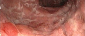
- Changes in the color of the pupil - its repainting into a grayish, yellowish, or even whitish color;
- Impaired photosensitivity - the patient sees the world around him as if through a dull glass, or, on the contrary, cannot tolerate bright lighting, and sees better in cloudy weather or at dusk;
- There is a violation of color perception, it is especially difficult for the patient to distinguish between purple and blue colors;
- Flashing before the eyes of stripes, spots, balls, reminiscent of the splashes of a waterfall;
- The development of strabismus is more common in children.
Cataracts of the eye: causes, symptoms
Good vision is the basis of a fulfilling life. Very often, with age, vision begins to decline. One of the most common causes of vision changes with age is the development of cataracts.
Cataract is an eye disease characterized by clouding of the lens, which in turn leads to decreased vision. As a rule, the disease develops simultaneously in both eyes, but one eye is more damaged in the initial stages. Cataracts can be congenital or acquired. Congenital cataracts are quite rare and occur in newborns. In this article we will mainly talk about acquired cataracts.
How do cataracts manifest (signs and symptoms of eye cataracts)
Lens cataract is dangerous because vision loss occurs gradually, and at the first, initial stage, when conservative treatment is most effective, signs of visual impairment are not critical. Unaware that the process of cataract development has already begun, people are in no hurry to see a doctor. Then, as the cloudy area of the lens increases, vision begins to decline. The main complaint is blurriness and blurriness of the image. It becomes difficult to read, watch TV, recognize friends on the street, or drive a car. Objects become fuzzy and blurry, as if looking through foggy glass. It may seem as if there is a spot in front of the eye that is making it difficult to see. Sometimes the so-called glare effect, or glare sensitivity, occurs (from the English glare - dazzling light, bright shine). This condition is characterized by discomfort and blurred vision in bright light and sunny weather. Doubling of objects and their distortion may also be observed. As the disease progresses, the pupil becomes milky white. Depending on the type of cataract and the stage of the disease, complaints may vary. It should be noted that visual impairment due to cataracts cannot be corrected with glasses and contact lenses.
How does lens cataract affect vision?
Cataracts affect the lens of the eye. The lens is one of the main refractive structures of the eye, focusing light rays on the retina. It is an elastic body shaped like a biconvex transparent lens. Essentially, the lens captures light as it passes through the pupil and focuses it onto the retina. Around the lens, the choroid forms the ciliary body, which contains a muscle that regulates the curvature of the lens, which ensures clear and distinct vision of objects at different distances.
The lens in the eye is “suspended” on thin radial threads that encircle it with a circular belt. The outer ends of the threads are attached to the ciliary muscle. When this muscle is relaxed (in the case of focusing the gaze on a distant object), the ring formed by its body has a large diameter, the threads holding the lens are tense, and its curvature, and therefore the refractive power, is minimal. When the ciliary muscle tenses (when viewing a nearby object), its ring narrows, the filaments relax, and the lens becomes more convex and, therefore, more refractive. This property of the lens to change its refractive power, and with it the focal point of the entire eye, is called accommodation.
Thus, we understand that the lens performs a very important function, and disturbances in its functioning directly affect vision.
Changes in the structures of the lens lead to diseases such as cataracts and presbyopia.
Cataracts and presbyopia
Cataracts (senile) and age-related farsightedness (presbyopia) are eye diseases that are typical for people over forty years of age. Both diseases are associated with the functioning of the lens.
Presbyopia is an age-related progressive decrease in vision, the development of so-called age-related farsightedness, when with age it becomes increasingly difficult to see clearly near, for example, to read. This is mainly due to a decrease in the elasticity of the substance and capsule of the lens, a change in its thickness and shape.
Cataract is clouding of the lens. Its transparency is impaired, which means it stops transmitting a sufficient and complete amount of light rays, and the picture ceases to be clear. The difference between the diseases is also that presbyopia can be corrected with near glasses and contact lenses, while cataracts cannot.
Causes of cataracts
What causes cataracts? For different reasons.
- The most common is the so-called senile (age-related) cataract. This type of cataract usually occurs in people over fifty years of age. The cause of its appearance is associated with chemical changes in the lens of the eye.
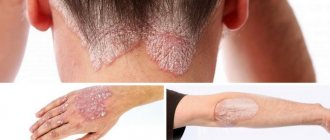
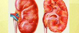
- Complicated cataracts are formed as a result of pathological processes occurring in the eye (for example, inflammation of the choroid, glaucoma) or diseases such as diabetes, kidney failure, skin diseases, etc.
- Traumatic cataract of the lens is usually caused by mechanical damage, as a result of which intraocular fluid penetrates into the lens capsule.
- Toxic cataracts occur due to the harmful effects of certain medications and chemicals.
Risk factors for developing lens cataracts also include:
- The presence of cataracts in close relatives;
- Prolonged exposure to ionizing or infrared radiation;
- Excessive insolation (prolonged exposure to the sun without sunglasses);
- Smoking;
- Hormonal changes.
Stages of cataract development
Cataracts usually develop gradually and have several stages of development (maturity).
Initial stage of cataract
As a rule, at the initial stage of the disease, vision does not decrease, but the first unpleasant signs of eye cataracts appear:
- Visual acuity decreases, a person sees the world as if through a cloudy glass;
- Decreased tolerance to bright light;
- The appearance of multi-colored circles around the light source;
- Color perception is impaired;
- Double image;
- The appearance of goosebumps, spots, circles before the eyes.
The severity of symptoms and their presence depend on the location of the cataract and the size of the opacities.
If you experience one or more of these symptoms, it is important to contact your ophthalmologist for an eye examination. At this stage, the doctor will identify the causes of cataracts and, most likely, will offer conservative treatment.
Conservative treatment
At the initial stage of cataracts, it is very important to stop or significantly slow down its development. The best drops for cataracts are those that contain vitamins, antioxidants and energy sources for the lens of the eye and some other substances, as they improve metabolic processes in the lens and slow down its aging and clouding.
Is it possible to treat cataracts with folk remedies? Traditional medicine will no doubt offer several recipes for treating cataracts, for example, with honey or onions, but will such treatment be effective? It is more advisable to use special ophthalmic drops, such as Oftan Katahrom. These combined drops contain Cytochrome C, which has a pronounced antioxidant effect, the energy source Adenosine and vitamin Nicotinamide, which help improve metabolism in the lens of the eye and slow down the development of cataracts, which is what is required at this stage.
Immature stage of cataract development
At the second stage of development, cataracts are called immature. The area of opacities in the lens expands, and visual impairment becomes more pronounced. At this stage, the image becomes increasingly blurry and unclear. The process of vision loss inevitably progresses; surgical intervention may be a way out of this condition.
But it is still better, if possible, to delay this stage by identifying cataracts in a timely manner and starting conservative treatment with the help of special drops “Oftan Katahrom”.
Mature stage of cataract
The third stage of cataract is called mature. At this stage, the lens becomes completely cloudy and dense. Vision is practically lost, only the perception of light remains. The changes are visible to the naked eye; the pupil seems to be covered with a white veil. In this situation, the only treatment is surgery.
Overmature stage of cataract
This is the most severe form of cataract. The masses of the lens are completely destroyed, it liquefies and becomes cloudy white. This can lead to further complications: glaucoma, the development of inflammation.
Thus, we can say that at the initial stage they try to stop the development of cataracts using conservative methods. Such treatment is recommended if vision is preserved to such an extent that it does not interfere with a person’s life as usual. Eye drops can be quite effective and help protect the eyes from the progression of cataracts. In this way, a person can delay surgery for a long period of time. In other cases, doctors recommend prompt replacement of the clouded lens with an artificial one.
How often should you visit an ophthalmologist?
Because vision changes occur in almost everyone as they age, do not wait to see an eye doctor until vision problems become obvious.
If you are between 18 and 30 years old and do not notice any visual impairment, visit an ophthalmologist once every 3 years; from 30 to 45 years - once every 2 years; from the age of 45 years - once a year.
More frequent eye exams are necessary if you notice signs of abnormalities, wear glasses, are over 45 years old, or have any of the following:
- Moderate or high myopia;
- Cases of glaucoma, myopia, retinal dystrophy in the family;
- Diabetes mellitus, hypertension, thyroid diseases, systemic diseases;
- Taking medications that have unwanted effects on the eyes;
- Work associated with visual stress or possible harmful effects on the eyes;
- Previous eye surgeries or injuries.
There are contraindications. Specialist consultation required
YOU MAY BE INTERESTED in: Eye cataract surgery Resorption cataract What is worse - glaucoma or cataract Literature on cataracts
Causes of cataracts
The causes of cataracts in most cases are:
- Age-related changes in the lens, so-called. “age-related cataract” - occurs in 90% of all cases of the disease;
- Eye injury;
- Exposure to radiation, x-rays;
- Exposing the eye to sunlight for a long time;
- The presence of certain diseases - glaucoma, uveitis, chorioretinitis, iridocyclitis, Fuchs syndrome, scleroderma, diabetes mellitus, hypothyroidism, metabolic disorders, hypovitaminosis, avitaminosis, malaria, varicella (chickenpox), typhus, anemia, eczema, neurodermatitis, Down's disease;
- Retinal detachment, which is possible with severe pain, for example during childbirth;
- Use of certain medications, especially for a long time - corticosteroids (hormones), antimalarial drugs and others;
- Bad habits – smoking, excessive consumption of alcoholic beverages;
- Poisoning of the body with naphthalene, thallium and other toxic substances;
- Intrauterine developmental deviation of the baby, which usually occurs when a pregnant woman develops infectious diseases - influenza, rubella, toxoplasmosis and others;
- Living in an unfavorable environmental environment.
It is also worth noting that age-related changes in the biochemical contents of the lens are a natural process caused by the denaturation of the protein that makes up the lens, which in turn occurs due to the aging of the body, however, the extent to which this process manifests itself or does not manifest itself largely depends on the person’s lifestyle .
Protein denaturation is a violation of the structure of protein molecules, leading to a loss of its transparency. During this process, the protein becomes white. Physically, this can be represented in the image of a protein being cooked.
Reasons for appearance
The disease can be congenital or acquired.
What causes congenital cataracts?
Congenital cataracts occur:
- with genetic predisposition;
- of alcohol by the expectant mother , prohibited medications, narcotic substances, smoking during the development of visual organs in the fetus;
- due to infectious diseases suffered by the mother in the first trimester of pregnancy (influenza, rubella, chickenpox);
- if the expectant mother has diabetes mellitus;
- as a consequence of autoimmune diseases of a pregnant woman.
How does acquired cataract begin?
Acquired cataract:
- Age . The disease is the result of general aging of the body, when decay processes prevail over regeneration processes. Moreover, these are natural phenomena for the human body.
- Traumatic , when the disease developed after injuries to the eye: Bruise , puncture, contusion.
- Chemical burn or exposure to high temperature.
- Radioactive or ionizing radiation.
- Occupational disease due to systematic exposure to unfavorable conditions (for example, a hot shop at a glass blower).
The lens is a fragile organ and therefore often suffers from injury. Violation of its integrity is diagnosed quickly.
Diabetic cataract: what is it?
Diabetic cataract is a complication of diabetes mellitus. As a result of an increase in blood sugar levels, it accumulates in the tissues of the eye . Low insulin levels cause a decrease in the transparency of the lens. Diabetes has a destructive effect on all tissues of the body.
Reference! The appearance of the disease is facilitated by low physical activity , a predominance of carbohydrate nutrition, and frequent exposure to a TV screen or computer monitor.
Types of cataracts
Cataracts are classified as follows...
By etiology (cause of disease)
- Congenital cataract;
- Acquired cataract, which is divided into the following types: - Age-related cataract; — Traumatic cataract; — Radiation cataracts that develop against the background of exposure to X-ray, infrared, radiation and other types of rays and irradiation on the body; — Toxic cataracts that develop while taking certain medications; — Complicated, developing against the background of other diseases of the organs of vision; — Cataracts caused by general diseases of the body - diabetes, hypothyroidism, metabolic disorders and others.
According to the location of the disease:
- Anterior polar;
- Posterior polar;
- Layered (zonular);
- Fusiform;
- Cortical (cortical);
- Nuclear (central);
- Posterior subcapsular;
- Total (complete) cataract.
Age-related cataracts are also divided according to the degree of maturity into the following stages:
- Initial cataract - characterized by the appearance of clouding of the lens at its edges, in the peripheral (non-optical) zones;
- Immature cataract - characterized by the advancement of opacification from the periphery to the central, optical zone of the lens, which is accompanied by a noticeable decrease in visual acuity;
- Mature cataract - characterized by complete clouding of the central (optical) part of the lens, while the patient sees only light, objects cannot be distinguished;
- Overmature cataract - characterized by liquefaction of crystallin, as well as repainting of the lens milky white.
What does a cataract look like: photo
According to the degrees of development, the stages of cataract are distinguished: initial, immature, mature and overripe.
At the initial stage, changes begin in the periphery, vision loss is absent or insignificant. Amenable to conservative treatment.
When immature, the degree of opacities increases and vision decreases. It is possible to develop swelling cataracts, leading to complications (phacogenic glaucoma).
Mature is characterized by compaction of clouded lens masses and a persistent decrease in visual acuity. Surgical treatment is required.
Overmature cataracts (milk cataracts, Morganian cataracts) are rare. In this case, the cortical substance disintegrates, the dense core separates from the capsule and “settles” at its bottom. It can be complicated by glaucoma (since inflammation occurs) and iridocyclitis when the capsule ruptures and the lens masses exit into the anterior and posterior chambers of the eye. Treatment is only surgical.
Diagnosis of cataracts
Diagnosis of cataracts includes the following types of examination:
- Measuring visual acuity using tests - Sivtsev and Golovin tables;
- Visual field examination (carried out using a computer perimeter);
- Measuring intraocular pressure (IOP);
- Ultrasound examination (ultrasound);
- Electrophysiological study of the retina and optic nerve;
- Biomicroscopy of the eye - using a slit (light) lamp, the degree of maturity of the cataract and lens opacification is measured.
Read also Hypothyroidism - symptoms, causes, types and treatment of hypothyroidism
Diagnosis of the disease
When diagnosing cataracts, both standard and innovative methods are used. Typical methods include:
- perimetry;
- visometry;
- determination by an ophthalmologist of binocular visual acuity. Also applicable:
- gonioscopy;
- ophthalmoscopy;
- color testing;
- biomicroscopy.
It is important to assess how damaged the lens is. Additionally, a fundus examination may be prescribed.
A collection of clinical tests is also prescribed. In particular, patients donate blood for biochemistry and glucose if they have a history of diabetes mellitus. Based on the results obtained, treatment is prescribed, which is selected individually for each patient.
Cataract treatment
How to treat cataracts? In many cases, the most effective treatment for cataracts today is surgery - replacing the clouded lens with an artificial one.
If the clouding of the lens is not stopped and everything is left as it is, a number of complications may develop over time, the most dangerous of which is atrophy (death) of the optic nerve, which is why even surgery will not help restore vision in the future, unless it is a higher power! In general, cataract treatment includes the following therapies:
1. Conservative treatment (drug therapy); 2. Surgical treatment of cataracts (surgery); 3. Rehabilitation after surgery.
1. Conservative treatment (drug therapy)
Important! Before using medications, be sure to consult your doctor!
Eye drops for cataracts. Drug treatment is aimed at improving metabolic processes in the lens and surrounding parts of the eye, thereby stopping the clouding process. The disadvantage of this method is the constant use of eye drops, because When the supply of drugs to the lens is stopped, the disease continues to progress again.
Among the eye drops for cataracts are:
"Oftan katachorm" - contribute to the normalization of metabolism, as well as activation of the processes of oxidation and reduction of crystallin in the lens;
"Quinax" - promote the resorption of clouded protein masses of the lens and normalize metabolism in the surrounding tissues;
"Taufon" - contribute to the normalization of metabolic processes in the eye, as well as the regeneration (restoration) of damaged cells;
“Visomitin” (“Skulachev Drops”) – are aimed at preventing dry eye syndrome, therefore they are often prescribed during the rehabilitation period after surgical treatment of cataracts. “Skulachev’s drops” help the eye produce its own tears, as well as improve the composition of the tear film.
Surgical treatment of cataracts
Surgical treatment of cataracts (surgery) is by far the most effective treatment method, which involves replacing the clouded lens with a new, artificial lens. The advantage of this method is that the artificial lens does not tend to wear out, so in the future this part of the eye will serve as efficiently as possible.
Among the modern methods of cataract surgery are:
Extracapsular cataract extraction - the operation is based on the complete removal of the clouded protein masses of the lens and their replacement with an intraocular lens (IOL), while the capsule (shell) of the lens remains intact;
Phacoemulsification - this modern type of operation is based on the removal of clouded masses of the lens using ultrasound, after which an intraocular lens is inserted in their place. Phacoemulsification is considered the least traumatic method of surgical treatment of Cataract disease. To replace the lens, local anesthesia is first administered using drops. Next, a micro-incision is made in the eye, 1.8-2.8 mm in diameter, into which the tip of the phacoemulsifier is inserted. With the help of ultrasound, crystallin is transformed into an emulsion, which is then removed from the eye, and in its place, through the same micro-incision, a folding intraocular lens is implanted. After installing the lens, the micro-incision seals itself. The entire treatment process takes no more than 10-15 minutes.
Another benefit of replacing the lens with an intraocular lens is vision correction. Thus, IOLs can have various properties - correcting astigmatism, improving visual acuity, and others, which allows a patient with other eye diseases to forget about devices such as glasses and contact lenses.
Femtolaser surgery is a technology for surgical treatment of cataracts similar to phacoemulsification, only femtosecond lasers are used instead of ultrasound.
Rehabilitation after surgery
After surgery, the ophthalmologist will usually prescribe the use of eye drops, as well as periodic examinations to prevent possible abnormalities.
Among the eye drops after surgical treatment of cataracts are:
Eye drops with an antibacterial effect aimed at preventing the impact of bacterial infections on the eye - Oftavix, Torbex, Floxal;
Eye drops for relieving inflammatory processes , aimed at calming the organs of vision after surgery - “Indocollir”, “Diclof”;
Eye drops for artificial irrigation of the organs of vision , if the eye experiences dryness, the so-called. “tear substitutes” - “Oxial”, “Systane”;
For severe pain in the visual organs, hormonal drops are prescribed - “Maxidex”, “Oftan-dexamethasone”.
Read also Brucellosis - first signs, symptoms, causes, treatment and prevention of brucellosis
Additional measures
After surgery, artificial lens substitutes are in the engraftment stage, so it is very important to follow doctors’ recommendations aimed at speeding up the rehabilitation process. Among these recommendations are:
- Do not neglect the use of eye drops prescribed by your doctor;
- Don't lift heavy things;
- Avoid hypothermia and chapped eyes;
- Do wet cleaning in your living space at least 2 times a week, and also avoid staying in dusty places;
- Try to eat foods enriched with vitamins and microelements (minerals).
Diagnostics
Anyone experiencing vision problems should see an ophthalmologist or optometrist.
Examination methods:
- Determination of visual acuity (from normal to light perception and blindness). With a normal retina, light perception with the correct projection. Otherwise, surgical treatment will not restore vision, but can save the eye as an organ.
- Perimetry to determine the condition of the retina and diagnose possible complications;
- Determination of intraocular pressure to exclude glaucoma. If necessary, tonography;
- Transmitted light research. With cataracts, against the background of a pink fundus reflex, shadows from a cloudy lens are detected;
- Biomicroscopy allows you to determine the location and degree of opacities;
- Examination of the fundus to exclude concomitant pathology.
Based on these studies, the doctor can make a diagnosis, but additional methods and consultations are needed to determine the causes:
- General clinical tests, including blood sugar;
- Consultations with a therapist, ENT, dentist to exclude concomitant pathologies and identify contraindications for surgical treatment (active inflammatory processes, severe decompensated diseases).
Cataracts - folk remedies
Important!
Before using folk remedies for treating cataracts, be sure to consult your doctor! Fir or pine resin. Make a mixture of 1 part fresh fir or pine (not spruce) resin and 3 parts sea buckthorn oil (if not, you can replace it with regular sunflower oil). The resulting mixture should be used as eye drops, instilling 1 drop into the damaged eye once a day for 30-40 days.
Bile. Before going to bed, once every 2 days, instill 1 drop of live pike bile into your eye. A total of 10 procedures need to be done, after which a 10-day break is taken and the course is repeated.
Herbal collection 1. Make a collection of 30 g of crushed dried horsetail, 20 g of knotweed grass and 10 g of fresh young stinging nettle. Mix everything thoroughly and pour 10 g of the collection with a glass of boiling water, then put the product on low heat for 3 minutes. Afterwards, the product should be set aside to infuse and cool for 1 hour, strain and take 100 ml before meals, 2 times a day, at within 14-21 days.
Herbal collection 2. Make a collection of 20 g of dried nettle, 20 g of high-salmon, 20 g of rhodiola rosea, 15 g of red hawthorn fruit, 8 g of rose hips and 1 teaspoon of dried St. John's wort. The resulting mixture is poured with 250 g of boiling water, infused for 40 minutes, filtered and taken for 21 days, 50 g before each meal.
Aloe, Kalanchoe and viviparus. Make a mixture of 2 parts aloe juice, 1 part Kalanchoe and 1 part viviparus. The prepared product is used as drops. You need to instill 2 drops into each eye 3-4 times a day.
Onion and honey drops. Dilute onion juice and clean, preferably distilled water in a 1:1 ratio. For greater effectiveness, add a little juice from dandelion leaves.
Additionally, make a mixture of 1 part honey, preferably acacia honey, diluted with propolis and 1 part clean water. The drops can be stored for no more than 3 days in the refrigerator.
You need to instill eye drops one by one, first with onion drops, and after 45 minutes with honey, 3 times a day, for a month, after which a 7-day break is taken, and if necessary, the course is repeated.
Signs of cataracts
Eye diseases
– one of the serious groups of pathologies that not only significantly reduce the quality of life of patients, but can also cause the development of disability. Perceiving the world using a visual analyzer is very important. Therefore, it is necessary to seek help from an ophthalmologist at the first signs of decreased visual acuity in order to understand the causes of unwanted symptoms of cataracts or other diseases. It is important to understand that some diseases appear due to irreversible changes in the internal structures of the organ of vision, and therefore cannot be treated with medications. However, timely consultation with a doctor and early diagnosis of eye diseases increases the chances of curing pathologies.
Some diseases of the organ of vision are associated with old age, although under the influence of internal or external factors they can occur at any period of life. One of the diseases that occurs mainly over the age of 60 years is cataracts. Cloudiness of the lens is the first sign of cataracts, which leads patients to suspect this diagnosis. However, in fact, the symptoms of the disease develop much earlier than a visible defect occurs. By paying close attention to your health or undergoing timely medical examinations, you can recognize the initial stage of cataracts.
What is cataract
Cataract is any pathological condition that is associated with a violation of the transparency of the structures of the lens of the eye. It entails a progressive deterioration of vision up to its complete loss.
It is not known for certain which factors contribute to the development of the disease; various theories are being studied:
- genetic;
- metabolic;
- exposure to aggressive environmental factors;
- taking certain medications;
- traumatic injuries;
- the influence of existing refractive errors;
- other pathologies of the organ of vision.
However, it has been proven that the direct cause of lens clouding is structural changes in its protein components - protein denaturation is irreversible and requires urgent correction in order to stop the pathological process.
Cataract is recognized as the most common disease in older people; the risks of its development increase after 40 years of age, reaching maximum values after 70 years of age. This is due to the fact that at a certain point the physiological density of the lens begins to change, adding unfavorable factors for the development of cataracts. Presbyopia or age-related farsightedness, which usually appears after 40-45 years, is considered a normal physiological age-related condition. However, older people are especially susceptible to adverse effects, which affects the development of any pathological processes in the organ of vision.
The opinion that cataracts are exclusively an age-related pathology has long been recognized as a myth. Traumatic damage to the organ of vision, ingestion of toxic substances, the consequences of diabetes mellitus, systemic diseases of the body, hereditary predisposition, genetic characteristics, environmental degradation and other unfavorable factors can cause clouding of the lens at any age.
The first signs of the disease
Recognizing the first symptoms of cataracts can be quite difficult; to do this, you need to treat your own health with increased responsibility and daily monitor changes in the process of visual perception of the world around you. Impaired transparency of the lens fibers or its capsule is a sign of disease progression, but the first pathological changes appear much earlier.
A person may notice signs of cataracts in the form of a cloudy sensation when looking at surrounding objects. At first, such changes will appear only during moments of eye fatigue, and after a 10-minute rest from work or reading, they will disappear. Further progression of symptoms leads to frequent episodes of difficulty looking at objects for a long time or concentrating on certain types of visual work. Letters become blurry when reading, causing diplopia, the so-called “double vision.” Rest and gymnastics for the eyes will eventually cease to bring the expected effect - vision will not be restored even after sleep.
Typically, at this stage, patients make an appointment with an ophthalmologist for the first time, since such symptoms significantly affect the usual rhythm of life and reduce the ability to perform daily work or household tasks.
Symptoms of cataracts
Further development of the pathology manifests itself in the form of clouding of the lens noticeable to the naked eye - the pathological process of loss of transparency affects its fibers or capsule. The onset of the disease manifests itself only in the form of small areas of cloudiness, and with the progression of denaturation of protein structures, the entire lens becomes cloudy.
Such disturbances inevitably affect visual functions - now the patient not only feels a change in the usual visual acuity, but also feels the appearance of a “film” and blurring when looking at any objects. Patients themselves describe such manifestations of cataract features as viewing the world around them, as if through foggy glass or film.
Twilight vision changes - a person experiences serious difficulties when trying to see surrounding objects in low light conditions. Relatives may notice that the patient often needs additional lighting while reading, and also increases the brightness of the monitor when working at the computer. In the evening, a person who develops cataracts may not recognize his acquaintances on the street, associating this with fatigue or overwork. It is important to pay attention to such changes and make an appointment with an ophthalmologist as soon as possible.
Other symptoms of the pathology include:
- deterioration in the perception of colors and shades;
- decreased contrast when looking at multi-colored objects;
- the appearance of blurred contours;
- double vision;
- flashing “flies”;
- light scattering, the appearance of halos - bright circles around light sources;
- the appearance of flashes of light, “glares”;
- difficulty working with small details, carelessness, absent-mindedness;
- lack of coordination - frequent hits on corners or furniture in rooms.
Significant deterioration in the functions of the visual system with cataracts cannot be corrected. Often people over 40 years of age associate such symptoms with manifestations of presbyopia - age-related farsightedness and difficulty viewing close objects. However, glasses with positive diopters to correct farsightedness do not improve the situation.
The processes of concentration in conditions of visual impairment are associated with a pain syndrome - unpleasant sensations can both arise in the eyeballs and manifest themselves in the form of tension headaches. Often there is a feeling of “sand” in the eyes, pressure inside the eyeball, as well as other unpleasant painful symptoms.
Features of cataracts in the last stages of the disease bring significant discomfort and cause difficulty in performing daily tasks - a person sees only bright colors or flashes of light, and ceases to clearly see the outlines of objects. This leads to the patient becoming virtually disabled. He is unable to care for himself and needs constant help and additional care. Externally, the stages of mature cataracts appear in the form of white spots, visible to everyone around. Older patients often wear dark glasses - bright sunlight irritates their eyes and causes unpleasant pain.
Prevention of cataracts
Cataract prevention includes following the following recommendations from ophthalmologists:
- Undergo a preventive examination by an ophthalmologist at least 1-2 times a year;
- To prevent ultraviolet (sun) rays from entering your eyes, wear sunglasses, but not made of cheap plastic, preferably glass;
- If there is a risk of developing cataracts, after consulting a doctor, use preventive eye drops - “Vicetin”, “Quinax”;
- Try to eat food enriched with vitamins and microelements;
- In the autumn-winter-spring period, take additional vitamin complexes;
- Do not leave various diseases to chance, especially infectious and endocrine ones;
- When working with high risks of eye injury, be sure to use protective equipment, for example, goggles, masks;
- Follow the rules of personal hygiene - do not touch your eyes with dirty hands;
- Stop smoking and drinking alcoholic beverages.
Methods and means of treatment depending on the type and stage of the disease
For therapy in the initial stage, the ophthalmologist prescribes drops with a complex of vitamins and microelements, for example: Taurine, Vitaiodurol, Oftan-katachrome, Cytochrome.
This therapy does not cure, but is a measure that reduces the progression of the disease.
To normalize glucose metabolism in age-related and diabetic cataracts, Pirenoxine .
Quinax drops are used to treat cataracts of various origins. The active ingredient of the drug destroys harmful compounds formed in the lens.
As part of complex treatment, injections with vitamin B2 and a course of electrophoresis with cysteine are used.
Is surgery necessary?
At more complex stages, treatment occurs only through surgery .
An artificial lens is implanted. Medicine is well developed in this area, transplantation is quick and painless.