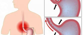
Emergency conditions always require resuscitation measures. One of these is hypovolemic shock. The pathology is accompanied by a sharp decrease in the volume of circulating blood and total fluid in the body, which leads to a decrease in blood pressure. As a result of changes in the functioning of the cardiovascular system, in the absence of the necessary treatment, death occurs.
What is hypovolemic shock?
Hypovolemia is a sharp, almost instantaneous decrease in the volume of blood or fluid in the body. This condition is provoked by a number of reasons and is very life-threatening. The disorder occurs when a patient loses more than 20% of the total volume of fluid or blood in the body. As a result of such changes, the heart is unable to deliver sufficient blood and oxygen to organs and tissues. Hypovolemic shock with blood loss always leads to the development of multiple organ failure, which requires immediate, qualified medical care in intensive care.
Hypovolemic shock - pathogenesis
The mechanism of development of the disorder directly depends on its type. According to this, in medicine the following types of hypovolemic shock are distinguished:
- Hemorrhagic
– is a consequence of acute blood loss (develops with the loss of 20-25% of the total blood volume). - Burn
- develops with the loss of a large volume of blood plasma, which is released through the surface of the burn. - Dehydration
– develops as a result of the body’s rapid loss of water and mineral salts. - Traumatic
- occurs as a result of acute blood loss and a decrease in plasma volume.
The pathogenesis of the development of hypovolemic shock is described by the following successive stages:
- Gradual inhibition of the midbrain, thalamus, which creates a barrier to the flow of afferent impulses into the cerebral cortex, reducing pulsation activity.
- A decrease in circulating blood volume leads to centralization of the general blood flow, a decrease in cardiac output, which impairs blood supply to tissues.
- Due to the development of shock, the concentration of some hormones and biological substances (cortisol, catecholamines, insulin, renin) decreases, but the hormone aldosterone actively accumulates. It interferes with the normal separation of urine, which increases intoxication of the body.
- All this leads to a deterioration in the properties of blood, an increase in its viscosity, agglutination of formed elements, activation of the hiding system, and the development of multiple organ failure.
Causes of hypovolemic shock
The disorder is a concomitant pathology and does not develop independently. As medical observations show, hypovolemic shock most often complicates the course of such conditions as:
- acute blood loss;
- burns;
- severe dehydration as a result of uncontrollable vomiting, severe diarrhea;
- gestosis during pregnancy;
- disruption of the endocrine system.
In all these conditions, a decrease in blood volume or plasma is observed. In some cases, the loss of water and minerals can also cause hypovolemic shock. This is more often observed with intestinal infections, when due to severe diarrhea the body loses a lot of free fluid.
Causes and mechanism of development
The main causes of hypovolemic shock are:
- Bleeding. Large blood loss is possible during surgery due to ulcerative lesions of the gastrointestinal tract, accompanied by damage to veins and arteries, injuries to the skull, abdominal organs, failure of the blood coagulation system due to hereditary pathologies, and so on.
- Plasma loss. In this case, the decrease in blood volume is due to the presence of extensive burns, accompanied by the release of plasma from the bloodstream. Liquid accumulates in a certain area or flows out of the affected area of the skin.
- Hemostasis. Inadequate functioning of blood vessels and other systems can lead to the deposition of blood in a certain part of the body, which will be accompanied by a lack of fluid in other vital organs.
- Digestive system disorder. Here we are talking about constant vomiting and diarrhea that develop with an infectious lesion (dysentery, cholera), with diseases of the central nervous system, peritonitis due to organ rupture, and other gastrointestinal diseases.
The pathogenesis of hypovolemic shock is quite complex. The entire blood volume is divided into two parts: that which circulates through the vessels and that which is located in the liver, spleen and bones. As, for example, bleeding from a wound progresses, the deposited blood leaves the listed organs in order to at least restore the bcc, that is, the compensation mechanism is activated. When this volume is not able to compensate for all losses, another mechanism is activated aimed at maintaining the functional activity of vital organs, namely the brain, lungs, and heart. The remaining vessels are spasmed and practically do not nourish the remaining areas of the body. If this does not help, paralysis of the vascular wall occurs, the lumen of the veins and arteries increases.
Taking into account the entire described development mechanism, three degrees of hypovolemic shock are distinguished:
- decrease in blood volume;
- activation of the sympathoadrenal system;
- hypovolemic shock.
Hypovolemic shock - symptoms
The clinical picture becomes more pronounced as the blood volume in the body decreases. At first, patients feel a sharp loss of strength, dizziness, and a feeling of thirst. The presence of open bleeding almost unmistakably indicates a high probability of developing hypovolemia. As the pathology progresses, patients experience various mental disorders associated with a malfunction of the nervous system: panic, depression of consciousness. Doctors record the following signs of hypovolemic shock:
- bradycardia;
- decreased blood pressure;
- loss of consciousness;
- coma.
Early sign of hypovolemic shock
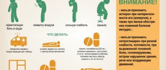
Hypovolemic shock is characterized by a gradual, step-by-step development. However, the rate of change of stages depends on the degree of blood loss and dehydration of the body. Initially, the attention of doctors is drawn to the semi-fainting state of the patient, who complains of the following symptoms:
- general weakness
- nausea;
- rapid breathing
- confusion (not always)
- feeling thirsty
- cardiopalmus.
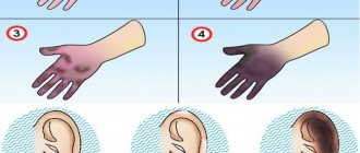
The skin acquires a pale tint, with a slight cyanosis. It gradually intensifies as oxygen deprivation increases. With the development of DIC syndrome, pinpoint microhemorrhages may appear on the surface of the skin, which are the result of disseminated intravascular coagulation, provoking thrombosis. In the absence of qualified medical care at this stage, late symptoms of hypovolemia appear:
- a sharp decrease in blood pressure;
- severe tachycardia;
- decreased body temperature;
- loss of consciousness.
Stages of hypovolemic shock
As the disorder progresses, doctors note characteristic clinical signs. In accordance with them, in the development of such pathology as hypovolemic shock, the following degrees are distinguished:
- The first or mild one is characterized by a loss of up to 15% of circulating blood volume.
In this case, breathing is not impaired, slight pallor of the skin and severe anxiety are observed. - Second degree (moderate severity) – blood loss reaches 30-40%.
Blood pressure drops to 100 mmHg. Art., the pulse is frequent, sweating increases, the skin is pale, the patient is very restless and irritated. - Third (severe) – the volume of circulating blood decreases by more than 40%.
The pulse is weak, frequent, blood pressure drops to 70 mm Hg. Art. The skin is white, cold sticky sweat, speech becomes incoherent, and loss of consciousness occurs.
Hypovolemic shock - complications
The consequences of shock directly depend on the rate of loss of blood and fluid by the body and the volume of loss. Hypovolemic shock with salmonellosis develops against the background of severe dehydration. Severe cases in the absence of emergency medical care in a hospital result in death. The lack of the proper volume of fluid in the body, blood, leads to the fact that the work of all internal organs and systems becomes difficult. The patient has the following disorders that can lead to death:
- acute heart failure;
- renal failure;
- liver failure;
- depression of the nervous system;
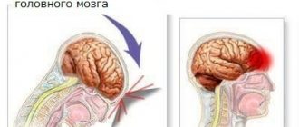
- cerebral infarction.
What is happening in the victim's body?
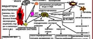
The pathogenesis of the shock state during hypovolemia begins with the body’s attempts to independently stop fluid loss and compensate for the deficiency:
- a reserve volume of blood enters the general channel from the depot;
- arterial vessels narrowing towards the periphery (arms and legs) in order to retain the required amount of blood for the brain, heart and lungs.
When the compensation mechanism is exhausted, spasm of peripheral vessels is replaced by complete paralysis, loss of tone and dilation. Because of this, most of the blood goes to the extremities and there is an insufficient supply of oxygen to vital organs, homeostasis is grossly disrupted, and metabolism suffers.
It is customary to distinguish 3 stages (phases) of shock development:
- Deficient - leading to the occurrence of acute fluid deficiency, a decrease in blood volume, which leads to a drop in venous pressure in the central veins, a decrease in blood flow to the heart. Fluid from the interstitial space passes into the capillaries.
- Stimulation of the sympathoadrenal system - receptors that control blood pressure signal to the brain and cause an increase in the synthesis of catecholamines (adrenaline, norepinephrine) by the adrenal glands. They increase the tone of the vascular wall, promote spasm in the periphery, increase the heart rate and increase stroke volume. Actions are aimed at supporting arterial and venous pressure for blood circulation in vital organs by reducing blood flow to the skin, muscles, kidneys, and digestive system. With prompt treatment, complete restoration of blood circulation is possible. If the period favorable for emergency interventions is missed, then a full-scale picture of shock develops.
- Actually hypovolemic shock - the volume of circulating blood continues to fall, the flow to the heart, lungs and brain sharply decreases. Signs of oxygen deficiency in all organs and changes in metabolism appear. The skin, muscles and kidneys are the first to suffer from the loss of compensatory protection, followed by organs located in the abdominal cavity, then life-supporting organs.
The mechanisms of shock development and the consequences for the body are described in detail in this video:
Diagnosis of hypovolemic shock
One of the main signs of the disorder is hemodynamic disturbances during hypovolemic shock. To detect them, a four-level classification is used, which is based on the following clinical manifestations:
- filling of the pulse;
- frequency of urination and volume of urine produced;
- condition of the patient's skin;
- mental status.
Depending on what caused the development of blood loss and dehydration of the body, a number of additional hardware studies and tests are prescribed:
- radiography;
- fibrogastroscopy;
- laparoscopy;
- blood and urine testing for biochemistry.
Symptoms
How can you tell if a person is in a state of hypovolemic shock? At an early stage of the disease, the following symptoms may be observed:
- Increased heart rate, tachycardia.
- The pressure may be slightly elevated, but often remains normal.
- A person's pulse may be jumping.
- There may be bloody mucous discharge.
- The mucous membranes of a person will be pale, and the skin will also appear pale.
Treatment of hypovolemic shock
Having previously established the cause, distinguishing hypovolemic shock from a bacterial intestinal infection with complications, they begin to actively treat the disorder. The main goal of the initial stage of therapy is to eliminate tissue hypoxia and restore normal blood circulation. This is necessary to normalize the vital activity of the myocardium, brain, and lungs, which are the first targets for hypovolemia. Along with stopping bleeding, if any, catheterization of the central veins is carried out for emergency administration of blood replacement solutions.
Hypovolemic shock - emergency care
First aid can only be fully provided in the intensive care unit. If hypovolemic shock has developed as a result of severe blood loss, trauma from a car accident, or injury, you can help the patient on the spot before the doctors arrive. By following a few simple steps, you can slow down hypovolemic shock.
Emergency assistance, the algorithm for its provision is as follows:
- The victim is placed on his back, his head is turned to the side (if there is no damage to the cervical spine).
- The legs are raised 30 degrees to unload the myocardium.
- To reduce the rate of heat transfer and prevent hypothermia, it is recommended to cover the patient to prevent cooling.
- If the patient is conscious, you can give him as much to drink as he can to replace fluids.
Therapy for hypovolemic shock
In a hospital setting, blood substitutes are administered intravenously to the patient. Infusion therapy for hypovolemic shock forms the basis of treatment, helps to quickly improve the patient’s condition and restore the functioning of internal organs. For this purpose, the following drugs are used for hypovolemic shock:
- Crystalloid solutions - saline, dextrose of varying concentrations (5, 10%). The goal is to increase systolic pressure to 70 mmHg. Art.
- Plasma substitutes - administered if there is no effect from the first solutions: Dextran, Gelatin are administered in a volume of 800-1000 ml. If there is no effect, move on to the next stage.
- Hydrocortisone at a dosage of 10-15 mg/kg.
- If there is no effect from corticosteroids, sympathomimetics are used - Phenylephrine, Dopamine, Norepinephrine.
Treatment
The main goal of treatment is:
- restoration of blood supply to the heart, brain and lung tissue, elimination of their oxygen deficiency (hypoxia);
- combating acid-base imbalance;
- replacement of lost electrolytes and vitamins;
- normalization of blood supply to the kidneys and daily diuresis;
- symptomatic support for the functioning of the heart and brain.
Mild symptoms of hypovolemia can be eliminated by slowly drinking plain water, or better yet, lightly salted water. At high temperatures, profuse sweating, and diarrhea, doctors recommend drinking more tea, juices, compote, and herbal decoctions. Avoid coffee, alcohol, and carbonated drinks that affect vascular tone and the surface of the stomach.
Sterile disposable catheters allow you to quickly set up a transfusion system
The emergency aid algorithm includes the initial actions of people around who can assist the victim.
We advise you to read: Causes of low red blood cells
- Therapeutic measures for hypovolemic shock should begin with the fight against bleeding if the victim has a wound: application of a tourniquet, tight bandaging, immobilization of the damaged area of the body (do not forget to record the time of application of the tourniquet).
- It is necessary to call an ambulance, and until it arrives, ensure the person’s peace and immobility. If unconscious, it is better to turn him on his side.
- Infusion therapy (intravenous administration of fluid) begins at the inpatient stage, the ambulance doctor installs an intravenous system and administers a saline solution containing a minimum of sodium. Small doses of glycosides are indicated to support cardiac activity.
- Hospitalization is carried out, depending on the reason, in the intensive care unit of a surgical hospital or the intensive care ward of an infectious diseases hospital.
- Due to the need for transfusion of a large volume of fluid, a catheter is placed in the subclavian vein.
- While the blood type of the victim is unknown, blood substitutes such as Poliglyukin or Reopoliglyukin are quickly administered by drip. The preparations are solutions of dextrans.
- In case of large blood loss, a jet infusion of up to 0.5 liters of single-group blood, plasma, Protein or Albumin solutions is indicated.
- To relieve peripheral vascular spasm, glucocorticoids are administered intravenously in large dosages.
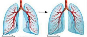
- Breathing with an oxygen-air mixture through nasal catheters is indicated.
Planned therapy
Planned measures include:
- correction of metabolic acidosis using sodium bicarbonate solutions (up to 400 ml per day);
- Panangin (a drug with potassium and magnesium) is added to the infused solutions.
Blood substitutes are able to maintain the biological and physical properties of blood, reduce viscosity, improve blood circulation in the vascular network, and maintain blood flow in the kidneys
The effectiveness of activities is judged by:
- sufficient stabilization of blood pressure levels;
- control of urine output (diuresis).
The discharge of 50–60 ml of urine per hour through the urinary catheter is considered normal. If the fluid loss deficit is considered to be replenished, and not enough urine is excreted, stimulation with Mannitol is necessary (daily slow drip of no more than 1 liter).
Measuring central venous pressure and increasing it to 120 mmH2O. Art. allows you to verify the achieved stabilization.
Further treatment
Therapy can be carried out either in an ambulance or in a hospital intensive care unit. Before arriving at the intensive care unit, the patient is given inhalations of pure oxygen, and, if necessary, artificial ventilation of the lungs is performed. Injections with drugs that stimulate blood circulation and, if indicated, painkillers are also administered.
- improving the functioning of the cardiovascular system;
- accelerated restoration of blood volume inside the vessels;
- replenishment of the number of lost red blood cells in the blood;
- correction of fluid deficiency;
- restoration of damaged homeostasis systems;
- elimination of dysfunction of internal organs.
Read about the diagnosis of Meniere's disease here.
Effective treatment methods include sympathomimetic agents and diuretics, oxygen exposure, and platelet transfusions.
Hypovolemic shock is a very dangerous condition, so it is necessary to know about effective preventive measures. A healthy diet, as well as taking iron supplements and other medications designed to increase hemoglobin levels, can help reduce the risk of developing shock as a result of injuries with blood loss.
32. Providing emergency care for hypovolemic shock
The cause of hypovolemic shock is profuse diarrhea and uncontrollable vomiting, leading to loss of tissue fluid, impaired tissue perfusion, hypoxia, and metabolic acidosis.
Emergency medical care for hypovolemic shock consists of primary rehydration in an amount corresponding to the calculated dehydration.
Patients with first-degree hypovolemic shock can be limited to oral rehydration; with more severe shock and preserved consciousness, with the ability to take fluid orally, they begin enteral rehydration, and then switch to intravenous fluid administration.
Enteral rehydration should be preceded by gastric lavage with 2% sodium bicarbonate solution. Gastric lavage is certainly indicated for foodborne toxic infections and reliable exclusion of myocardial infarction or acute surgical diseases of the abdominal organs.
Prioral rehydration involves drinking slowly in small sips of 1 liter of warm (38-40° C) water with 20 g of glucose, 3.5 g of sodium chloride, 2.5 g of sodium bicarbonate, 1.5 g of potassium chloride. Glucose can be replaced with table sugar, sodium chloride with table salt, sodium bicarbonate with baking soda. For oral rehydration, you can use polyionic infusion solutions with the addition of 40% glucose.
For infusion rehydration, polyionic solutions are used with the addition of 20-40 ml of a 40% glucose solution to replace the estimated fluid loss at a rate of 100-120 ml/min. After normalization of the pulse and stabilization of blood pressure, the rate of intravenous fluid administration is reduced.
Pressor amines and cardiovascular drugs are contraindicated. Antibiotics are not prescribed.
Rehydration for hypovolemic shock, which has complicated food toxic infection against the background of alcohol withdrawal syndrome, alcoholic delirium and convulsions, is carried out in the same volume, but must be supplemented by intravenous administration of 4-6 ml of 0.5% seduxen solution or 20-30 ml of 20% sodium hydroxybutyrate solution and 4-6 ml of 1% furosemide intravenously.
Main dangers and complications.
Late diagnosis of dehydration syndrome and erroneous interpretation of seizures in severe hypovolemic shock.
Providing emergency care for infectious-toxic shock
The causes of the development of infectious-toxic shock can be, first of all, meningococcal, fungal and intestinal infections, as well as other acute infections with an unfavorable course of the disease. In its development, infectious toxic shock sequentially goes through 3 stages - compensation (shock of the 1st degree), subcompensation (shock of the 2nd degree), decompensation (shock of the 3rd degree).
Treatment.
1. In adults, compensated infectious-toxic shock does not require infusion therapy, and upon admission to the hospital, treatment is limited to the use of antipyretics, analgin 50% - 2 ml and diphenhydramine 1% - 2 ml intramuscularly; for agitation and convulsions, seduxen 0.5% - 2-4 ml intramuscularly (intravenously) and magnesium sulfate 25% - 10 ml (15 ml) intramuscularly.
2. In case of subcompensated shock, 400 ml of polyglucin (reopolyglucin) and glucocorticoid hormones (prednisolone 90-120 mg, or equivalent doses of other drugs - dexamethasone, methylprednisolone, etc.) are administered intravenously.
3. In case of decompensated shock, polyglucin is administered in a stream followed by a transition to a drip infusion, and if there is no effect, 200 mg of dopamine per 200 ml of 5% glucose solution is prescribed intravenously.
4. Excitement and convulsions are controlled by intravenous administration of 2-4 ml of a 0.5% solution of diazepam (seduxen) or 10-20 ml of a 20% solution of sodium hydroxybutyrate.
5. If a diagnosis of meningitis is established, levomecithin sodium succinate is administered at a dose of 25 mg/kg, and 2-4 ml of a 1% solution of furosemide (Lasix).
6. Infectious-toxic shock with influenza requires additional administration of 5.0 ml of anti-influenza (donor, anti-measles) gamma globulin intramuscularly, as well as 5-10 ml of 5% ascorbic acid solution and 10 ml of 10% calcium gluconate solution intravenously.
Main dangers and complications:
Late diagnosis of infectious-toxic shock as a result of erroneous interpretation of a decrease in body temperature to subnormal and normal numbers and the cessation of psychomotor agitation as indicators of improvement in the patient’s condition.
Incorrect diagnosis of influenza in a patient with meningitis, and sore throat in a patient with diphtheria.
Incorrect diagnosis of a convulsive syndrome not associated with infectious-toxic shock and refusal to administer infusion therapy at the prehospital stage when the patient is delivered to the hospital under the guise of anticonvulsant therapy only.
Source: https://studfile.net/preview/4081721/page:24/