Trichinosis is an acute disease caused by roundworms of the Trichinella class - nematodes. Both people and animals can become infected. The main carriers of the disease and sources of infection are wild animals. Infection occurs when eating poorly cooked meat.
Trichinosis
Characteristics of the disease
Not many people know what trichinosis is. Meanwhile, trichinosis in humans is a fairly serious disease. It is characterized by severe clinical manifestations and leads to serious complications. In some cases, the risk of death is high.
This disease is widespread throughout the world. First of all, the disease affects residents of rural areas. Infection with trichinosis occurs when eating meat containing capsules of Trichinella larvae.
The cause of the disease is Trichinella - these are small worms, their body length is no more than 2 mm. Encapsulated larvae enter the human body when they eat contaminated, poorly cooked meat. Particularly dangerous are pork and wild boar meat, as well as bear meat.
The disease develops in several stages:
- After entering the body, the larvae settle in the duodenum, where young individuals hatch from the larvae. This process takes about 60 minutes. Over the next two days, further development of Trichinella occurs, after which they begin to multiply. This process can last a little over a month, after which the parasites die. During this period they are able to lay 2,000 eggs. This is the first stage of the disease - intestinal.
- The newly hatched larvae begin their migration journey. This stage is called migration.
- The third stage is muscular. The larvae reach muscle tissue through the circulatory system. Most often, parasites are found in the muscle tissues of the respiratory system, as well as in the facial and chewing muscles. The larva grows in the muscle tissue, it increases significantly in size and takes on a spiral shape. Then the larva is encapsulated and fenced off from the muscle tissue. After one year, the capsule begins to become covered with lime. Worms can remain in this state for several decades. From 10 to 40 years, the larva in the capsule can maintain its viability.
Trichinosis parasites are most dangerous to health in the first and second stages of the disease, when the larva turns into a capsule, it becomes harmless to humans. But the person himself will become a carrier of the disease.
You may also be interested in: Where can you get scabies?
It is worth noting that the disease does not pass from person to person. Each stage of the disease has characteristic symptoms. Signs of trichinosis depend on the severity of the disease.
What is trichinosis?
Trichinosis is a massive infection of the human body by parasites called Trichinella. This is a filamentous helminth of microscopic size. As a rule, a mature female does not exceed 5 mm. The helminth in the mature stage parasitizes in the small intestine, but its larvae can be found in all muscles except the heart muscle.
Once in the human body, Trichinella parasitizes there for 56 days, giving birth to 20,000 live larvae during this time. The larvae penetrate the lymphatic and blood vessels and spread throughout the host’s body. 18-20 days after infection, the parasite lengthens to 0.8 mm and begins to curl up.
The peculiarity of this helminth is that it forms a capsule around itself, which complicates the treatment process. In encapsulated form, the larva can live in the body for more than 25 years, disrupting the functioning of most organs and causing death.
Symptoms of the disease
With trichinosis, symptoms in humans can be expressed with varying intensity. At the first stage of the disease, symptoms are mild or may not exist at all. If a few parasites have entered the body, the disease will be asymptomatic. In some cases, diarrhea, abdominal pain, nausea, and abdominal cramps may occur.
After a week, new larvae begin their journey through the body. This stage is accompanied by the following symptoms:
- fever;
- peripheral blood eosinophilia;
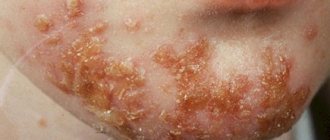
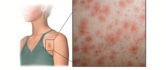
- skin rash.
The larvae release allergens into the blood, so skin rashes may occur at this stage. This is how an allergic reaction manifests itself. A severe version of the disease can cause myocarditis, lead to pneumonitis, and affect the functioning of the central nervous system.
The third stage of the disease is muscular, accompanied by weakness and pain in the muscles. Signs of the disease increase and become more complex. At this stage they become more obvious. The patient is concerned about the following ailments:
- headache;
- swelling of the eyelids, face;
- eye infectious diseases;
- fever;
- increased body temperature;
- increased light sensitivity.
The intensity of symptoms depends on the number of helminths in the body and the state of the immune system. After the encapsulation process is completed, the symptoms of the disease will begin to subside .
Pathogenesis
Clinical signs of the disease are due to the body’s increased susceptibility to allergens and irritants, the reasons for this being irritation caused by metabolic products and the death of helminths. Substances that are released during the growth of the larva cause local inflammation, swelling, affect joints, and disrupt the blood clotting process. In muscles the larvae cause myositis, in other organs they die. The intensity of the signs of the disease depends on the scale of the infection - the more larvae that have entered the body, the more intense the symptoms appear.
The first 2–3 weeks are the incubation period when there are no signs of the disease. The shorter this period, the more severe trichinosis will occur. During the incubation period, a person may experience allergic manifestations of helminthiasis, diarrhea, and abdominal pain. With massive infection, the larvae can infect the intestinal mucosa. A month after infection, the second stage of the disease begins, which is manifested by complications of the disease: myocarditis, inflammation of the lungs, meninges, hepatitis, thrombus formation in the veins and arteries.
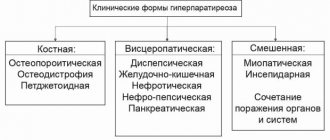
Forms of the disease
Depending on how pronounced the symptoms of the disease are, three forms of trichinosis are distinguished:
- The mild form lasts about two weeks. It is accompanied by an increase in body temperature no higher than 38.5 degrees. There is slight swelling in the face and eye area.
- The average form of the disease lasts two to three weeks. This stage is characterized by an increase in body temperature to 39 degrees. Pain begins to develop in the back of the head and in the muscles. The eyes are affected by conjunctivitis.
- Severe form. This form of trichinosis is characterized by intense symptoms. The patient develops a high temperature, it can reach 40 degrees. Severe complications affect internal organs: heart, lungs, brain. The central nervous system suffers. A skin rash appears. The patient is bothered by intense muscle pain. Sometimes there is a restriction of mobility or complete immobilization of a person.
It will also be interesting: Bull tapeworm: structural features of the life cycle
Severe forms of trichinosis are treated in a hospital setting. This form of the disease can be fatal. If trichinosis is diagnosed, symptoms and treatment are closely related. The course of treatment that the attending physician will develop will depend on what symptoms accompany the disease.
How can you become infected with trichinosis?
A person can become infected with trichinosis from poultry or wild animals. There are many natural foci registered in the world that provide excellent conditions for maintaining the population of worms.
In nature, helminthic infestation is common among some mammal species:
- Wolves.
- Foxes.
- Badgers.
- Boars.
- The Bears.
Self-infection and the actuality of the disease are ensured by eating carrion by animals. Sometimes worms are recorded in rodents (rats and mice), which suggests that the cat family can also spread this type of helminthic infestation among the city population.
The moment of feeding food waste and slaughter products to pets is especially important. This food is rarely sufficiently processed thermally, so it can become a source of infection for animals.
Taking into account the mechanisms of spread and the characteristics of the life cycle of trichinosis, it is highly likely that the disease is a non-contact helminthiasis. This infection is not transmitted from person to person.
The full life cycle of a helminth can occur in one person without much difficulty. For puberty and the birth of offspring, Trichinella does not need an intermediate host.
Currently, isolated cases of morbidity and, more rarely, group cases are being recorded. Familial cases are very common among the population of countries such as Ukraine and Russia. The mechanism for the development of this type of morbidity is quite simple: infected meat appears in the family, which was purchased at a spontaneous market or brought from a hunt. A dish is prepared from it, which is then eaten by the whole family during a family dinner. The meat contains encapsulated Trichinella larvae, which are the infectious factor.
There is practically no option of not developing the disease when eating a larva, since the human body is very highly sensitive to this type of helminthiasis.
Interesting and important! A convalescent (a person who has recovered from an illness) develops a weak and short-lived immunity, so it is very easy to become infected again, although the course of the disease will be much easier.
Complications
The complication of the disease depends on the muscles of which organ the encapsulation occurred in. Parasites can settle in the heart tissue, in which case there is a high risk of inflammation of the heart muscle. If the parasites reach the brain, encephalitis or meningitis may develop. When Trichinella settles in the lung tissues, the patient develops bronchopneumonia. If the liver is damaged - nephritis, hepatitis.
The risk of death is low, but possible. Between 10 and 30% of those infected die. Death occurs only in severe forms of trichenellosis. Death is possible at the initial stage of the disease. If the patient is diagnosed with a mild form, then after 5 - 6 weeks after infection there will be a complete recovery.
Trichinosis: causes, symptoms and treatment in humans
Trichinosis is an acute parasitic disease (helminthiasis) that affects not only humans, but also other mammals.
Clinical manifestations of the disease are severe, complete loss of work activity and death are possible. When infected with trichinosis, severe fever, pain and aches in the muscles, swelling of the face, and rashes appear. In case of complications, damage to the respiratory and central nervous systems occurs.
Infection occurs due to invasion, that is, the entry of a parasite (helminth) into the body. The helminth in the muscular system lays larvae.
The incubation period is about four weeks, after which the larvae actively develop and become covered with a capsule. The main causative agent of trichinosis is a helminth, which reaches up to 3 mm and has the shape of a spiral.
Causes of trichinosis
The source of infection for humans are different types of adult animals infected with Trichinella - wild animals: bears, foxes, nutria, wolves, wild boars.
Infected marine species include: seals, sea lions, beluga whales, walruses, and whales. No exception is livestock, pets and rodents: dogs, pigs, cows, goats, mice, rats, cats. Trichinosis is not transmitted from person to person.
The most common causes of infection with trichinosis are:
- good adaptation of the pathogen to temperatures;
- the human body is extremely sensitive to trichinosis;
- collective outbreaks of the disease. This often happens in groups of people, families who have consumed meat with helminths;
- relapses of trichinosis occur due to instability of the immune system that arose after the first invasion.
Provoking factors for the development of the disease
In the photo above, pork meat affected by trichinosis
A person becomes infected by consuming food, that is, orally. Helminth larvae enter the human body and begin active development. The most dangerous type of meat is considered to be dried, raw, improperly prepared (boiled or fried) and smoked lard with streaks.
Symptoms of trichinosis
Initially, trichinosis in humans does not have any specific symptoms. There are no visible changes in the muscular system.
Proteins, which are the main component of the helminth’s body, harm humans. Proteins are very strong allergy triggers and foreign elements for the human body. When infected, a severe allergic reaction occurs.
The latent period lasts from 1 week to 1 month. With trichinosis, symptoms are not pronounced. The prolonged development of symptoms indicates that the disease is becoming severe.
Symptoms of mild and moderate forms of trichinosis are as follows:
- hyperthermia. Body temperature is relatively not high (37.5 C), the daily deviation is 1C;
- swelling of the legs, arms or whole body (see photo above). This occurs due to the body’s sharp reflex to the ingress of foreign protein;
- muscle pain. This symptom is one of the signs of a severe form. Acute pain manifests itself in the calf muscles, which affect the entire muscular system. When moving and touching, the pain worsens. Symptoms and signs begin to bother a person from 4 days after infection;
The following symptoms are skin rashes, which have the following forms:
- urticaria - pinkish blisters of different sizes;
- utricarial rash - very itchy blisters that are slightly raised above the skin;
- papular rash - multiple plaques connecting to each other.
The severe form of trichinosis has its own complications:
- meningoencephalitis – pathological processes in the membranes of the brain;
- eosinophilic pneumonia. The occurrence of the disease is provoked by a high content of eosinophils (foods that cause allergies) in the lungs. Against the background of pneumonia, pleurisy and symptoms of bronchial asthma may develop;
- myocarditis – inflammation of the heart muscle (myocardium), due to allergies and a sharp reaction of the immune system. The complication can lead to the death of the patient;
- nephritis – pathology of kidney tissue;
- hepatitis is an inflammatory disease of the liver.
The mortality rate is about 30% in severe cases. Death occurs 8 weeks from the beginning of the invasion. In mild cases, the prognosis is favorable and complete recovery occurs after 6 weeks.
Trichinosis affects not only adults, but also children. Trichinosis in children can occur when eating meat contaminated with Trichinella. For a child to become infected, 15 grams of the product is enough.
The period from the onset of infection ranges from one week to 1 month. If there is insufficient or no treatment, a severe form of the disease can develop.
The mild stage of the disease lasts about 15 days, but after recovery, minor symptoms may remain for a week.
Symptoms of the mild stage look like this:
- hyperthermia. The temperature reaches 39 C;
- slight swelling of the face;
- mild muscle pain;
- high eosinophil content (13%).
Symptoms are at a moderate stage. The duration of the illness lasts more than three weeks, rehabilitation takes place within two weeks after recovery. The symptoms are:
- hyperthermia reaches 40 C. Taking antipyretic drugs reduces the temperature by only 1 C and for a short time;
- the child’s muscles and stomach begin to ache;
- rashes;
- swelling of the face;
- the content of eosinophils reaches 40%.
Treatment of the disease at a severe stage is carried out in an inpatient setting in the infectious diseases department. Without treatment, the child may die. Symptoms of the severe stage:
- temperature more than 40 C;
- the liver becomes enlarged;
- the central nervous system is disturbed - hyperactivity, seizures, seizures, convulsions;
- pain in the stomach;
- diarrhea, vomiting;
- muscle pain accompanied by convulsions;
- subcutaneous hemarthrosis;
- the content of eosinophils is up to 90%.
Therapeutic gymnastics, massages, baths with various medicinal substances are used as rehabilitation.
Stages of trichinosis
At the onset of the disease, symptoms depend on the number of larvae. Further progression depends on the proliferation of parasites and the general state of the patient’s immunity.
Severe complications of the disease are caused not by the vital activity of helminths, but by the body’s non-standard reaction to the ingestion of a foreign protein.
The stages of trichinosis are:
- Trichinosis infestation (infestation of parasites). Trichinella penetrates the human body and is located in the small intestine. Fixation occurs on the membrane, causing severe inflammation. Over the course of several months, helminths develop into adults in the intestines, fertilization occurs, and further spread of more parasites occurs. One Trichinella can produce more than 1,500 new individuals.
- Dissemination (reproduction of parasites throughout the body). Helminths begin to migrate into the tissues of the whole body, entering the muscles. The larvae spread through blood vessels. After entering the blood, helminths attach to muscle fibers. The development and growth of Trichinella begins, and allergens are released. The intoxication process and allergies progress in the body.
- Encapsulation stage. At this time, the tissues are regenerated. Trichinella reaches 0.5 mm in size and takes the shape of a spiral. The larva is enveloped in a capsule and temporarily stops development. Symptoms and discomfort cease to be pronounced and practically disappear. After a certain time, the capsule with the larva calcifies and the released salts can kill the helminth. Trichinella can maintain its vital activity for many years.
Trichinosis in children is divided into three stages - early, moderate and severe.
Diagnosis of trichinosis
Similar symptoms to other muscle pathologies indicate that this causes difficulties for specialists to establish a diagnosis.
Diagnosis of trichinosis is carried out using laboratory tests. A stool test is taken from the patient in order to identify the presence of the main pathogen - Trichinella. In addition, the following diagnostic measures are carried out:
- General blood analysis. Trichinosis provokes the appearance in a person of too high a level of eosinophils in the blood. makes up 80% of the total number of leukocytes;
- Serological diagnosis. Study of the reaction of erythrocytes to the addition of antigens taken from parasites. This diagnostic method is divided into five types: 1. Complement fixation reaction (CFR). When antibodies are detected in the patient’s blood, they group with the antigen and bind to complement (an element involved in immune reflexes);
- 2. Indirect hemagglutination reaction (IRHA). This type of diagnosis is based on the ability of blood cells to connect when antigens and antibodies are located on the surface of red blood cells;
- 3. Enzyme-linked immunosorbent assay (ELISA). The interaction of antigen with antibody is established. For the mark, which allows you to see the result, special enzymes are used;
- 4. Immunofluorescence reaction (RIF). There is a special mark in the material used that leads to luminescence if the antibody reacts to the antigen;
- 5. Enzyme-labeled antibody reaction (EMA). The result is assessed by a label that acts as a special enzyme;
Treatment of trichinosis
Since the symptoms and course of trichinosis are severe, treatment should be stopped in a short time. Treatment takes place in a hospital setting under the supervision of doctors.
During the period of illness, bed rest is recommended. One of the methods of stopping the disease is etiotropic therapy, that is, taking various drugs.
Drug treatment of trichinosis (medicines)
Relief and treatment of trichinosis involves taking anthelmintic drugs that fight the main causative agent of the disease.
Such means include:
- Mebendazole. The drug disrupts the metabolism of helminths and their absorption of glucose. Due to disruptions in synthesis, the parasites die. The drug is contraindicated for pregnant and breastfeeding women;
- Albendazole. The effect of the drug is almost the same as that of Mebendazole. It is known to be effective against maggots. It comes in the form of tablets of 0.2 grams. Pregnant women and people with retinal diseases are contraindicated;
- Vermox. The drug is 90% effective. The active ingredient in the composition is mebendazole;
- Thiabendazole. 90% effective.
To eliminate the symptoms of trichinosis, the following drugs are prescribed:
- anti-inflammatory medications - Diclofenac, Ortofen, Voltaren, Diclogen . The drugs neutralize inflammation caused by allergic reactions;
- antipyretic drugs - Paracetamol, Aspirin, Nurofen, Ibuprofen . Reception is necessary at temperatures above 38 C;
- glucocorticoids (hormonal drugs that suppress immunity and reactions to allergens).
Surgical intervention for trichinosis
Surgeries are not performed for the disease; it has been established that this is impossible. Trichinosis is treated with drug therapy.
Complementary and alternative treatments for trichinosis at home
Self-medication at home is ineffective compared to medical methods carried out in a hospital setting.
The patient must be under the supervision of specialists, observe bed rest and take medications. As recovery progresses, doctors may reconsider the medical strategy and discharge.
At home, the patient must carefully follow the doctor’s prescriptions and control the dosage of medications taken.
Source: https://tvojajbolit.ru/parazitologiya/trihinellez-prichinyi-simptomyi-i-lechenie-u-cheloveka/
Diagnostics
Symptoms alone will not be enough to make a diagnosis. Other research methods will be required. Trichinosis is detected using the following methods:
- serological analysis for trichinosis, the essence of which boils down to the determination of antibodies to Trichinella;
- a general blood test will show an increased content of eosinophils, which is typical for this disease;
- a muscle biopsy will help find encapsulated parasites;
- allergy test.
These animals are the main carriers of trichinosis.
Diagnosis of trichinosis will also help differentiate the disease from other diseases with similar symptoms.
At the initial stage of the disease, trichinosis can be confused with other parasitic diseases, such as:
- ascariasis;
- enterobiasis;
- trichocephalosis.
But trichinosis has a significant difference: the disease is characterized by increased fever and muscle pain, and in other diseases these symptoms are not clearly expressed.
Some symptoms of trichinosis are similar to those of other infectious diseases: brucellosis, malaria, typhus, pseudotuberculosis, mononucleosis.
Diagnostics will help determine the severity and period of trichinosis.
Trichinosis in the world
Trichinosis is a common disease characterized by parasitic infection of the muscular system of a person or animal. The disease is manifested by severe symptoms of intoxication and unexplained allergic manifestations.
According to reports of sanitary and epidemiological conclusions, it can be concluded that the pathogen is widespread in all climatic zones. The pathogen is especially often found in Belarus, Ukraine, the North Caucasus, and the central regions of Russia. Stationary foci of trichenellosis have been identified in the Primorsky Territory.
Factors contributing to the spread of the disease:
- Uncontrolled grazing of pigs.
- Sale of meat products without inspection by the sanitary and veterinary service in spontaneous markets.
- Uncontrolled consumption of meat products from wild animals.
All meat products must be purchased only after presenting recent documents confirming that the meat has been tested for trichinosis and other parasitic diseases.
Treatment
After diagnosis, you will need to begin treatment. The sooner it is started, the higher the chance of clearing the body of parasites before they begin to migrate and settle in muscle tissue.
At the initial stage, antihistamines are taken: Albendazole, Vermox and Mebendazole. The drugs are toxic, so when using them you need to strictly follow the dosage. Medicines will help get rid of trichinella before they have time to lay larvae. Once the larvae settle in muscle tissue, it will be impossible to get rid of them.
It will also be interesting: Symptoms of toxoplasmosis in humans
In most cases, it is not possible to make a diagnosis in time, since at the first stage of infection, symptoms may not appear and it is not possible to identify the disease.
To reduce the symptoms that accompany the disease, take glucocorticosteroids such as Prednisolone and Dexamethasone.
To help cope with inflammatory processes:
- Ortofen;
- Diclofenac;
- Voltaren.
If the illness is accompanied by an increase in temperature, antipyretic drugs are prescribed. Drugs that strengthen the immune system are also needed.
A patient with trichinosis is usually treated at home, and only when the disease is severe will hospitalization be required. Thus, for trichinosis, treatment depends on the intensity of the symptoms.
A parasitologist will tell you how to treat the disease; after diagnosis, he will develop a course of therapy.
Life cycle of a parasite
In order to understand the nature of the disease, you should know the development cycle of Trichinella. These parasites are very resistant to external environmental irritants. Therefore, their larvae continue to exist, despite severe cold and snow. Thus, starting their life cycle in the soil, they enter the body of cattle along with contaminated grass. The larvae begin active life in the stomach of the animal, as a result of which they spread throughout the body and enter the muscle tissue.
A person becomes affected after consuming affected beef meat. Trichinella eggs enter the stomach, after which they migrate throughout the body.
Important to remember! Having the disease once gives you immunity for the rest of your life! But you shouldn’t neglect proper consumption of meat.
Trichinosis - symptoms and treatment in humans
Trichinosis (lat. trichinellosis) or trichinosis is an acute parasitic disease caused by roundworms (nematodes) of the genus Trichinella (lat. Trichinella).
Several types cause diseases in humans. The most famous and widespread among them is Trichinella spiralis. The infection at the first stage often occurs without symptoms, which depends on the number of parasites that have entered the body. But it may cause diarrhea, abdominal pain or vomiting.
Larval migration into muscle tissue (one week after infection) may be accompanied by swelling of the face or area around the eyes, conjunctivitis, fever, muscle pain, hemorrhagic hemorrhages under the nails, rashes and peripheral blood eosinophilia. More severe cases may result in myocarditis, central nervous system dysfunction, and pneumonitis.
The larvae encyst in the muscles causing pain and weakness, followed by a slow progression of symptoms.
Omnivores and carnivores are considered possible sources of infection. Meat of individuals raised specifically for slaughter in rural farms, subject to compliance with standards, can very rarely be infected. This is especially true for developed countries. Trichinosis is very common around the world, especially in rural areas.
Trichinelloscopy is a test used to analyze meat from animals that are suspected of being contaminated or for the purpose of complying with legal regulations for subsequent sale. Its essence is to separate a certain number of pieces of fabric, apply strong pressure under pressure, and examine the samples under magnification.
Mechanism of infection
Trichinosis mainly occurs due to the consumption of undercooked meat containing cysts - encysted (in a special capsule) Trichinella larvae. In the stomach, they are exposed to gastric acid and pepsin, which release them from the membrane.
The parasites then begin to penetrate the wall of the small intestine, where they develop into adult worms. Females are 2.2 mm in length, males are 1.2 mm. Life expectancy in the small intestine is about four weeks.
The male dies after mating, and after 1 week the females begin to secrete a large number of larvae, which migrate into striated muscle tissue, where they are encapsulated and live for decades.
Vectors and pathogens
The most common pathogen is T. spiralis (found worldwide in many carnivores and omnivores). But there are several other species of Trichinella that infect a variety of animals. A person can become infected by eating their meat.
| Species (causative agent of trichinosis) | Main hosts |
| T. spiralis | Many animals around the world, often domestic |
| T. pseudospiralis | Mammals and birds around the world |
| T. Nativa | Arctic bears |
| T. Nelsoni | African predators and scavengers |
| T. Britovi | Carnivores of Europe and western Asia |
| T. Papuae | Wild and domestic pigs in Papua, New Guinea and Thailand |
| T. zimbabwensis | Crocodiles in Africa. But human infection has not yet been recorded |
| T. Murrelli | Carnivores in North America. |
The most common pathogens
Symptoms
With primary infection, symptoms usually do not appear until the larvae begin to appear. They become especially obvious when they reach muscle tissue. After this, the symptoms definitely become obvious.
When larvae are in the intestines
If a small number of Trichinella larvae are ingested, there is a high probability that a person will not pay attention to changes in well-being. But as you grow (the first 2 weeks), symptoms may begin to appear:
- diarrhea;
- abdominal cramps;
- fatigue, decreased activity;
- nausea, vomiting.
When larvae enter muscles
At this stage, the disease will definitely manifest itself. But how much will depend on the number of parasites. The following symptoms are possible:
- muscle pain and weakness;
- heat;
- swelling of the face and eyes;
- sensitivity to light;
- persistent eye infections;
- unexplained rashes;
- headache;
- chills.
The symptoms of this stage (migration of parasites into the muscles) depend on the severity of the disease.
Light form
- the temperature remains at 38.5 degrees for 7-10 days;
- swelling of the face, eyes;
- moderate muscle pain.
The mild form of the disease lasts no more than two weeks.
Medium form
- fever with a temperature of 38-30 degrees;
- conjunctivitis;
- intense pain in the back of the head, in the muscles.
Without special treatment, most symptoms go away after 3 weeks - when the adult female stops producing new larvae and dies.
Severe form
- damage to organs: heart, lungs and brain;
- digestive disorders;
- central nervous system disorders;
- high temperature of about 40 degrees for two to three weeks;
- skin rashes;
- maculopapular rash (dark hemorrhages under the skin).
- stiffness in the joints combined with muscle pain.
Photo of symptoms of trichinosis in humans (click to view).HivesSwelling around the eyes and faceMaculopapular rash can occur on different parts of the bodyHemorrhagic hemorrhage under the nails in the form of vertical stripes
Diagnosis in humans
The diagnosis is usually made on the basis of symptoms, and is confirmed by a serological blood test (for antibodies to helminths) or by finding encysted or non-encysted larvae through biopsy or even autopsy, i.e. autopsy in case of death.
A subcutaneous allergy test is also performed, with the help of which a diagnosis is established after two weeks from the onset of infection.
Interestingly, the allergy test will be positive five and even ten years after recovery.
During initial diagnosis, it is important to distinguish trichinosis from other parasitic infections, such as ascariasis, trichuriasis, and enterobiasis. The main differences between the diseases are that in the latter, fever and muscle pain are mild.
It is important to distinguish trichinosis from influenza, typhoid or typhus, pseudotuberculosis, blood poisoning, brucellosis, malaria, and infectious mononucleosis.
Treatment
Once trichinosis has been confirmed, you should immediately start taking antiparasitic drugs such as Albendazole or Mebendazole.
Initiating treatment quickly can help kill the adult worms and thus stop further larvae from appearing.
Once the larvae have established themselves in the muscle tissue (usually 3 to 4 weeks after infection), treatment may not completely eliminate the infection or symptoms. They can remain in the muscles for decades, forming a special capsule around themselves.
Treatment will be most effective in the first three days after infection. But in most cases the diagnosis is made later than this time. Mebendazole (200-400 mg three times daily for three days) or Albendazole (400 mg twice daily for 8-14 days) are used to treat trichinosis. They kill adults and newly emerged larvae.
Both of these drugs are considered safe but are associated with side effects such as toxic bone marrow suppression. Therefore, patients with a relatively long course should be checked for signs of it.
Regular blood tests will help identify such side effects in time and stop taking them. Both of these medications should be used with caution if you are pregnant, breastfeeding, or younger than 2 years of age.
At the same time, WHO gives priority to the treatment of trichinosis over possible risks.
In addition to anthelmintic medications, treatment with steroids is sometimes used in more severe cases to reduce symptoms. Drugs such as prednisone are used to relieve muscle pain associated with larval migration.
Severe forms of the disease require hospitalization. Drugs for trichinosis are quite effective only in the first two weeks after infection. In the first three days of Albendazole therapy, there is an increase in clinical symptoms due to the death of parasites inside the body. In hospitals, doctors give patients IVs with colloidal solutions to eliminate intoxication.
Vaccine
Currently, there is no vaccine for trichinosis, although experimental studies in mice have partially confirmed the possibility of its creation.
In one study, microwave-attenuated Trichinella larvae were used to develop immunity in mice, which were then infected. Depending on the dose and frequency of such immunization, results ranged from a decrease in the number of larvae inside infected mice to complete protection against trichinosis.
Another 2006 study used extracts and secretory products from first-stage larvae to produce an oral vaccine. To prevent such antigens from being dissolved by stomach acid before reaching the small intestine, the scientists placed them in copolymer microcapsules.
This vaccine significantly increased CD4 cells and immunoglobulins (antibodies) IgGq and IgA, resulting in a significant reduction in the average number of adult worms in the small intestine of mice. The significance of this approach is that if the immune system sees Trichinella (via a vaccine) before infection, it will begin to produce antibodies in advance.
Therefore, during an infestation with worms, it will react faster and prevent the female from producing larvae.
In 2008, a DNA vaccine was tested on mice, which made it possible to reduce the number of larvae during subsequent infection with T. spiralis by 29%.
Complications
Trichinella larvae settle and encapsulate mainly in striated muscle tissue. Possible complications depend on the location of these capsules.
- Myocarditis is inflammation of the heart muscle.
- Encephalitis is inflammation of brain tissue.
- Meningitis is an inflammation of the lining of the brain and spinal cord.
- Bronchopneumonia is inflammation of the bronchi and respiratory tract.
- Nephritis is a disease that causes inflammation of the kidneys.
- Sinusitis is inflammation of the sinuses.
Prevention
To prevent people from becoming infected with trichinosis, many recommendations have been developed around the world, some of them are approved in regulations. This primarily concerns the living conditions of farm animals and meat quality control. On the consumer side, we can only hope that manufacturers adhere to the norms. It is also important to adhere to the rules of prevention at home.
Food processing
The larvae can be killed by heating or irradiating (a food processing procedure) raw meat. Freezing usually only helps protect against the T. spiralis species that most often infects humans. Other Trichinella species such as T. Nativa are very frost resistant and can tolerate low temperatures for long periods of time.
- All meat (including pork) is made safe when cooked at 74°C for 15 seconds or more;
- Meat from wild animals must be subjected to more thorough heat treatment. Freezing game does not kill all trichinosis pathogens. This is because the types of worms that usually parasitize wild animals may not be afraid of low temperatures.
- Freezing pork pieces no thicker than 15cm for 20 days at -15°C or 3 days at -20°C will kill Spiralis larvae. But it may not be effective against other trichinella if they are in pork (which is unlikely).
But in order to disinfect meat, it is not necessary to process it at a temperature of 74 °C. Can be cooked at lower temperatures, but longer. The relationship between time and temperature can be seen in the table below, which was developed by the US Department of Agriculture.
| Internal temperature, ° C | Minimum processing time, minutes |
| 49 | 1260 |
| 5 o'clock | 570 |
| 51,1 | 270 |
| 52,2 | 120 |
| 53,4 | 60 |
| 54,5 | 30 |
| 55,6 | 15 |
| 56,7 | 6 |
| 57,8 | 3 |
| 58,9 | 2 |
| 60,0 | 1 |
| 61,1 | 1 |
| 62,2 | Instantly |
Microwaves, drying, smoking and salting are considered unsafe and unreliable methods of cooking meat, as these methods are difficult to standardize and control.
Source: https://gelmintoz.net/gelmintozy/trixinellez.html