| Hyperparathyroidism | |
| Microscopic specimen: parathyroid adenoma on the left, unchanged parathyroid tissue on the right. | |
| ICD-10 | 21. |
| ICD-9 | 252.0252.0 |
| OMIM | 145000, 145001, 610071, 610071, 145001 and 145000 |
| DiseasesDB | 20710 |
| MedlinePlus | 001215 |
| eMedicine | emerg/265 med/3200 med/3200 |
| MeSH | D006961 |
Hyperparathyroidism
(
Bernet's syndrome
,
fibrous generalized osteodystrophy
,
fibrocystic osteitis
) is a disease of the endocrine system caused by excessive production of parathyroid hormone due to hyperplasia of the parathyroid (parathyroid) glands or their tumor lesions[1][2] and characterized by a severe disturbance in the metabolism of calcium and phosphorus.
Content
- 1. History
- 2 Epidemiology
- 3 Classification
- 4 Etiology
- 5 Pathogenesis 5.1 Osteoporosis
- 5.2 Nephro- and urolithiasis
- 5.3 Gastrointestinal disorders
- 5.4 Neuropsychiatric disorders
- 5.5 Cardio- and myopathies
- 5.6 Vascular disorders
Story
The term “parathyroid gland” was first introduced by Swedish professor Ivar Sandstrom from the city of Visby. In 1891, Von Recklinghausen, studying various lesions of the skeletal system using autopsy material, identified a peculiar combination of symptoms and called it “osteitis fibrosa cystica generalisata.” The pathogenetic connection of the specific bone lesion discovered by Recklinghausen with a tumor of the parathyroid glands was finally established by 1925, at which time the first operation to remove the parathyroid glands was performed. Its author, Julius von Hocheneg Felix Mandl, became the founder of parathyroid surgery. The success of the operation aroused great interest throughout the world in the problem of primary hyperparathyroidism. Many clinical forms of this disease have been described, fundamental research has been carried out, but primary hyperparathyroidism has long been considered a relatively rare disease, characterized by high rates of disability and mortality, and diagnosis is possible only when severe complications occur.
In the 1970s, routine screening testing of serum ionized calcium concentrations became available. This contributed to the rapid development of the diagnosis of primary hyperparathyroidism and allowed some authors to introduce the term “epidemic of hyperparathyroidism” - most patients with primary hyperparathyroidism were not previously identified, and only patients with clear symptoms of the disease received treatment. The development and implementation of a radioimmunological method for determining blood parathyroid hormone into the diagnostic algorithm made it possible to differentiate between diseases occurring with hypercalcemia syndrome. The active widespread implementation of laboratory diagnostics makes it possible to identify patients suffering from primary hyperparathyroidism at the stage of minimal signs that are difficult to clinically recognize.
Hyperparathyroidism in pets
Hyperparathyroidism in cats is similar to that in humans. The symptoms are almost the same as in humans, namely aching bones and joints, lethargy and lameness. The animal sleeps a lot and eats little.
Hyperparathyroidism in pets is also divided into three stages. It is treated by surgical removal of damaged areas of bone tissue, as well as by the use of medications before and after surgery. During the recovery period, the animal is limited in movement using knitting needles and splints.
Hyperparathyroidism in pets can occur from the moment of birth, as well as with improper care and nutrition. The disease is diagnosed by X-ray and laboratory tests.
Epidemiology
Currently, primary hyperparathyroidism is one of the most common endocrinopathies, ranking third after diabetes mellitus and thyroid diseases. Primary hyperparathyroidism affects approximately 1% of the adult population. The risk of its occurrence exceeds 2% over the age of 55 years; women suffer from this disease 2-3 times more often than men (men 1:2,000, postmenopausal women 1:500). The incidence of primary hyperparathyroidism is 20-200 new observations per 100,000 population. In the United States, there are about 100,000 cases annually, a rate of 15.4 per 100,000, and among older adults the rate is 150 per 100,000. In Europe the rate is higher, with an average of 300 cases per 100,000 people. According to a 2001 report by the United Nations Population Division, population aging is virtually irreversible. The proportion of elderly people was 8% in 1950, 10% in 2000 and is projected to reach 21% by 2050. Thus, as the population ages, the incidence of primary hyperparathyroidism will steadily increase.
Classification
There are hyperparathyroidism[3]:
- primary (Recklinghausen disease),
- secondary (secondary hyperfunction and hyperplasia of the parathyroid glands with prolonged hypocalcemia and hyperphosphatemia),
- tertiary (autonomously functioning adenoma(s) of the parathyroid gland, developing against the background of long-term secondary hyperparathyroidism).
In addition, there are forms of hyperparathyroidism[2]:
- bone,
- visceral and
- mixed.
Diagnostics and necessary tests
Diagnosis of hyperparathyroidism is quite complex due to the variety of forms of manifestation of the disease.
To do this, use the following methods:
- Functional tests;
- Anamnestic data, collection;
- Bone biopsy;
- Fibrogastroscopy, if necessary;
- Ultrasound examination, which will determine what complications the disease caused;
- Ultrasound;
- MRI;
- Computed tomography of the neck and mediastinum;
- Blood tests, which include: biochemistry, hormone levels;
To make a correct diagnosis, it is necessary to exclude similar diseases:
- Lack of vitamin D;
- Tumors that are malignant in nature;
- Thyrotoxicosis;
- Addison's disease.
Etiology
Additional information: Primary hyperparathyroidism
Additional information: Secondary hyperparathyroidism
Additional information: Tertiary hyperparathyroidism
Depending on the cause of development, hyperparathyroidism is divided into [1]:
Primary hyperparathyroidism
- pathology of the parathyroid glands themselves.
Called:
- solitary adenoma (80-85%),
- multiple adenoma (2-3%),
- hyperplasia of glands (2-12%),
- parathyroid carcinoma (less than 5%).
Secondary hyperparathyroidism
- a pathology caused by a compensatory increase in parathyroid hormone production in response to prolonged hypocalcemia.
Causes:
- lack of calcium in food,
- malabsorption syndrome,
- chronic renal failure,
- pathology of the skeletal system (osteomalacia, deforming osteodystrophy),
- hypovitaminosis D.
Tertiary hyperparathyroidism
- a condition that occurs with the development of an autonomously functioning parathyroid adenoma against the background of long-existing secondary hyperparathyroidism.
Pseudohyperparathyroidism
- a condition that occurs when parathyroid hormone is overproduced by ectopic tumors.
Secondary form of the disease and its features
Secondary hyperparathyroidism is a disease that develops in initially healthy glands. Increased secretion of parathyroid hormone occurs against the background of a decrease in calcium levels in the blood, which is usually associated with other pathologies.
In most cases, hypocalcemia is associated either with severe chronic kidney disease or with impaired absorption of nutrients (including calcium) by the intestinal walls. The level of parathyroid hormone increases after gastric resection, as well as against the background of hemodialysis. Causes include rickets and severe liver damage, which are accompanied by impaired vitamin D metabolism.
Pathogenesis
The mechanism of formation of hyperparathyroidism is the excessive production of parathyroid hormone and disruption of calcium-phosphorus homeostasis. Excess parathyroid hormone increases bone resorption and the mobilization of calcium and phosphorus from them, reduces the reabsorption of phosphate in the kidneys, which leads to hypercalcemia (an increase in the concentration of calcium in the blood above 2.57 mmol/l), hypophosphatemia, hypercalciuria and phosphaturia. Excessive secretion of parathyroid hormone causes the release of calcium from the depot into the blood, which leads to hypercalcemia. These changes contribute to renal dysfunction, the formation of stones and calcifications in the renal parenchyma. Hyperparathyroidism provokes increased formation of 1,25(OH)2D3, which enhances the absorption of calcium in the intestinal lumen, exacerbating hypercalcemia, and predisposes to the development of gastric and duodenal ulcers, pancreatitis[1].
Osteoporosis
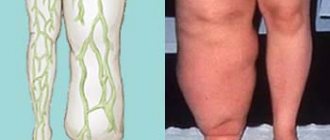
Parathyroid hormone releases calcium from the main depot - bones, which leads to bone degeneration with a decrease in its density. Bone decalcification is often accompanied by pain and fractures. This phenomenon is called parathyroid osteodystrophy.
Nephro- and urolithiasis
Additional information: Urinary tract
Additional information: Urolithiasis
Calcium is excreted from the body by the kidneys. With its excess, stones form in the urinary tract. Accompanied by a clinical picture of urolithiasis.
Gastrointestinal disorders
Additional information: Gastrointestinal tract
Additional information: Gallstone disease
An increase in calcium in the extracellular fluid leads to a transmembrane imbalance of ions, the development of degenerative processes in tissues, and an increase in the activity of sympatho-adrenal effects on the gastrointestinal tract. Which leads to nausea, vomiting, weakened peristalsis of the stomach and intestines, constipation, abdominal pain, peptic ulcers and anorexia are often detected.
Increased calcium concentrations also lead to the formation of stones in the gallbladder and pancreatic ducts.
Psychoneurological disorders
A decrease in the content of intracellular calcium leads to disruption of the formation of the resting membrane potential, disruption of cellular metabolism and plastic processes. Which is manifested by a decrease in intellectual activity, fatigue, lethargy, stupor, coma.
Cardio- and myopathies
A decrease in intracellular calcium levels leads to disruption of the formation of the resting potential, which is accompanied by muscle hypotension. The heart pumps blood worse, and arrhythmia may develop.
Vascular disorders
An increase in extracellular calcium concentration leads to increased rigidity of red blood cell membranes and reduces their ability to deform when passing through capillaries, which leads to their damage. In addition, the aggregation ability of platelets in the bloodstream increases and vascular tone increases.
What is hyperparathyroidism?
Hyperparathyroidism is a disease characterized by excess parathyroid hormone in the bloodstream.
The parathyroid glands are located in the neck on the surface of the thyroid gland and secrete a hormone called parathyroid hormone (parathyroid hormone). The main task of the parathyroid glands is to regulate the level of calcium and phosphorus in the body. Each person has four small parathyroid glands, which are usually the size of a grain of rice ().
Typically, when calcium levels drop, the body produces more parathyroid hormone (PTH) to restore levels. And when calcium levels increase, the body produces less parathyroid hormone, so levels decrease. People with hyperparathyroidism have too much calcium in their blood and less than normal (or sometimes near-normal) amounts of phosphorus.
Parathyroid hormone performs the following important functions ():
- Stimulates bones to release calcium and phosphate into the bloodstream.
- Causes the kidneys to release less calcium into the urine.
- Causes the kidneys to release more phosphate into the blood.
- Stimulates the digestive tract to absorb more calcium.
- Causes the kidneys to activate more vitamin D, which allows for increased calcium absorption.
There are two main types of hyperparathyroidism:
- Primary hyperparathyroidism : Occurs when one or more parathyroid glands become enlarged. This causes excess production of parathyroid hormone and high levels of calcium in the blood (hypercalcemia).
- Secondary hyperparathyroidism : Occurs as a result of another medical condition, such as kidney disease or vitamin D deficiency. This results in low calcium levels. Low levels of calcium in the blood cause an increase in the production of parathyroid hormones.
- Normocalcemic primary hyperparathyroidism : This is when parathyroid hormone levels are higher than normal, but blood calcium levels are normal. Many patients with normocalcemic primary hyperparathyroidism will continue to develop classic primary hyperparathyroidism.
Clinical picture
- Microscopic specimen of parathyroid adenoma.
- Microscopic specimen of parathyroid adenoma
.
- Microscopic specimen of parathyroid adenoma
.
- Microscopic specimen of parathyroid adenoma
.
- Microscopic specimen of parathyroid adenoma
.
Often, hyperparathyroidism occurs for a long time without significant clinical symptoms. There are five main signs of primary hyperparathyroidism, different combinations of which form its multifaceted clinical picture.
- Osteopathy (osteoporosis).
- Nephropathy (nephrocalcinosis and nephrocalculosis).
- Visceropathy (cholelithiasis, calculous pancreatitis, gastric ulcer).
- Psychopathy.
- Myopathy.
Since there are no specific signs of the disease, the diagnostic search is based on a comprehensive assessment of complaints, anamnesis and objective examination data.
- General symptoms
- Weakness
- Apathy
- Dehydration
- Signs of kidney damage
- Polyuria
- Nephrolithiasis and/or nephrocalcinosis
- Decreased renal excretory function
- signs of gastrointestinal damage
- Loss of appetite
- Nausea
- Constipation
- Abdominal pain
- signs of central nervous system damage
- Depression
- Cognitive decline
- Changes in psycho-emotional status
- Psychosis
- Signs of damage to the cardiovascular system
- Arterial hypertension
- Shortening of the QT interval
- Hypersensitivity to digitalis preparations
The most striking example of the clinical picture of primary hyperparathyroidism is a hypercalcemic crisis, in which the above signs of hypercalcemia take on a life-threatening nature. This complication usually develops when serum calcium concentration rises above 14 mg/dL.
Prediction of hyperparathyroidism
The prognosis of hyperparathyroidism will be favorable with timely diagnosis and surgical treatment. If the bone tissue was mildly affected, the recovery period takes about 4 months.
When a severe case of hyperparathyroidism has been identified, normal work capacity will return only 2 years after the operation. If the disease has been neglected, then often the full functionality of the organs is not restored.
With kidney damage, a less favorable recovery is predicted. It also depends on the size of the lesion. If surgery is not performed in a timely manner and medications are taken incorrectly, patients become bedridden and may die from kidney failure.
Hypercalcemic crisis
Manifested by nausea, uncontrollable vomiting, paralytic intestinal obstruction, abdominal pain, anuria, and dehydration. The skin becomes dry, its turgor decreases, severe muscle weakness is noted, and patients are unable to care for themselves. Constant bone pain becomes debilitating, and arterial hypertension appears in the first hours of the crisis. Tendon reflexes are reduced. Sometimes intravascular thrombosis and disseminated intravascular coagulation syndrome occur.
Neuropsychiatric disorders during a hypercalcemic crisis are manifested by confusion, depression, psychosis, or psychomotor agitation. When the serum calcium concentration reaches 15-18 mg/dL, depression of the central nervous system is possible with inhibition of the function of the respiratory and vasomotor centers and the development of irreversible shock. Patients in a state of hypercalcemic crisis require emergency hospitalization and intensive care.
Symptoms of hyperparathyroidism
Most often , the disease develops slowly and the symptoms are varied , so the diagnosis is made late. The main symptoms are as follows:
- Muscle weakness and lethargy: Patients feel tired from ordinary physical activity, for example, after climbing stairs or getting out of a chair, it becomes difficult to enter public transport. Often people feel pain in their feet and joints, and a “duck” gait develops;
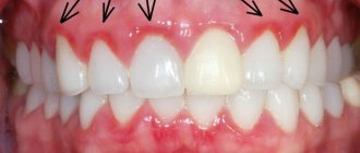
- An early symptom will be tooth loss due to osteoporosis of the jaws;
- Extreme thirst and excessive urination – quite often these symptoms lead the patient to an endocrinologist, but the doctor’s lack of awareness leads to an incorrect diagnosis of “Diabetes Insipidus”. The hallmark of hyperparathyroidism will be the lack of response to antidiuretic drugs ;
- Gastrointestinal disorders will manifest themselves as nausea, vomiting, and loss of appetite, which leads to a person’s sudden weight loss. Abdominal pain, bloating, and constipation are also common. Often an ulcerative lesion of the gastrointestinal tract is formed with a picture corresponding to the lesion;
- Arterial hypertension, the appearance of arrhythmias
- Pathological fractures - the patient’s bones break in the most unexpected places from previously familiar loads. There are even fractures of vertebral bodies under the weight of the human body. These changes lead to a change in the appearance of such patients in the later stages of hyperparathyroidism : the person’s posture is deformed, pathological curves of the spine (hump) appear, and height decreases due to compression fractures; in the area of bone fractures, false joints form (areas where the bone has not fused), with muscle atrophy and disruption of the limb.
Diagnostics
- Laboratory diagnostics:
Routine methods:
- Increase in serum concentration of calcium and its ionized form.
- Hypercalcemia in combination with a decrease in the level of inorganic phosphates.
- A sharply positive Sulkowicz test (urinary calcium excretion exceeds 200 mg/day), however, if renal function is impaired, this may not be observed [2].
- Differential diagnosis of hypercalcemia:
- Positive test with thiazide diuretics.
- Negative test with hydrocortisone.
- Instrumental diagnostics:
- Ultrasound examination is the simplest and cheapest method of instrumental diagnosis of formations of the parathyroid glands; ultrasound can be used intraoperatively to search for glands. The results very much depend on the experience of the ultrasound diagnostic doctor, since thyroid nodes, lymph nodes, muscles, blood vessels, and so on are often mistaken for parathyroid glands.
- Computed tomography with intravenous contrast is a very sensitive research method; its advantage is that it can detect the retrosternal parathyroid glands.
- Radionuclide diagnostics - assessment of the functional activity of parathyroid gland formations.
Tertiary form of the disease
Tertiary hyperparathyroidism occurs in patients who have undergone a successful kidney transplant.
As already mentioned, kidney disease is often accompanied by an increase in parathyroid hormone levels. The fact is that such pathologies are accompanied by increased excretion of calcium from the body. Prolonged hypocalcemia can lead to permanent changes in the parathyroid glands. Even after complete restoration of renal parameters, patients still experience disruption of the glands and increased secretion of parathyroid hormone.
Treatment
To date, conservative treatment methods (forced diuresis, infusion of sodium chloride or sulfate solutions, administration of bisphosphonates and glucocorticosteroids) have not shown effectiveness as an independent method of treating hyperparathyroidism.
The “gold standard” in the treatment of hyperparathyroidism is the surgical method. In the hands of experienced surgeons, its effectiveness reaches 95-98%.
The surgery is performed under anesthesia (general anesthesia). To gain access to the parathyroid glands, a 5-7 cm long incision is made on the anterior surface of the neck. Searching for the parathyroid glands is most often difficult, and therefore, for topical diagnosis of the parathyroid glands and monitoring the effectiveness of the surgical intervention, intraoperatively the following is performed: a study of the level of calcium and parathyroid hormone in the blood, Ultrasound and gamma detection of the parathyroid glands. A serious and rare complication of parathyroidectomy is damage to the recurrent laryngeal nerve (unilateral or bilateral). Clinical manifestations of damage to the recurrent laryngeal nerve are: hoarseness or lack of voice, stridor breathing. It is important that the surgical intervention is carried out in a specialized clinic by an experienced endocrine surgeon.
Treatment methods for hyperparathyroidism
We have already covered questions about what constitutes hyperparathyroidism. Symptoms and treatment in this case are closely related. If we are talking about the primary form of the disease associated with the formation of a tumor, then surgical removal of the tumor is possible. The operation is not always performed. The fact is that the disease can develop over decades without causing any particular inconvenience to the patient. And it mainly affects older people, which creates additional difficulties.
The decision about the need for surgical intervention is made by the doctor. It is believed that surgery is necessary if there is a strong increase in the level of calcium in the blood (more than 3 mmol/l) and severe disturbances in the functioning of the kidneys. Indications for the procedure are stones in the excretory system, significant loss of calcium in the urine, a history of hypercalcemic crises, as well as severe osteoporosis.
If the doctor decides not to remove the tumor or gland (if it is hypertrophied), then patients still need to undergo regular examinations - it is important to conduct examinations of the kidneys and bone apparatus at least 1-2 times a year. Constant monitoring of blood calcium levels and blood pressure is important.
As for the secondary form, treatment of hyperparathyroidism comes down to eliminating the primary diseases. Calcium deficiency in the blood can be eliminated with medication - patients are prescribed medications containing this mineral, as well as vitamin D. If taking the drugs does not give the expected effect, surgical excision of parts of the gland can be performed.
Notes
- ↑ 1 2 3 Efimov A. S.
Small encyclopedia of an endocrinologist. — 1st ed. - K.: Medkniga, DSG Ltd., Kyiv, 2007. - P. 78-81. — 360 s. — (“Library of a practicing physician”). — 5000 copies. — ISBN 966-7013-23-5. - ↑ 123
Handbook of pediatric endocrinologist / Ed. M. A. Zhukovsky. — 1st ed. - M.: Medicine, 1992. - P. 42-45. — 304 p. — 20,000 copies. — ISBN 5-225-02616-8. - Clinical endocrinology. Guide / Ed. N. T. Starkova. — 3rd ed., revised. and additional - St. Petersburg: Peter, 2002. - pp. 187-200. — 576 p. — (“The Doctor’s Companion”). — 4000 copies. — ISBN 5-272-00314-4.