Hello! My retired father was diagnosed with atrophy of the leg muscles. The treatment prescribed to him does not produce results. What else can you do?
If atrophy of the leg muscles develops, treatment is prescribed taking into account the cause of the disease. Elastic muscle tissue is replaced with connective tissue that does not have the ability to contract. Such changes can lead to paralysis.
Atrophy is a dangerous pathology that can cause loss of the ability to move. Manifestations:
- weakness in the legs, worsened by physical activity;
- feeling of trembling;
- gait disturbance;
- crawling sensation;
- change in the volume of the sore muscle;
- difficulty going up and down stairs.
Symptoms and treatment
The onset of the disease is accompanied by rapid leg fatigue and an increase in the size of the calf muscles. More often there are atrophied proximal leg muscles. There is a limitation of motor functions; it is difficult for the patient to get up from a lying position.
Atrophy of the leg muscles can last for several years. The disease affects one or both sides at once, symmetrically or asymmetrically. Symptoms depend on:
- state of the body;
- age;
- forms of the disease;
- the reasons that caused the pathological process.
When choosing treatment, they are guided by the patient’s age, the causes of the disease, and the degree of severity.
Prescribed medications:
- Galantamine to improve the conduction of nerve impulses.
- Pentoxifylline for better blood supply to the legs (Papaverine, No-Shpa have a similar effect).
- B vitamins normalize the functions of the peripheral nervous system, accelerate metabolic processes in tissues, which helps to quickly restore muscle volume.
The treatment is long-term; the course must be repeated over several months. During this period, physiotherapy and massage, therapeutic exercises are carried out. All prescriptions are made by the doctor, and only after consultation with him can the chosen regimen be changed. Traditional medicine uses calcium tincture (prepared from fresh eggs with lemon juice), medicinal herbs, and grains.
It is important to eliminate the cause. Attention should be paid to neurological and infectious diseases, atherosclerosis. Unfortunately, there are no drugs yet that are guaranteed to eliminate muscle atrophy. With proper treatment, the process slows down, muscle fibers are restored faster, and lost capabilities gradually return.
Atrophy of the muscles of the arm, hand, and forearm develops mainly as a secondary disease against the background of impaired innervation (nutrition, blood circulation) in a certain area of muscle tissue and less often as a primary disease (usually with myopathy), when motor function is not impaired.
Causes of muscle atrophy in the arm, shoulder and hand
For the development of muscle atrophy, the predisposing reasons are: occupational factor (constant overexertion during heavy physical labor), wrist joint, endocrine pathology - obesity, and thyroid disease, acromegaly; cicatricial processes after injuries, metabolic and systemic diseases (lupus erythematosus), of various origins, congenital pathologies of the lower limb.
Symptoms of muscle atrophy in the arm, shoulder and hand
Muscle atrophy is a serious disease that primarily affects muscle fibers. The main typical symptom is the symmetry of the lesion (except for myasthenia gravis) and the slow development of the disease (except for myositis), atrophy of the affected muscles and weakening of tendon reflexes with preserved sensitivity.
Most peripheral nerves have a mixed structure, and when damaged, the work of sensory, motor and autonomic fibers is disrupted. It happens that one of the fibers is most affected.
If motor fibers are involved in the process, then paresis of the muscles innervated by this nerve occurs. The patient complains of muscle weakness, low muscle tone. Atrophy does not develop immediately, but 2-3 months after the lesion. In the absence of proper treatment, after a year and a half, the muscle will completely atrophy.
If sensory fibers are involved in the process, the clinic manifests itself as parasthesia - patients feel a tingling sensation, goosebumps. Neurological symptoms are expressed in the form of hypersthesia (increased sensitivity) or hypoesthesia (decreased sensitivity). A feeling of numbness in the affected limb appears with extensive damage to the nerve fibers.
In most cases, there is a decrease in pain sensitivity while maintaining tactile sensitivity. In the later stages of the disease, deep hypoesthesia occurs, up to a complete lack of sensitivity. Redness or paleness of the skin, the appearance of a marble pattern indicates vascular disorders that occur when the vegetative fibers are directly damaged.
As a rule, increased or decreased sweating of the affected limb is associated. The concern is a burning pain of a hyperpathic nature, radiating to the entire limb involved in the process. The trophism (cellular nutrition) of tissues is disrupted due to deep vegetative disorders.
Atrophy of the arm muscles begins, as a rule, from the most remote or distal parts of the upper limbs. The hand takes on the appearance of a “monkey hand” due to damage to the interosseous muscles and fingers. There is a complete loss of tendon reflexes, but sensitivity is retained in the affected limb. As the disease progresses, the muscles of the neck and torso become involved in the process.
Diagnosis of atrophy of the muscles of the arm, shoulder and hand
Making a diagnosis does not currently cause any particular difficulties due to the introduction of electromyography and biopsy of the affected muscles into the clinical examination. The patient is required to undergo biochemical and general blood tests, and a urine test; The activity of muscle enzymes is determined in blood serum (mainly by the CPK method). Creatine and creatinine levels are calculated in urine. According to indications, the patient is sent for a CT or MRI of the cervicothoracic spine and brain, and examination for endocrinological pathology.
Treatment of muscle atrophy in the arm, shoulder and hand
When choosing a treatment method, the following factors are taken into account: the form of the disease, the severity and extent of the process, the age of the patient. Along with drug treatment, great importance is attached to proper nutrition, physiotherapeutic procedures, courses of therapeutic massage and gymnastics, and electrotherapy. In some cases, it is appropriate to prescribe psychotherapy sessions for the patient.
Currently, there is no drug that can completely cure muscle atrophy, but the correct choice of treatment method and timely diagnosis can slow down the pathological process, restore muscle regeneration and return lost abilities to the patient. The main thing is to strictly adhere to the doctor’s recommendations.
Expert editor: Mochalov Pavel Alexandrovich | Doctor of Medical Sciences general practitioner
Education: Moscow Medical Institute named after. I. M. Sechenov, specialty - “General Medicine” in 1991, in 1993 “Occupational diseases”, in 1996 “Therapy”.
Muscle atrophy is a weakening of muscle fibers, a decrease in their size, or degeneration due to prolonged immobility. Even minor atrophy entails a decrease in muscle mobility and strength. In other words, the disease leads to thinning and even disappearance of muscle fibers, which means long-term immobility or limitation of the patient’s motor activity.
Preventive measures
To prevent muscle atrophy, the following rules must be followed:
- eat a balanced diet;
- avoid heavy loads;
- consult a doctor in a timely manner;
- wear comfortable shoes;
- lead a healthy lifestyle.
Muscle atrophy is a complex pathology that cannot be completely eliminated. If you follow all the recommendations, visit doctors and eat right, you can significantly improve your quality of life.
Malakhov Yuri
Cardiovascular surgeon of the highest category, phlebologist, ultrasound specialist, Honored Doctor of the Russian Federation, Doctor of Medical Sciences
Varicose veins and all problems associated with human hips.
- Varicose veins of the lower extremities.
- Postphlebitic syndrome.
- Acute thrombophlebitis.
- Trophic ulcers.
- Deep vein thrombosis.
- Lymphedema of the lower extremities.
- "Spider veins."
- Obliterating atherosclerosis of the vessels of the lower extremities.
- Diabetic foot syndrome.
- Stenosis of the carotid arteries.
Higher education:
- 1985 — Military Medical Academy named after S. M. Kirov (therapeutic and preventive care)
- 1986 - Military Medical Academy named after S. M. Kirov (internship in the Northern Fleet, specialty: “surgery”, Murmansk)
- 1991 — Military Medical Academy named after S. M. Kirov (clinical residency at the Department of Naval and Hospital Surgery)
Training:
- 1992 – Training in angiography and vascular surgery in Hamburg, Germany
- 1992 — Vascular surgery
- 2003 — Cardiovascular surgery
- 2004 - Internship at the University Clinic of Nuremberg (Clinic of Vascular Surgery) Professor D. Raithel; Germany
- 2006 — Lymphedema and venous edema: European treatment experience
- 2006 - Internship at the University Clinic of Nuremberg (Clinic of Vascular Surgery) Professor D. Raithel; Germany
- 2008 — Cardiovascular surgery
- 2008 — Dornier Medilas D MultiBeam laser system
- 2009 — “Ultrasound research methods in the diagnosis of surgical pathology of the vessels of the lower extremities”
- 2009 — Cardiovascular surgery
- 2009 — Training in a phlebology clinic; Wiesbaden, Germany.
- 2012 — “X-ray endovascular diagnosis and treatment”
- 2013 — “Cardiovascular surgery”
- 2016 — “Ultrasound diagnostics”
Experience:
- 1985-1989 Large nuclear submarine of the Northern Fleet
- 1989-1991 Military Medical Academy named after S.M. Kirov
- 1991-1994 Central Naval Clinical Hospital
- 1994-1998 Central Naval Clinical Hospital
- 1998-2015 Central Naval Clinical Hospital
- 2016-present V. Multidisciplinary clinic CELT (Center for Endosurgery and Lithotripsy)
Muscle atrophy is a serious disease that leads to a decrease in volume and transformation of muscle tissue. Thinning of muscle fibers can lead to limitation of motor activity, and even to complete immobilization of the patient.
Most often, the disease develops with an inactive lifestyle, various injuries, joint pathologies, and nervous system disorders. If there are clear signs of muscle atrophy, such as rapid fatigue, decreased muscle tone, or characteristic twitching of the limbs, the patient should seek medical help to avoid further complications. As an additional treatment, you can use traditional medicine recipes, but only after consultation with a neurologist.
Treatment of muscle atrophy with tincture of eggshells, lemon, honey and cognac
It is believed that muscle atrophy can develop due to a lack of calcium in the body. It would seem that it is quite easy to replenish its reserves, because pharmacies sell all kinds of preparations with this macroelement, but only organic calcium is useful for the human body.
An effective calcium tincture can be prepared using this recipe. Take 6 fresh eggs with white shells, wash them thoroughly and carefully place them in a glass jar. Squeeze the juice from 10 lemons and pour it over the eggs. The neck of the jar is tied with gauze, and the jar itself is wrapped in dark paper, after which it is placed in a warm place for 5-8 days until the eggshell is completely dissolved.
The eggs are carefully removed, and 300 g of heated linden honey mixed with 200 ml of cognac is poured into the jar. To avoid decomposition of calcium, the drug is stored in a dark, cool place and consumed 1 dessert spoon 3 times a day immediately after meals.
Treatment of muscle atrophy with herbal infusion
The herbal mixture for the treatment of muscle atrophy includes: calamus root, sage, flaxseed, corn silk and knotweed, taken 100 g each. To prepare the infusion, 3 tbsp. spoons of mixed crushed herbs should be poured into a thermos with 0.7 liters of boiling water and left overnight. After filtering, the resulting liquid is divided into 4 equal portions, each of which is drunk an hour before meals.
Treatment of muscle atrophy with oat kvass
Oatmeal kvass for muscle atrophy can be drunk all year round, because it is not only healthy, but also tasty. To prepare an amazing drink, you should take a half-liter jar of high-quality oat grains (in the shell, but not in the husk), rinse them 3 times and pour them into a 3-liter jar filled with purified water. You should also add 1 teaspoon of citric acid and 3 tbsp. spoons of sugar. In 3 days the kvass will be ready. With one serving of oats, kvass can be prepared 3 times, adding sugar and water (without citric acid).
It is also recommended to eat sprouted wheat grains as often as possible, eat corn, millet and oatmeal porridge, and use foods high in vitamin B.
Treatment of muscle atrophy with reed panicles
The most popular traditional medicine recipe for muscle atrophy is considered to be a recipe for the preparation of which reed panicles are used. They are used fresh, collected from October to March. Two bunches of panicles are poured with boiling water for 45 minutes, after which they cover the limbs, securing them to the body with bandages and wrapping them warmly. When the compress has cooled, the patient’s arms and legs are massaged, thoroughly rubbing the muscles, starting from the fingertips.
Forms
The initial stage of the disease is characterized by symptoms such as pathological fatigue of the leg muscles when running and walking, and, less commonly, spontaneous muscle twitching and an increase in the volume of the calf muscles. The disease is initially localized in the lower extremities, pelvic girdle, hips, which leads to atrophy of the leg muscles. This causes difficulty walking, especially on stairs, and leads to a change in gait.
Primary muscle atrophy
The main symptom is direct muscle damage. This type of pathology may be due to poor heredity or bruises, injuries, or physical stress. In this case, the patient quickly gets tired, his muscle tone decreases, involuntary muscle twitching is noted, which indicates motor neuron disorders.
Secondary muscle atrophy
This type of pathology develops most often after infections and severe injuries. Patients experience atrophy of the muscles of the legs, hands and forearms, which leads to partial or complete paralysis of the limbs. In most cases, the disease progresses slowly, but exacerbations are accompanied by severe pain.
The secondary form of the disease is divided into several types:
- neural myotrophy, when atrophy of the leg muscles with deformation of the legs and feet is observed. When walking, a person has to raise his knees high, and his gait is greatly impaired. Gradually, the feet lose reflexes, and the disease progresses to the remaining limbs;
- Arand-Duchenne muscular atrophy, localized in some parts of the arms. In patients, the fingers and interosseous muscles atrophy, as a result of which the upper limbs take on the appearance of a so-called “monkey hand”. Sensation in the hands is preserved, but tendon reflexes are absent. The process progresses steadily and reaches atrophy of the muscles of the neck and torso;
- progressive muscle atrophy first appears, as a rule, in children. The disease is extremely severe and is characterized by loss of tendon reflexes, hypotension and twitching of the limbs.
Anatomy of the pectoral muscles
The following muscles are directly related to the pectoral muscles:
- Intercostal muscles. Located between adjacent ribs. The main function is to reduce the intercostal spaces, which ensures the act of exhalation.
- Serratus anterior muscles. Located directly above the skeleton of the chest. They stretch from ribs 1–9 to the inner edge of the shoulder blades on both sides. The main function of the muscles is to create the so-called muscle ring, which presses the shoulder blades to the ribs from the back. Thus, the serratus anterior muscles indirectly participate in the movements of the upper limb girdle.
- Pectoralis minor muscles. Also start from the ribs. But they go over the serratus muscles and are attached to the acromion process of the scapula. When it contracts, the scapula is pulled forward and downward. Muscles can also take part in inhalation.
- Large pectoral muscles. With their wide part, the muscles are attached to the lower edge of the collarbone, the cartilage of the ribs and the tendons of the latissimus abdominis muscle. The other edge of the muscle is attached to the tuberosity of the humerus. The muscles are involved in the movement of the arms and the act of breathing.
It is important! Thus, all the muscles of the chest (with the exception of the intercostal muscles) are involved in the movements of the upper limbs or its belt, and also contribute to breathing. These circumstances determine the list of reasons leading to their dystrophic changes. Also, these circumstances are leading in the development of the clinic of muscle lesions.
Treatment
The choice of methods and means of treating muscle atrophy depends on the patient’s age, the severity of the process and the form of the disease. Drug therapy usually involves subcutaneous or intramuscular administration of the following drugs:
- Atrifos or Myotrifos (adenosine triphosphoric acid);
- vitamins E, B1 and B12;
- galantamine;
- Prozerin.
Proper nutrition, therapeutic exercises, massage and physiotherapeutic procedures, and psychotherapy are also of great importance in treatment. When muscle atrophy occurs in a child who lags behind his peers in intellectual development, neuropsychological sessions are prescribed.
Muscle atrophy is a symptom of a certain pathological process that leads to thinning of muscle fibers, and as a consequence, to immobility of the patient. It should be noted that the development of this pathology is quite long in time - from several months to several years. In fact, muscle tissue is replaced with connective tissue, which entails disruption or complete loss of motor function in a person. Treatment of such a disorder should be carried out strictly under the supervision of a specialized medical specialist.
Etiology
Clinicians distinguish two types of etiological factors of muscle atrophy - primary and secondary. The primary form of the disease is hereditary, and any neurological pathologies can only aggravate the pathology, but will not become a provoking factor.
Secondary etiological factors include the following:
- constant physical stress, which is a consequence of excessive physical exertion in sports or due to the nature of work;
- infectious pathologies;
- nerve ending injuries;
- pathologies of motor cells of the brain;
- infectious diseases with typical etiology.
In addition to pathological processes that can lead to muscle atrophy, general predisposing factors for the development of this pathological process should be identified:
- disturbances in the functioning of the peripheral nervous system;
- paralysis;
- mechanical damage to the spine;
- lack of proper nutrition and rest;
- damage to the body by toxic substances;
- disruption of metabolic processes in the body;
- long bed rest.
It should be noted that quite often this symptom can be observed after severe trauma to the musculoskeletal system or being immobile. In any case, rehabilitation after such pathologies should be carried out only by a qualified medical specialist. Self-medication (in this case we are talking not only about taking medications, but also about massage, exercise therapy) can lead to complete disability.
Why does atrophy develop?
The circumstances due to which muscle atrophy occurs are of two types. The first type refers to burdened heredity. The condition is aggravated by neurological disorders, but they do not provoke atrophy. The secondary type of pathology is associated with external root causes: pathologies and trauma. In an adult, the muscles will begin to atrophy first in the arms.
In children, myofibers atrophy due to:
- Neurological disorders, for example, autoimmune pathology causing muscle paresis (Guillain-Barré syndrome).
- Benign pseudohypertrophic myopathy (Becker myopathy). Appears due to complicated heredity in adolescence and in young people 25–30 years old. This is a mild degree of atrophic changes with damage to the calf myofibers.
- Injuries during childbirth, difficult pregnancy.
- Spinal paralysis in a child caused by infection (poliomyelitis).
- Stroke in a child. Microcirculatory processes in the vessels of the brain are disrupted due to blood clots or hemorrhage.
- Abnormal development of the pancreas.
- Chronic inflammation of muscle tissue.
The main causes of muscle atrophy in adults:
- A job in which a person experiences constant overexertion.
- Incorrectly selected physical education classes, when the load is incorrectly calculated according to the person’s weight.
- Endocrine dysfunctions. If a person is sick, for example, with diabetes, then metabolic processes are disrupted and polyneuropathy occurs.
- Poliomyelitis infection or other infectious pathologies that cause movement disorders.
- Oncological processes of the spinal column, causing compression of the spinal nerve fibers. Their nutrition and conductivity are disrupted.
- Paralysis after injury, infarction changes in the brain.
- Vascular disorders and disorders of the central nervous system, PNS. Oxygen deficiency occurs, muscles starve.
- Chronic intoxication syndrome, which occurs during prolonged contact with chemical toxins, alcohol, drug intoxication.
- Physiological aging, due to which muscles atrophy.
Symptoms
At the initial stage of development, atrophy of the muscles of the back or other parts of the body manifests itself only in the form of increased fatigue from physical activity. As a consequence of this, the patient may experience pain.
As symptoms develop, the clinical picture may be supplemented by the following symptoms:
- with atrophy of the muscles of the limbs it is observed;
- restriction of movements of arms, legs, torso;
- change in habitual gait;
- loss of sensation in the limbs;
- low blood pressure.
If the cause of atrophy of the thigh muscles or other parts of the body is an infectious process, then the clinical picture may be supplemented by the following symptoms:
- – insomnia during the day and increased sleepiness at night;
- , especially at night;
If the cause of spinal muscle atrophy is damage to the nervous system, then the overall clinical picture may be supplemented by the following signs:
- impaired motor function, up to complete paralysis.
The severity of symptoms in spinal atrophy will depend entirely on the severity of the injury or the degree of deterioration in muscle tone. Therefore, you should consult a doctor at the first signs of damage to muscle tissue. In this case, serious complications can be avoided.
Features of manifestations in children
Since atrophied muscles in children are in most cases caused by genetic factors, the first symptoms can be detected in utero - late and weak movements of the fetus are noted; after birth, such children usually die in the first few weeks of life due to paralysis of the respiratory muscles.
With the development of atrophy in infancy, the so-called flaccid child syndrome is formed; in such children, a characteristic “frog pose” is noted - with widely separated hips and a flat stomach, a pronounced decrease in general tone and motor activity, with some diseases there is a disorder of sucking, swallowing and breathing.
Muscle dystrophy in an older child is manifested by disturbances in motor activity and specific deformities of the limbs.
Diagnostics
If you suspect the development of a muscle atrophy process, you should immediately seek medical help. The doctor's specialization during the initial examination will depend on the current clinical picture and general condition of the patient.
The diagnostic program consists of the following:
- physical examination with general history;
- clinical and biochemical blood test;
- electromyography;
- hormonal studies;
- Ultrasound of the thyroid gland;
- muscle tissue biopsy;
- nerve conduction testing;
- CT and MRI.
Additional diagnostic methods will depend on the current clinical picture and the patient’s condition at the time of seeking medical help. It is important that if the patient took any medications to eliminate symptoms, the doctor should be notified about this before the diagnosis begins.
Treatment
Basic therapy will depend entirely on the identified underlying factor. Treatment, first of all, will be aimed at eliminating the underlying disease, and only then the symptoms.
In this case, it is impossible to single out a single treatment program, since muscle atrophy is a nonspecific symptom and therapy will depend not only on the etiology, but also on the age of the patient. In any case, exercise therapy, massage and optimal physiotherapeutic procedures are almost always included in the complex of therapeutic measures.
Prevention
There are no targeted preventive measures, since this is a symptom and not a separate ailment. In general, you should adhere to the rules of a healthy lifestyle and prevent those ailments that can cause such a disorder.
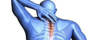
A disease such as atrophy of the lower leg muscles is a fairly rare pathology that develops for a number of reasons related to impaired nutrition and innervation of muscle tissue, as well as when the internal metabolic processes of the muscle are disrupted.
It is important!
The disease is a destructive process of striated muscles, accompanied by a decrease in organ volume, replacement of muscle fibers with fatty and connective tissue, as well as a significant decrease in the functionality of the affected limb.
Muscle atrophy most often develops in young men, as well as among people who have impaired motor function of the legs and move in a wheelchair. In addition, the development of the atrophic process can be influenced by pronounced vascular atherosclerosis or other factors affecting their capacity.
Atrophy of muscle tissue, including tissue of the lower leg, can be primary or secondary. The primary form develops as an independent disease, often associated with metabolic disorders directly in the affected muscle. Secondary atrophies are the result of other existing somatic and neurological diseases.
Brief description of the disease
In medical practice, several types of atrophy are distinguished, but we will talk about them a little lower, but for now we will dwell in more detail on the reasons for the appearance of this pathology. Atrophy of the muscles of the legs and other limbs of the body occurs due to:
- decreased metabolism and aging of the body;
- chronic diseases of the endocrine glands, intestines, stomach, connective tissue;
- infectious and parasitic diseases;
- disorders of the nervous regulation of muscle tone associated with poisoning, polyneuritis and other factors;
- congenital or acquired enzyme deficiency.
Reasons for the development of atrophic phenomena in the lower leg muscles
Depending on the form of the disease (primary or secondary), leg muscle atrophy can develop for several different reasons. Thus, primary atrophy often occurs due to the following pathological processes:
- Hereditary disorder of metabolic processes directly in the muscle itself
- Infectious processes in striated muscles
- Traumatic injuries associated with long-term disruption of muscle nutrition processes
- Frequent physical overexertion without an appropriate recovery regimen
Secondary atrophies, as mentioned above, are the result of generalized somatic diseases. Moreover, the factors that influence the development of atrophy do not always have a direct effect on the leg muscles.
Diseases such as:
- Damage to the nerve trunks innervating the lower limbs and the anterior horns of the spinal cord. Pathology can occur due to exposure to infectious, traumatic, and trophic factors.
- Circulatory disorders accompanied by insufficient blood supply to the lower extremities
- Long-term inactivity of muscle groups associated with the patient’s disability and his movement in a wheelchair.
- Genetic pathology of innervation processes of the lower extremities in the spinal cord
Atrophy of the muscles of the thigh and lower leg can begin with the influence of certain provoking factors. Thus, long-term smoking, alcohol abuse, and a diet that includes large amounts of unhealthy fats and contributes to the development of atherosclerosis contribute to the development of the disease.
It is important!
Atrophic phenomena, if there are prerequisites for them, can develop with low physical activity, a sedentary or recumbent lifestyle. The other extreme is also important - excessive physical activity, not provided with a sufficient amount of proteins and carbohydrates to restore muscle tissue.
Common Causes of Contracture Deformity
The most common causes of contracture are inactivity and scarring from injury or burn. People who have other conditions that prevent them from moving are also at higher risk of contracture strain.
For example, people with severe osteoarthritis (OA) or rheumatoid arthritis (RA) often develop contractures. Because their muscles and joints do not move within their normal range, contracture may occur in these tissues.
For example, joint contractures are common in patients discharged from intensive care units or after long hospital stays. They are also very common in people who have suffered stroke and paralysis.
Other causes include diseases that are inherited or develop in early childhood, such as:
- Muscular dystrophy. People with this condition often experience muscle tension because significantly weaker muscles impair their ability to move.
- Cerebral paralysis. This disease causes muscle tension and limits movement.
- Diseases of the central nervous system. These include polio, multiple sclerosis (MS) or Parkinson's disease.
- Inflammatory diseases. Having rheumatoid arthritis (RA) puts you at greater risk of contracture deformity.
Clinical signs and diagnosis of muscle atrophy of the lower extremities
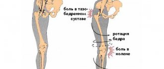
The development of the disease occurs slowly, with a gradual increase in the severity of the clinical picture. The first symptoms of the disease are the appearance of muscle weakness, changes in the patient’s gait, and rapid fatigue. The gait that occurs at the initial stage of development of atrophy is swaying, reminiscent of a duck.
Over time, a significant decrease in volume of the affected muscles becomes visually visible. They lose tone and become flabby. The patient cannot walk for a long time and gets tired quickly. He needs rest.
With atrophy of the lower leg muscles, deformation and atony of the lower limb occurs. In this case, raising the patient's leg leads to limp sagging of the foot. With disturbances in nerve conduction, which is the cause of atrophy, the limb may lose pain sensitivity and lose previously existing reflexes.
Atrophy itself is extremely rarely accompanied by the development of pain. However, such sensations may occur due to the underlying disease that caused atrophic phenomena (intermittent claudication due to atherosclerotic lesions of the vessels of the legs).
Diagnosis of the disease is carried out on the basis of visual examination, palpation of the muscle formations of the legs, as well as after assessing their functional state. At the same time, it is important to remember about the possible phenomena of pseudohypertrophy, when the subcutaneous tissue volume can be preserved due to fat and connective tissue deposits that arise in the place of the atrophied muscle.
Therapeutic exercise and physiotherapy
A set of exercises for restoring atrophied muscles is designed so that the load is applied in doses, with a gradual increase. At the same time, muscle mass increases and functionality returns.
At the first stage, exercises are carried out to prevent fatigue or pain. All movements are performed in slow mode. It is important to understand that at the moment the body is weakened and does not have the strength it had before the defeat.
It is recommended to start with exercises in water. This allows you to make all muscle groups work with minimal load. In addition, water will reduce pain. Regular exercise in the pool helps restore tone in the affected areas of the body, and the memory of functioning returns to the muscles. Damaged muscle tissue relaxes. It is advisable to carry out all exercises with an instructor, who will tell you the correct execution and can advise additional movements for better results.
While in the pool, perform the following actions:
Walking in the pool does not pose a threat to health; on the contrary, the muscles develop and strengthen. The lower body also becomes significantly healthier. At this time, objects taken to maintain balance are used to train the upper half.
Raising your legs to hip height gives good results. Push-ups from the pool floor allow you to develop the muscular system of the upper limbs. To do this, you need to go to the wall of the pool, put your hands on the floor and lift your body out of the water to the position of outstretched arms, then smoothly return to the water. If this exercise cannot be performed, then a simpler version is used: place bent elbows on the floor and lean towards the wall.
After sufficient strengthening of the muscles, they begin to perform exercises without using water space. In this case, the first classes are carried out under the supervision of a doctor or an experienced instructor, then the second stage is allowed to be performed independently.
The movements are performed from a lying position. This includes raising your legs, training your arms, and stressing the whole body.
Treatment of muscle tissue atrophy
It is important! The existing treatment of atrophy of the muscles of the thigh and lower leg is a complex measure, including both pharmacological and physiotherapeutic measures. In addition, some traditional medicine recipes and physical therapy can be used as an auxiliary measure.
In addition, an important point in the treatment of atrophy is the elimination of the factor that caused the disease. Thus, active treatment of atherosclerosis, infectious and neurological diseases is carried out.
Drug treatment of muscle atrophy
Drug treatment of muscle atrophy is aimed at improving blood flow in the affected limb, improving the conduction of nerve impulses, and increasing the activity of anabolic metabolic processes.
Thus, the following medications are most often prescribed to patients:
- Nivalin (galantamine). This drug helps to significantly facilitate the conduction of nerve impulses. It is used for a long time, with a gradual increase and decrease in dose. Can be used exclusively as prescribed by a doctor in the form of subcutaneous, intramuscular or intravenous injections.
- Pentoxifylline (trental). Used to dilate peripheral blood vessels and improve blood flow in the lower extremities. Antispasmodics (papaverine, no-spa) can also be used for the same purpose.
- B vitamins (cyanocobalamin, thiamine, pyridoxine). Vitamins help improve the conduction of nerve impulses and the functioning of the peripheral nervous system as a whole. In addition, they significantly increase the intensity of metabolism in organs and tissues, which contributes to a more rapid restoration of lost muscle volume.
Physiotherapeutic techniques
It is an indication for prescribing a course of electrotherapy to patients. In this case, the affected tissues are exposed to currents of low voltage and strength, which is a stimulating regenerative factor. The procedure is painless and does not bring any discomfort to the patient. However, electricity is not used as an independent method of treatment due to the low effectiveness of the method.
In addition to electrotherapy, patients are prescribed massage treatments. Massage for atrophy of the leg muscles helps improve blood flow, which ensures accelerated regeneration of muscle tissue due to the normalization of the processes of its nutrition and cellular respiration.
Physiotherapy
Restoring muscle mass after atrophy is impossible without some physical activity, the intensity of which directly depends on the patient’s capabilities. As a rule, after severe atrophy, the beginning of physical exercise occurs in bed or within the confines of the room. Subsequently, exercises take place in a gym or playground.
It is important!
Physical education should be combined with good nutrition. Thus, restoration of muscle mass requires obtaining 2 grams of protein per 1 kg of the patient’s body weight per day. In addition, the patient must receive the required amount of carbohydrates and fats. Otherwise, there is a risk of worsening the patient's condition.
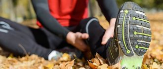
How long does it take for an ankle sprain to heal?
Three bones—the tibia, fibula, and talus—form the ankle joint. Its functionality is ensured by the ligamentous apparatus - three groups of connective tissue cords that fix the bones to each other. Ligaments maintain the stability of the joint, somewhat limiting the mobility of its components. They also “control” the rotation, abduction and adduction of the foot.
When the range of motion of the foot exceeds the permissible range, the ankle ligaments can become torn. Popularly, such an injury is called a sprain.
The ligaments that run along the outer ankle are most susceptible to it. Why does ankle sprain occur, how long does it take to heal, and what measures will speed up recovery?
Victims of beauty and more
Doctors call the leading cause of sprains a twisted leg, when the ankle suddenly moves outward and the heel turns inward. This situation is familiar to almost all lovers of high heels. If an unsuccessful “start” in high-heeled shoes is accompanied by a crunching or cracking sound, a complete rupture of the ligament or bone damage should be suspected.
Sometimes the tendon-ligamentous system is weakened already from birth, or there are congenital changes in the configuration of the foot - then the slightest traumatic impact is fraught with spraining the ligaments.
And only in 6 patients out of 1000, damage to the ligamentous apparatus of the ankle joint occurs during sports battles. Most often, emergency assistance is required for skaters and skiers who, during sudden braking at high speed, experience an inward rotation of the foot with subsequent spraining of the ligaments.
Other causes of “tear” of the ligamentous fiber include excess weight, carrying heavy loads, excessive physical activity caused by professional needs, as well as a number of diseases – flat feet, arthrosis and inflammatory phenomena in the joint.
Aw, it hurts!
Depending on the extent of damage - complete or incomplete rupture - there are 3 degrees of ankle sprain, each of which is characterized by symptoms:
- Degree 1 – delamination of ligament fibers. Swelling and tenderness in the ankle, slight. the victim limps;
- When the ligaments of the ankle joint are sprained grade 2, swelling of the soft tissues is pronounced, the victim experiences severe pain, and a hematoma forms in the joint area. Movement is difficult;
- Symptoms of grade 3 ankle sprain indicate a complete rupture of the ligaments: the injury is accompanied by severe swelling with extensive hematoma and hemarthrosis; local hypothermia or increased body temperature. Sharp pain makes it impossible to stand on your foot or take a step, but the foot is unnaturally mobile.
What to do if you sprain your ankle?
In the event of a significant injury - if the victim cannot lean on his leg - an ambulance should be called and the victim should be provided with first aid, which, in the case of a sprained ankle, consists of maximum immobilization of the joint by applying a splint from "improvised" means.
For this purpose, a narrow board is used, which is bandaged, covering the lower leg, ankle and knee.
For mild sprains of the ankle joint, a figure-eight bandage is applied. It is secured by making a coil at the level of the ankles, and gradually, in eight-shaped coils, it is lowered onto the foot. Be sure to apply ice to the injured area and give the victim an anti-pain medication. Sometimes a complete tear requires a cast.
How to treat a sprained ankle?
Having received qualified assistance from a traumatologist or surgeon, the victim continues treatment for a sprained ankle at home.
How to fix your foot
Within 7 days, the injured ankle needs to be fixed and completely unloaded. Fixatives - a bandage or tape (an elastic bandage held in place by a bandage) - should not be left in place for more than 2-3 hours. They should not be too tight, otherwise the vessels adjacent to the injured tissues will be compressed and the cyanosis will increase.
Taping the ankle joint when spraining the ligaments is correctly performed as follows:
- The patient is laid down; feet at an angle of 90 degrees;
- The instep of the foot is lubricated with Vaseline and 2 shock-absorbing pads are placed on it, covering the entire damaged area;
- 3-4 strips of tape are wrapped around the shin. The strips must be overlapped;
- From the next 3 strips, a “stirrup” is formed that covers the bottom bandage;
- Close the “stirrup” by making 7-8 turns from top to bottom towards the foot. The strips are laid overlapping;
- The foot is wrapped with 2-3 strips of tape and, having secured it on the instep, it is moved across the foot to the sole and placed under the heel;
- Place the tape around the heel and secure it in the part of the ankle where you began to fix the instep. The strip is torn off;
- The heel is fixed by placing a new strip of tape across the foot and passing it through the sole;
A more affordable and universal means of fixation is an elastic bandage - indispensable when treatment of an injury is accompanied by the application of a plaster.
How to apply an elastic bandage to an ankle:
- Moving in a circle, form the beginning of the bandage slightly above the ankle;
- Having covered the heel with a bandage, grab the foot twice;
- Next, the bandage is moved in a cross-shaped motion: from the foot to the shin and again to the foot;
- Once again returning to the shin, wrap the ankle several times and secure the bandage.
You will learn how to apply a bandage correctly from the video:
In addition to an elastic bandage and tape for the ankle, special fixatives are used for sprained ligaments - elastic therapeutic ones that stop the inflammatory process, or hard ones that provide maximum immobilization in case of a serious injury.
For basketball fans, special sneakers with a lock are also produced.
How to relieve swelling
A bruise and sprain of the ankle with swelling puts the victim to bed for several days. To reduce swelling of the feet, it is recommended to lie with your legs raised up.
On the first day, ice compresses are applied to the injured area covered with gauze. They are done by holding for 15-20 minutes, then a half-hour break and the sore spot is cooled again.
The next day, they move on to warming procedures. A hot heating pad and warm baths relax muscles, speed up blood flow, promoting healing. In the absence of bone damage, swelling will subside by the 3rd day, and a hematoma, even a large one, will go away after 4 days. If, after 14 days after an ankle sprain, the swelling still does not go away, you should visit a doctor, who will recommend detailing the consequences of the injury through an X-ray examination and MRI.
How to relieve pain
Anti-inflammatory therapy is an essential component of treatment for ankle sprains.
On the first day, severe pain will be relieved by injections of ketanov, analgin, and renalgan. On the second day they are replaced with tablets.
From the third day, the treatment is made more intense by adding ointments - for sprained ankles, these are drugs that promote pain relief - Nicoflex, Finalgon, as well as blood outflow and “resorption” of the hematoma - Troxevasin, Dolobene, Lyoton, Indovazin.
Is it possible to walk with a sprained ankle? You can walk if it doesn't hurt. If there is pain, orthopedic doctors and traumatologists do not recommend “loading” the ankle, or advise moving using crutches.
How to restore “stretched” ligaments?
A minor injury allows you to begin rehabilitation within 3-4 days.
This early start will prevent joint stiffness and muscle atrophy.
Exercise therapy
Simple gymnastics will speed up the recovery of ankle ligaments after a sprain:
- Flexion and extension of the ankle is very effective, incl. with little weight; spinning it;
- Carefully move around the room, bending and unbending your fingers and fingering them;
- Shift from heel to toes and back;
- Hook your toes on the leg of the chair and pull it towards you.
You will learn more exercises for ankle sprains in the video:
Massage
It is started with a favorable course from 2-3 days. When spraining the ankle joint, the massage begins with stroking and kneading the area just above the injury site. Then, grasping the adjacent muscle tendons and trying to penetrate the fingers deep into the joint, they stroke and rub the ankle itself. The massage effect promotes the outflow of exudate and the resorption of subcutaneous hemorrhages, and accelerates the fusion of ligaments.
Physiotherapeutic treatment
It is prescribed to relieve pain, in combination with exercise therapy and massage. For ankle sprains, physiotherapy includes laser and paraffin treatment, diadynamic therapy and UHF. Magnetic therapy is also used to accelerate lymphatic drainage and increase vascular tone.
Read more about physiotherapeutic treatment methods in this article...
Folk remedies for ankle sprains
With the permission of the doctor, it is useful to supplement the treatment of ankle sprains at home with natural remedies.
The best of them - various lotions and compresses - will help you quickly cope with swelling when you sprain your ankle and relieve pain:
- Soak a bandage or gauze in vodka and apply to the damaged area. Cover the top with cellophane film and cotton wool (you can use a woolen cloth). Leave for 6-8 hours. Compresses are made from warm milk in the same way. Already on the second day the swelling subsides;
- Pass 2 onions through a meat grinder, mix the pulp with 1 tbsp. salt. Place a layer of gauze on the ankle, apply the mixture on top, and cover with gauze. You can mix onion gruel with a mixture of two grated potatoes, cabbage leaves and sugar;
- Pain and inflammation can be significantly relieved with a compress made from dimexide diluted to 50%: if the ankle sprains, apply it for an hour for 15-20 days;
- An excellent effect is obtained by combining 50 ml of a diluted drug with 30 ml of water and a 2% solution of novocaine with the addition of 1 ampoule of hydrocortisone solution. Leave the compress on for 40 minutes.
You will learn more recipes for folk remedies for the treatment of sprains in the video:
Instead of a conclusion
The most unpleasant consequences of an ankle sprain are that if you do not pay attention to it, the joint may become “loose” and there is a high probability of repeated injuries in the future.
Therefore, do not neglect the doctor’s advice, and be healthy!