Norm
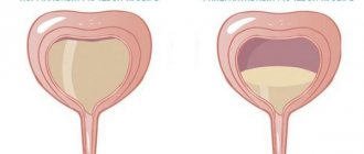
What stream of urine is considered normal? Normally, the process of urination does not cause a person any discomfort or difficulty. The stream of urine should be wide and fast, flow out on its own and not require straining. The elimination process should not be intermittent, painful, and after completing urination, the person feels relief and a completely emptied bladder.
The next urge to visit the toilet occurs after 1-2 hours, depending on the person’s drinking regime and nutrition.
However, it is difficult to talk about urination disorders based on general characteristics, because each organism has its own individuality and structural features. Also, urine pressure depends on a person’s ability to visit the toilet on time.
Where does urine come out in women photo
The human urethra, or urinary tract, is a tubular organ that runs from the bladder to the external genitalia. In men and women, it differs in its structure and colonization by microflora.
The organ in representatives of both sexes looks like a soft, elastic tube. Its walls consist of 3 layers:
- external, consisting of connective tissue;
- middle, formed by the muscle layer;
- internal (mucous membrane).
In men, the urinary tract passes through the penis to the outlet and serves to drain urine and release ejaculate during orgasm. In women, it goes from the bladder to the external opening, which is located between the clitoris and vagina, and is needed only for the removal of urine.
The external urethral sphincter is formed as paired muscles. It compresses part of the urethra. In the female body, these muscles are attached to the vaginal area and are capable of compressing it.
The muscles of the urethra in men are connected to the prostate. The internal sphincter has a fairly strong muscle located near the exit from the bladder.
Microflora in the organ
The channel for excreting urine in representatives of different sexes differs in microflora. Immediately after the birth of a child, various microorganisms enter his skin. They gradually penetrate into the body and settle on the mucous membranes and internal organs.
Bacteria cannot penetrate further than the mucous membranes; this process is hampered by the internal secretions of the body, urine, and ciliated epithelium, so they become attached to them. Pathogenic organisms that remain on the mucous membranes become the innate human microflora.
The female urethral mucosa contains several times more bacteria than the male urethral mucosa. It is dominated by lactobacilli and bifidobacteria. They produce acid, creating an acidic environment. If there are few bacteria, then the acidic environment is replaced by an alkaline one, which allows inflammatory processes to develop.
As we grow older, the beneficial microflora in the female urethra becomes coccal. The microflora of the male urethra is represented by streptococci, corynebacteria, and staphylococci; it does not change throughout life.
The composition of the microflora may change depending on the large number of sexual partners. Frequently changing partners introduces dangerous microbes into the body that can cause serious illness.
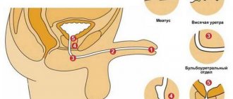
Men's channel
The male urethra during the embryonic period is similar to the female one, as it consists of the same structures. And once formed, it begins to differ significantly, becomes longer and smaller in diameter, is located inside the penis, and its functions, in addition to urine output, also include ejaculation.
The redistribution of these functions of the male body completely depends on the degree of filling with blood of the cavernous bodies and the spongy body, which surround the male urethra. With erection, the blood supply to the penis occurs, ejaculation occurs, and in the absence of blood supply to the penis, the process of urination occurs.
The male urinary canal has a length of 18-22 cm. In a state of excitement, the length becomes a third longer, in boys before puberty it is a third less.
The urethra in men is divided into posterior (the distance from the internal opening to the beginning of the corpus cavernosum) and anterior (the remotely located part of the canal).
It has two bends in the shape of the letter S:
- The upper (infrapubic) bend bends around the pubic symphysis (half-joint) from below as it passes from top to bottom of the membranous part of the urethra into the cavernous one.
- The lower one (prepubic, prepubic) is located at the point of its transition from the fixed part of the urethra to the mobile one.
When the penis is raised, both curves form one common one, the concavity of which is directed forward and upward. Throughout the male urethra, the diameter of the lumen is not the same, narrow parts alternate with wide ones.
The extensions are located in the prostatic, bulbous part and at the end of the urethral canal (where the scaphoid notch is located). The narrowings are located at the internal opening of the urinary canal, in the area of the urogenital diaphragm, at the external opening of the urethra.
Conventionally, the male urethra is divided into 3 parts:
- Prostatic (prostate). It has a length of 0.5-1.5 cm. It consists of tubules for ejaculate release and 2 ducts (prostatic and sperm excretory).
- Sponginous (spongy). The urethral part is located along the penis in its lower part and has a length of 13-16 cm.
- Cavernous (membranous). The longest section of the male urethra, the length of which is approximately 20 cm. The spongy section contains ducts of numerous small tubules. It is located deep in the perineum, passes through the urogenital diaphragm, which has a muscular sphincter.
The male urethra originates from the urinary sac. Smoothly passing into the prostate area, it crosses this gland and ends at the head of the penis, from where urine and seminal fluid exit. The average size of the urethral lumen in men along its entire length is 4-7 mm, in boys 3-6 mm.
The female urethra is an anteriorly directed, straight tube passing close to the elastic vaginal wall and pubic bone. Its length is 4.8-5 cm, and its diameter is 10 - 15 mm, while it is easily stretched.
Inside, the urinary canal is lined with a mucous membrane, which has the appearance of longitudinal folds, due to which the urethral lumen appears smaller. In the female urethra there is a special obstructing pad consisting of connective tissue, veins, and elastic threads. It closes the duct of the urinary canal.
The female urethra does not perform reproductive functions, although substances are excreted through it, which can be used to determine whether a woman is pregnant or not. The female urethra is surrounded by tissues that are similar in structure to the corpus spongiosum of the penis, and the corpus cavernosum of the clitoris, which is similar to the corpus cavernosum of the penis, is located in front of the urethra.
The urethra itself is hidden in the tissues of the pelvis and therefore does not have mobility. Its anterior surface is adjacent to the tissues that cover the pubic symphysis, and in distant places to the legs of the clitoris. The posterior surface of the external urethral outlet is adjacent to the anterior wall of the vagina.
It is closely connected to the anterior wall of the vagina and is firmly attached to the lower branches of the pubic bones, as well as partially to the ischial bones.
Since it is short and wide in women, located next to the vagina and anus, the danger of bacteria, microbes and other pathogenic microflora entering it is much higher in women than in men. Therefore, they are more susceptible to genitourinary infections.
External hole
In the male half of humanity, the main part of the urethra passes inside the penis, and the outlet is located at the top of its head. If it is not there, the disorder is called hypospadias. If there is partial or complete clefting of the anterior wall of the urethra, the disorder is called epispadias.
The external urethral canal in the fair sex is located between the clitoris (slightly below it by about 3 mm) and the entrance to the vagina.
The location of the external opening may vary. If the lower wall is underdeveloped, it will be located on the anterior wall of the vagina, distant from the entrance.
This process is called hypospadias. The outer hole has a diameter of approximately 0.5 cm, its shape can be round or star-shaped.
Reasons for deviation from the norm
Impaired urine flow occurs in both women and men. Pathological disturbances in the urination process can be caused by:
- inflammatory phenomena in the urinary organs (cystitis, pyelonephritis);
- neoplasms;
- narrowing of the lumen of the urethra;
- blockage of the ureter or urethra with a stone;
- prostate adenoma in men;
- prostatitis in men;
- cervical cystitis in women;
- pelvic inflammation;
- consequences of injuries.
Symptoms
When can one suspect obvious disturbances in the urination process? If you notice the following symptoms, you should consult a specialist:
- thin and weak stream;
- in order for urine to begin to come out, you need to push;
- intermittent stream of urine;
- the process of urine excretion is accompanied by pain;
- frequent urge to go to the toilet, but there is no feeling of complete emptying of the bladder;
- leakage of drops of urine before and after urination;
- jet splitting and splashing;
- urine flows for a long time and with low pressure.
The signs listed above indicate the development of disturbances in the normal process of urination. To find out the cause of this phenomenon, you need to consult a doctor as soon as possible.
Diagnostics
Diagnosis of urination disorders includes the following studies:
- General urine analysis;
- General blood analysis;
- Biochemical blood test for the renal complex;
- Urine culture tank for sterility and sensitivity to antibiotics;
- Ultrasound of the urinary system;
- PSA analysis for men, digital examination of the prostate, TRUS.
Research helps the specialist identify the underlying disease that leads to disruption of the urine excretion process. If necessary, the doctor prescribes additional instrumental diagnostic methods to clarify the diagnosis.
It is especially worth noting the problem of urinary disorders in men after 40 years of age. Every year, it is recommended to undergo routine examinations with a urologist in order to identify prostate diseases that can be effectively treated at the initial stage of the disease. Men are recommended to take a prostate-specific antigen test once a year, which allows identifying pathological disorders in the prostate.
How urine is formed in the human body
There is a so-called theory of urine formation. After entering the body, all food and liquid begins to be processed and its constituent elements enter the blood. During circulation, blood passes through almost all organs, the last of which is the kidneys. The process of urine formation consists of blood passing through three successive stages:
- Filtration. At this stage, primary urine is formed. First, the blood passes through the vessels, and the kidneys filter it (for this, there is a three-layer purification system that thoroughly processes it). The kidneys are responsible for clearing the body of protein, which is why if a person’s urine tests show increased protein, we can safely assume that there may be problems with the kidneys,
- Reabsorption. All proteins and particles that enter the kidneys after filtration go through this stage. During the process of reabsorption, they return back to the blood. The body also receives back the fluid it needs,
- Secretions. The opposite stage of reabsorption consists in the formation of secondary urine and its entry into the bladder through the ureters.
If there is a disruption in the way urine is produced, a person may experience serious consequences and illnesses.