Types and forms of abdominal hernias
A hernial protrusion is understood as the exit of the peritoneum with viscera through a defect in the anterior wall.
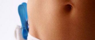
The structure of the abdominal wall is such that some “weak spots” can become hernial orifices - this is the inguinal ring and the medial inguinal pit. Over time, the hernial sac may descend into the scrotum or labia majora. Pathology education (classification)
- inguinal – protrusion of the seminal canal, intestinal loop, ovary, bladder;
- perineal – in the pelvic floor area, protrudes into the perineal pit, outer labia, rectum;
- femoral - develops in women after 30 years, an omentum or a loop of intestine can get inside;
- umbilical - the insides come out of the umbilical ring;
- linea alba – bulging through the gap in the midline;
- Spigelian line - protrusion of internal organs into the area of the rectus vaginalis muscle;
- ventral at the site of surgery.
A hernia is never safe. A hole in the soft skeleton (peritoneal wall) leads to complications. The most dangerous thing is strangulation of the hernial sac in the ring. If there are vital organs in it, then every minute counts. Not in all cases, surgery completely solves the problem - often the organ turns out to be non-viable and has to be removed.
Do not ignore a hernia with the risk of strangulation. The hernial ring has a tendency to grow and “tighten” more and more organs (omentums, intestines, stomach), which leads to its growth. This completely eliminates the possibility of reduction and complicates treatment.
- reducible – can be reduced, pain does not occur when pressed;
- not reducible - there is no way to return the contents to their place, causes pain;
- strangulated - part of the intestine extends beyond the walls of the abdomen and compression of the vessels occurs, which disrupts the nutrition of the tissues and provokes their death.
Video
In the video, the head physician of the multidisciplinary clinic N.I. Pirogova will talk about the pathology, its dangers and modern methods of treatment.
Advantages and disadvantages of hernia meshes
Advantages of mesh endoprostheses:
- are not rejected by the body;
- low probability of recurrence of a hernia;
- does not interfere with the patient after surgery and does not cause discomfort;
- short rehabilitation period;
- fuses with connective tissue and reliably fixes the hernial orifice.
Disadvantages of mesh endoprostheses:
- slight inflammation, as the body’s reaction to a foreign body, which quickly passes.
Concept and classification
An abdominal hernia is a disease characterized by the protrusion of abdominal organs onto the surface of the abdomen or their direction through the hernial orifice into the abdominal cavity. Hernial orifices are clearings that appear in the wall of the abdomen. This defect is of natural origin or can form due to injury or surgery.
Statistics show that approximately 5% of the population suffers from various types of abdominal hernias. The vast majority of them (80%) are men, and the remaining 20% are women and children. As a rule, abdominal hernia is diagnosed in preschool children and people over 50 years of age.
There are different types of diseases, including abdominal hernias. The classification is very extensive and includes a huge number of subspecies. For clarity, let's make a table.
| Signs | Types of hernias |
| Location |
|
| Localization |
|
| Strangulated hernia |
|
| Volume |
|
| Special types |
|
Internal abdominal hernias are diagnosed in 25% of cases. In all other situations, patients suffer from external symptoms. The ventral hernia also deserves special attention. Lately she has started to meet more often. Ventral hernia occurs after surgery.
Types of mesh for hernia
According to the degree of resorption, medical meshes for hernia treatment are:
- Absorbable . Over a certain period of time they dissolve in the human body. When using these endoprostheses, it is necessary to take into account that the time for complete wound healing and patient recovery is less than the time for resorption of the mesh endoprosthesis. There is a risk of relapse after complex operations.
- Non-absorbable . They do not dissolve in the human body at all. Such an endoprosthesis stays in the human body all his life and does not interfere with it in any way. Over time, they become overgrown with muscle tissue, as they have sufficient porosity. Most commonly used in medicine. The most reliable, with minimal risk of re-development of the hernia. They are produced, for example, by the Volot company.
- Partially absorbable . After muscle tissue builds up, it is partially resorbed in the body.
By form:
- Simple one-dimensional ones . They represent a grid that is in the same plane.
- Complex three-dimensional . A mesh consisting of 3 flaps located in different planes. The structure of the three-dimensional mesh is such that one flap is applied on the inside of the hernial orifice, and the other on the outside, and they are connected by a flap rolled into a cylinder, which fills the hole itself.
By coverage:
- Drug coated . Coated with a substance that prevents infection and promotes rapid wound healing.
- No drug coverage . No drug coverage.
By thickness:
- Lungs . Meshes with a thickness of 0.32-0.44 mm with a monofilament diameter of 0.09±0.01 mm. Intended for reconstructive surgical plastic surgery after removal of hernias of various locations, in cases where the tissues do not experience increased stress and/or their own tissues are in satisfactory condition, as well as for strengthening wounds.
- Classic . Meshes with a thickness of 0.45-0.70 mm with a monofilament diameter of 0.12±0.01 mm. Intended for reconstructive plastic surgery of the anterior abdominal wall after removal of a hernia, for damage to the diaphragm and chest wall, for soft tissue defects, as well as for strengthening traumatic and surgical wounds.
- Heavy . Meshes with a thickness of 0.65-0.99 mm with a monofilament diameter of 0.14±0.02 mm. Used to treat giant ventral hernias. They are used on areas of soft tissue that have greater mobility and are subject to increased stress.
- Superheavy . Meshes with a thickness of 0.65-0.99 mm with a monofilament diameter of 0.16±0.02 mm. Used in the most severe cases when the highest strength is required.
Reasons for the development of pathology
- congenital
- acquired.
An umbilical hernia develops due to improper muscle development. It appears in the first months in children, more often in girls. Intra-abdominal pressure from crying or bloating causes the ring to expand, into which the intestine protrudes.
READ MORE: How to prepare for umbilical hernia surgery
In adults, an increased risk of developing pathology appears after 50 years. Numerous births and obesity lead to weakening of the abdominal muscles.
Additional Information!
Pathology in oncology becomes a consequence of surgery, prolonged ascites, weakness of connective tissues, and sudden weight loss.
- persistent cough;
- ascites due to problems with the heart, kidneys, liver;
- peritoneal dialysis;
- weight loss;
- problems with urination, constipation;
- injury.
Postoperative hernias occur as a result of surgery when the doctor did not connect the incision site well. Other causes may include inflammatory reactions in the wound, prolonged use of tamponade and drainage.
How is a hernia treated?
Treatment options depend directly on the type of hernia and your overall health. First of all, your doctor may recommend monitoring your condition for possible changes (in any direction). This may include changes to your diet, physical activity and habits. Accompanying medication is sometimes prescribed to reduce symptoms.
If the condition does not improve or even worsens over time, the doctor will suggest surgery. There are 2 main types of operations. Laparoscopic surgery is less invasive because the surgeon will only make a few tiny incisions in the affected area. But open surgery involves a larger incision for reconstructive surgery.
As part of the operation, the doctor may use a special medical device - a surgical mesh, the purpose of which is to support weakened muscles and tissues. The mesh can be either synthetic or made from natural fabric, and they come in two types. The temporary mesh is gradually broken down by the body, and the permanent mesh provides long-term muscle support.
Life with a hernia
The vast majority of hernia cases are treatable, but hernias in general are characterized by recurrence. In some cases, the chances of such an outcome are reduced by installing the already mentioned nets. Be that as it may, in each specific case you need to consult with your doctor about the most optimal treatment options and prevent possible relapses.
People with a congenital diaphragmatic hernia may have other health problems, so this disorder requires increased attention.
Symptoms of the disease
The main sign of a hernia is a protrusion behind the abdominal wall. At first it is small and can be easily adjusted, but there may be a feeling of fullness and discomfort in this area. Education increases with increasing internal pressure (coughing, defecation). Pain increases after exercise and eating.
Complications develop when the omentum or part of the intestine gets in contact. Infringement of the organ occurs, especially when pressure increases. Severe pain, vomiting, intestinal obstruction, fever and other signs of intoxication develop. Without surgery, peritonitis develops and there is a high risk of death.
With an umbilical hernia, the size of the ring does not exceed 10 cm, but the protrusion itself can be large. Infringement, intestinal obstruction and fecal stagnation appear.
Note!
The pain syndrome is more pronounced in a sitting or standing position. When a person lies down, the pain may disappear - the abdominal muscles relax and tissue compression does not occur.
A hernia of the linea alba begins with penetration of fatty tissue. But a distinctive feature will be a sharp pain in the upper abdomen, similar to an attack of peptic ulcer or cholecystitis.
According to the clinical classification, hernias can be single or multiple. Uncomplicated hernias are divided into reducible, or free, and irreducible.
The most typical complaints of patients are pain and the presence of protrusion in the abdominal wall. Pain caused by a hernia of the abdominal wall occurs predominantly in the area of the hernial orifice, but may also have a different localization in the epigastrium, lumbar region, according to the projection of the localization of the root of the mesentery of the small intestine, or be of the nature of diffuse abdominal pain without a specific localization.
The nature of pain and its severity are very individual and varied, and the severity of pain is not proportional to the increase in the size of the hernial protrusion. Typical is increased pain when walking, after physical activity, lifting weights, or straining the patient. Pain may be the only complaint in the initial stages of hernia formation. With large hernias, especially irreducible ones, sometimes there are complaints of dysuria and dyspeptic disorders (urinary and digestive disorders), constipation.
Complications of hernias include strangulation, coprostasis, inflammation of both the contents of the hernia and the hernial membranes, damage and neoplasms.
An abdominal hernia does not appear spontaneously. It takes time and several pathological factors to occur. Causes are divided into 2 types: disposing and fulfilling.
Those available include:
- hereditary factor;
- congenital weak muscles;
- changes resulting from injuries, surgical interventions, exhaustion, after which weak points appear on the body.
The underlying causes provoke an increase in intra-abdominal pressure and the development of a hernia of the anterior abdominal wall at weak points. Among them are:
- regular heavy physical activity;
- excess weight;
- tumors of organs located in the abdominal cavity;
- persistent cough that occurs with chronic lung diseases;
- impaired urination;
- constant constipation;
- pregnancy, difficult childbirth;
- some diseases (tuberculosis, cirrhosis, enlarged prostate, paralysis of the legs, polio, etc.).
All of the above reasons that cause the appearance of pathology must continue for a long time. Only then does a hernia of the anterior abdominal wall form.
Symptoms of an abdominal hernia manifest themselves in different ways.
All types are characterized by a feeling of discomfort, pain and protrusion, which occurs in a horizontal position. If you observe these symptoms, you should see a surgeon. He will conduct the necessary examinations and make the correct diagnosis.
When a hernia forms in the abdominal cavity, symptoms depend on its location and severity. Signs of an abdominal hernia are:
- A protrusion in the form of a tumor that appears during any physical stress.
- Aching and nagging pain in the hernia area.
- Urinary disorders.
- Various digestive disorders - bloating, diarrhea, constipation, vomiting, nausea, constant belching.
What are the symptoms of a hernia?
Depending on the type of hernia, symptoms may or may not be present. The only sign common to all types of hernia is a visible protrusion. Other symptoms include a feeling of pressure, cough, heartburn and difficulty swallowing. Severe symptoms of a hernia include stabbing pain, vomiting, and constipation. If the hernia suddenly becomes soft or you cannot feel it, call an ambulance immediately. In general, hernias can lead to infection, blockage, or damage to organs and tissue.
Diagnosis of the disease
A tumor-like formation in the groin, along the linea alba, at the site of a scar or near the navel should be a reason to visit a surgeon. Only a doctor can determine the correct diagnosis, the nature of the hernia and the method of treatment.
READ MORE: Reasons why the leg is pulled under the knee from behind
Inspection
The presence of pathology can be determined by routine examination and palpation. Be sure to check the gastrointestinal tract, liver, bladder and chest. Intestinal obstruction is diagnosed using x-ray or CT scan. Barium may be used to determine precise localization.
A hernia is diagnosed by visual examination
If in doubt, the doctor inserts a finger into the interhernial canal and asks the patient to cough. A push at this moment will indicate the presence of a hernia.
Ultrasound can reveal inflammation in tissues, which are accompanied by the formation of adhesions in internal organs. This causes dangerous complications and complicates treatment.
The study is carried out in a vertical position to determine the thinning of the tissue in the area of the hernial orifice. In more complex cases, some organs will be found in the hernial sac.
Ultrasound examination helps to exclude other diseases with similar symptoms - oncology, peptic ulcer, cholecystitis. The data will help to study the state of the pathology for further selection of the removal method.
If you suspect the pathology of an “abdominal hernia” and the corresponding symptoms are observed, you need to contact a specialist for a comprehensive examination of the body.
If the tumor forms in the usual places for hernias (groin, navel and thigh), the disease is easily diagnosed. A ventral hernia is recognized by a “cough impulse.” You need to put your hand on the protrusion and ask the patient to cough, while clear shocks should be felt. Diagnostic methods include palpation of the hernial orifice, feeling and tapping the tumor.
For some types of hernias of the anterior abdominal wall, additional methods are used:
- gastroscopy;
- radiography;
- herniography (with this procedure, a contrast agent is injected into the abdominal cavity, which makes it possible to examine the hernia);
- Ultrasound of the protrusion itself.
The diagnosis is established on the basis of complaints and objective examination data. The patient must be examined in vertical and horizontal positions, at rest and with straining.
In a vertical position, the hernial protrusion increases, in a horizontal position it decreases or disappears. An increase in hernial protrusion is observed with coughing and straining, which depends on the movement of the contents of the hernia from the abdominal cavity into the hernial sac and back. When inserting a finger along the protrusion of the hernia to the location of the hernial orifice in case of coughing (a jerk-like pressure is felt on the fingertip, caused by a sharp short-term increase in intra-abdominal pressure).
A characteristic objective sign of an external abdominal hernia is the presence of protrusion of the anterior abdominal wall in the area of the hernial orifice. In the initial stages, a hernial protrusion may be absent, and then survey data become of great importance in recognizing a hernia.
Femoral hernias often occur in old age; on the right side they are often sliding. Femoral hernias are prone to strangulation. Irreversible hernia often develops. The hernial swelling in these hernias is usually small, oval or spherical in shape, located below the Pupart ligament.
Femoral hernias can manifest as abdominal pain radiating to the leg and be accompanied by digestive disorders. When pinched, acute cutting pain in the leg on the affected side may occur - Howship's symptom. With small protrusions, establishing an accurate diagnosis can be difficult. In rare cases, with a femoral hernia, swelling of the lower limb may develop as a result of compression of the hernial protrusion of the femoral vein.
Differential diagnosis should be carried out with lymphadenitis, metastases of a malignant tumor in the lymph nodes, edema abscess in spinal tuberculosis, varicose veins in the oval fossa, aneurysm of the femoral vessels, inguinal hernias.
There are hernias of the supra-umbilical section of the white line of the abdomen, the so-called epigastric or epigastric, but peri-umbilical and hypogastric hernias, located below the navel, can also occur. Hernias of the white line of the abdomen are rarely large; palpation is usually painless. With a reducible hernia, a defect in the linea alba can be identified. When the abdominal muscles are tense, the presence of diastasis of the rectus abdominis muscles, which is often found in epigastric hernias, is determined.
Diagnostic methods
Surgical treatment is prescribed in the absence of contraindications.
Tension plastic
Plastic surgery of the hernial orifice is carried out using the patient’s tissues (autoplasty). This method involves suturing the hernia defect using surgical threads. Suitable for young patients with small hernias. With this type of treatment there is a high risk of relapse.
The protrusion is closed with a prosthesis, which looks like a mesh. This technique is called alloplasty. Implantation of a synthetic prosthesis occurs directly under the fascia or skin.
Laparoscopy
The most atraumatic type of treatment. A mesh prosthesis is inserted into the abdominal area in the same way as with the previous method. However, this does not require an incision in the hernia area. The operation is performed by inserting medical instruments through punctures in the anterior abdominal wall. Relapses after such treatment are practically not observed, complications are kept to a minimum.
Abdominal hernia symptoms, causes and treatment
Abdominal hernia: inguinal hernia, umbilical hernia, femoral hernia and postoperative hernia
After the operation, the patient remains in the hospital for 1 to 7 days, depending on the type of surgery. The healing process of the wound edges lasts about 2 weeks, during which time a scar is formed. Temporary disability depends on the size and location of the hernia and the age of the patient.
It is recommended that you avoid physical activity for several weeks after surgery. Dietary recommendations must be followed:
- do not overeat;
- eat small meals, 5-6 times a day;
- exclude foods that cause constipation and gas formation: cabbage, legumes, mushrooms, yeast baked goods, grapes;
- Alcohol and carbonated drinks are contraindicated;
- preferred methods of culinary processing are stewing, boiling, baking in the oven;
- Food should be taken in puree or liquid form.
Mushrooms need to be removed from the diet.
It is recommended to abstain from alcohol for several weeks after surgery.
It is preferable to stew food.
The absolute indications for surgical intervention are complicated forms of hernia - strangulation, and for recurrent and postoperative hernias - adhesive intestinal obstruction.
Ventral hernia is treated surgically. Small defects are repaired by covering with local tissue. Large defects are difficult to eliminate in this way due to the possibility of circulatory impairment and increased intra-abdominal pressure, which may result in respiratory failure.
Therefore, when treating a large ventral hernia during surgery, a mesh implant is used, which makes it possible to close large defects without fear that the mesh may fuse with the internal organs.
To carry out the intervention, the surgeon makes several punctures in the abdominal wall. To do this, small incisions are made that do not injure the muscle part, but only the skin. This fact has a beneficial effect on the postoperative period, significantly shortening it, and does not cause pain, which is especially important for all patients.
Punctures are made by a surgeon using a special instrument - a trocar. It is used to insert a laparoscope into the abdominal cavity, to which a light source and an image transmitter are connected to the monitor. Thus, the specialist observes his movements from inside the body.
Using additional trocars, instruments are inserted to perform the operation. A clamp is passed through one trocar to capture the tissue, with which a mesh implant is installed. For others, the synthetic mesh is secured with sutures or special staples.
The operation lasts about 1 hour under general anesthesia. The patient is in the hospital for 1-2 days under the full supervision of medical personnel to avoid unforeseen complications and reactions of the body.
Many patients are interested in whether it is possible to eliminate the pathology without surgery. Treatment of a ventral hernia without surgery is not possible.
Conservative therapy involves wearing a special bandage that prevents the abdominal organs from coming out. Using such a bandage is very inconvenient and labor-intensive, so it is indicated in exceptional cases.
For some patients, surgical intervention may be contraindicated due to advanced age, pathologies of the cardiovascular system, or pregnancy. It is in such cases that doctors suggest using a special bandage for ventral hernias.
Postoperative recovery
Rehabilitation is about restoring the strength of the patient’s body. Recommendations depend on the method of surgery and must be prescribed by a doctor. After examining the patient, he prescribes diet, postoperative therapy and determines the intensity of exercise.
The patient must go to the hospital for 10 days to get dressings done. In addition, drug treatment with painkillers and antibiotics is prescribed. Physiotherapy courses will help speed up the recovery process.
You can't play sports for several months. You also need to adhere to proper nutrition. In the first days after surgery, you need to eat liquid foods: broths, light soups, jelly. Gradually, you need to add porridge, lean meats and fish, eggs, vegetables, fruits, and seafood to your diet. It is necessary to give up salty and spicy foods, smoking, and drinking alcoholic beverages.
The stitches are removed after a week. After this, it is recommended to wear a bandage to restore tone to the abdominal muscles. After 3 months, you can do light physical exercise, but the bandage cannot be removed. Breathing exercises and regular massage contribute to rapid recovery.
An abdominal hernia is a serious disease that requires treatment. Otherwise, serious complications may occur in the form of peritonitis, intestinal obstruction, and intoxication.
- re-development of the hernia;
- wound infection;
- problems with urination.
READ MORE: With an umbilical hernia, there is a fever
. Final recovery occurs after several months. A very important component of the postoperative period is rehabilitation to prevent relapse and complications.
At first, patients use a bandage. The wound is isolated with a sterile bandage to prevent friction. They begin to walk on the second day after surgery. They must take a course of antibiotics, and doctors prescribe painkillers.
Discharge occurs after 8-10 days, when normal healing of the wound begins. Sutures with regular threads are removed on the tenth day.
At home, dressings are made twice a day using sterile gauze and adhesive tape. The wound is treated with brilliant green. You can only take a shower after 2 weeks.
Rehabilitation period
With open surgery, the patient's recovery period lasts longer than with laparoscopy, since the operation affects more tissue.
After open surgery:
- The patient remains in the hospital for one to two weeks.
- For the first 5-7 days, the patient is treated with stitches and bandaged.
- The patient wears stitches for 2 weeks.
- For the first six months, the patient is prohibited from physical activity, standing for long periods of time, or lifting and carrying loads of more than 5 kilograms.
- After 8 months or later, the patient is prescribed physical therapy to restore and strengthen the abdominal muscles.
After laparoscopic surgery:
- The patient remains in the hospital for 3-7 days.
- Within 3-7 days, the patient’s wounds are treated and bandaged.
- The stitches are removed after a week.
- For the first six months, the patient is prohibited from physical activity, standing for long periods of time, or lifting and carrying loads of more than 5 kilograms.
- After 8 months, the patient is prescribed physical therapy to restore and strengthen the abdominal muscles.
Conservative methods
Conservative treatment of abdominal hernia is prescribed to prevent complications, tumor growth and alleviate symptoms. It is used in relation to patients for whom surgical intervention is contraindicated due to age, pregnancy, or serious illness.
Conservative methods include:
- drug therapy;
- eliminating the causes that increase intra-abdominal pressure;
- diet;
- gymnastics;
- wearing a special bandage;
- massage.
Rehabilitation
Physiotherapeutic procedures are prescribed to speed up healing. For the first 2 months, you should not carry more than 2 kg and strain your abdominal muscles. Sports activities are postponed for 3 months, during which a postoperative bandage is worn.
- dairy products;
- light soups and cereals;
- boiled eggs, meat, fish;
- processed vegetables and fruits.
Canned and spicy foods and baked goods are completely excluded. Eat food 5 times a day in small portions. It is better to steam meat dishes, and boil the rest.
Surgical treatment
The only way to cope with the pathology is to remove the abdominal hernia through surgery.
Only one type of hernia can disappear on its own - umbilical hernia in children under 5 years of age. Other types, including ventral hernia, will not disappear on their own; moreover, over time they will increase in size and pose a serious threat to human health.
When the first symptoms appear, you should immediately contact a surgeon. A timely operation gives a better chance of a quick recovery without various complications. Before the operation, the patient must undergo an examination and pass all the necessary tests. An analysis of the patient’s health will allow the surgeon to prescribe an appropriate treatment option.
As a rule, an abdominal hernia is removed using hernioplasty. There are 3 ways to do it:
- Tension (the hole at the site of the removed hernia is closed with its own tissues).
- Without tension (to close the hole, polypropylene mesh implants are used).
- Combined (both mesh and own fabrics are used).
Usually they resort to the second method. It does not stretch the fabric, and the polypropylene mesh provides reliable protection that can withstand significant loads.
Also, surgery to remove a hernia can be performed using open, laparoscopic and endoscopic methods:
- Open surgery is considered classic and is performed by making an incision in the peritoneum, examining, directly eliminating or reducing the hernia, installing a mesh and suturing.
- Endoscopic and laparoscopic methods of surgery do not require large incisions. Several small punctures (5 mm) are made. To create a working space, carbon dioxide is injected into the peritoneum. Then a device with a flashlight and a video camera and special manipulators are introduced, with the help of which the operation is performed.
Treatment
Treatment of this category of patients presents particular difficulties. On average, 10-15% of patients operated on for ventral hernias experience relapses of the disease, and after operations for recurrent hernias, the frequency of new relapses increases by another 2-3 times. Postoperative hernias of any size are best operated on under general anesthesia with intubation anesthesia. In elderly patients and patients with severe concomitant somatic pathology, the method of choice is the use of epidural anesthesia.
Surgical access to the hernial sac and its treatment. Surgical intervention begins with wide bordering incisions with complete excision of postoperative scars, excess skin and subcutaneous fat. When a hernia is localized in the epigastric region, longitudinal approaches are usually used, and in the presence of a hernia formation in the mesogastric region, transverse incisions can be used.
When a hernia is localized in the hypogastric region in obese patients, transverse or T-shaped approaches with complete removal of the skin-subcutaneous “apron” are considered the most optimal. The surgical approach is completed by isolating the hernial sac and the edges of the muscular aponeurotic defect of the anterior abdominal wall.
Treatment of the edges of the hernial orificeHernial orifices in postoperative ventral hernias usually have an irregular shape and are often separated from each other by scar tissue into separate fragments. During surgical intervention, it is mandatory to separate all scar septa with economical excision of their edges, as a result of which the hernial orifice takes on an oval shape.
From the outside, the edges of the hernial orifice are cleaned of fatty tissue, and from the side of the abdominal cavity they are separated from the soldered organs for a distance of 4-6 cm in all directions. When performing hernia repair, there is often a need to eliminate the consequences of the adhesive process in the abdominal cavity. In cases where deformed and scarred loops of the small intestine and areas of the greater omentum are identified, the latter sometimes even have to be resected.
Plastic surgery of the anterior abdominal wall The choice of method for plastic surgery of the anterior abdominal wall for postoperative ventral hernias is determined by the size and location of the hernial orifice, the condition of the tissues of the anterior abdominal wall, as well as the volume of the hernia formation and the degree of reduction in the volume of the abdominal cavity.
Simple restoration of the anatomical relationships of the anterior abdominal wall with its subsequent suturing is indicated only in the early stages after primary surgical intervention and only for small hernial orifices. In such cases, with good adaptation of the muscular aponeurotic edges of the wound without tension, autoplasty is most often used with the formation of a duplicative layer of the aponeurosis in the area of the hernial orifice.
When performing plastic surgery of the anterior abdominal wall, synthetic explants are often used. All types of prosthetics of the anterior abdominal wall using synthetic explants, depending on the degree of restoration of its functionality, are divided into reconstructive and corrective.
Reconstructive types include combined methods of plastic surgery of the anterior abdominal wall with tissue tension, with the help of which, when the hernial orifice is closed, the lost functions of the anterior abdominal wall are restored. These methods include “onlay” and “sublay” types of plastic surgery of the anterior abdominal wall.
Corrective types of prosthetics do not provide for complete restoration of the function of the anterior abdominal wall and are performed without tension on its tissues, while plastic surgery of the anterior abdominal wall is carried out using the “inlay” technique or with an intra-abdominal location of the explant (“intraabdominal”).
A postoperative hernia is a pathology that can only be completely eliminated through surgical treatment.
Sometimes the patient himself categorically refuses surgical intervention. In these cases, conservative treatment is prescribed.
Physiotherapy
You need to know that it is prohibited to carry out any physiotherapeutic procedures directly on the hernia area, since these methods can lead to serious complications.
To strengthen the abdominal muscles, you can massage: knead, rub, stroke and tap the abdominal area.
Folk remedies
Increased intra-abdominal pressure aggravates the course of the disease, so it is important to eliminate the factors that provoke flatulence and constipation. To do this, you can use traditional medicine:
- 1 tbsp. l. pour boiling water over chamomile and leave for 30 minutes. Strain the resulting broth. Take 100 ml 2 times a day.
- 2-3 tbsp. l. pour dill seeds with 1 glass of water and simmer over low heat for 15 - 20 minutes. Remove seeds from the broth. Take 100 ml before breakfast, lunch and dinner.
1 tbsp.
l. pour boiling water over chamomile and leave for 30 minutes. Herbal compresses on the hernia area are not effective.
Physiotherapy
Exercise therapy is used to restore the tone of the intra-abdominal muscles. This method does not eliminate an existing hernia, but is used to prevent its occurrence. The possibility of physical activity is agreed upon with the attending physician.
The exercises are aimed at working the rectus and oblique abdominal muscles: bending and turning the body to the sides, bending the torso towards the legs, protruding and relaxing the abdomen.
Bandage
The bandage is a temporary means of preventing complications. It is selected individually by the doctor and can only be used for reducible hernias. You need to know that due to constant friction, the bandage can cause skin infection. In addition, when constantly worn, the abdominal muscles weaken, which aggravates the course of the disease. It is recommended to wear the bandage for several hours a day during periods of active physical activity.
The bandage is a temporary means of preventing complications.
Removal of a ventral hernia is the only way to eliminate the pathology. Based on the patient’s health condition, the size of the protrusion and its location, the surgeon chooses the most optimal option for the operation:
- Tension plastic. Used to remove small hernias. The operation involves suturing a defect in the anterior abdominal wall using non-absorbable threads. This procedure is distinguished by its simplicity, but complications cannot be ruled out (relapses, breathing problems).
- Tension-free hernioplasty. Using this method, large hernias are eliminated. During the procedure, the surgeon removes the protrusion that occurred during the postoperative period and installs a mesh implant. After hernioplasty, the mesh is overgrown with the patient’s own tissues, which helps strengthen the abdominal wall.
- Laparoscopy. This is a technically advanced way to eliminate a hernia. The operation is performed using small punctures in the affected area. In the area of the hernial orifice, the doctor inserts a special mesh graft that strengthens weakened areas of muscle tissue.
Tension-free hernioplasty is characterized by a number of advantages, including rapid recovery and almost complete absence of complications. Among the main disadvantages are the high cost of the operation and the high likelihood of developing adhesions.
Many patients are interested in whether it is possible to eliminate the pathology without surgery. Treatment of a ventral hernia without surgery is not possible.
Conservative therapy involves wearing a special bandage that prevents the abdominal organs from coming out. Using such a bandage is very inconvenient and labor-intensive, so it is indicated in exceptional cases.
For some patients, surgical intervention may be contraindicated due to advanced age, pathologies of the cardiovascular system, or pregnancy. It is in such cases that doctors suggest using a special bandage for ventral hernias.
Without surgical intervention, it will not be possible to get rid of the protrusion of the ventral hernia. Treatment and correction of the peritoneal wall will be required, with removal of the resulting sac and subsequent plastic surgery.
Tension plastic
The protrusion is sutured with threads. This procedure can be performed only in young patients whose hernia size is small. In addition, the absence of concomitant diseases is important. The fact is that this method of intervention has a high relapse rate, which reaches 30%.
The advantages of the tension method:
- ease of implementation from a technical point of view;
- low price for materials and equipment used.
Disadvantages of the tension method:
- risk of relapse up to 30%;
- respiratory dysfunction due to too much tension on the wound;
- pain due to strong tension.
The place where the protrusion was is closed with a synthetic prosthesis. It is made of polypropylene mesh and is inserted directly under the skin or along the fascia.
The advantages of this technique:
- Low relapse rate.
- Absence of pain after surgery or its insignificant severity.
- No respiratory problems.
- When complementing the tension-free method with abdominoplasty, it becomes possible to plastically restore the peritoneal wall even with gigantic protrusions.