Harlequin ichthyosis. Causes
Harlequin ichthyosis is an inherited disorder. Researchers have already established that harlequin ichthyosis is caused by mutations in the ABCA12 gene, which is located on chromosome 2 (2q34).
Scientists' opinions on this matter are divided. Most of them believe that the main cause of the pathology is a gene mutation. Other researchers think that the reason lies in malfunctions of the thyroid gland and hormonal disorders.
Children most often suffer due to heredity. The current mutation prevents the formation of a normal stratum corneum, which is covered with scales, cracks, and formations. Fat and protein metabolism are disrupted, cholesterol increases, and the number of amino acids increases.
The disease can be inherited as an autosomal dominant trait. The initially dry, whitish dermis is covered with whitish, gray-black scales. With other types of illness, the baby’s body becomes bright red.
With a mild degree of the disease, the so-called abortive course, the symptoms are mild. This is only a slight peeling and dryness on the extensor limbs. But even here it is necessary to intensively care for the affected areas.
At birth, all babies are covered, as it were, with plates, a kind of shell. After a short time, peeling begins, the top, thick layer comes off. After exfoliation, doctors determine the exact diagnosis. Particularly severe cases appear immediately.
Is skin ichthyosis dangerous for children? Severe illness can be fatal. With this diagnosis, 2-3% of babies survive, but only thanks to careful care. After birth, they are immediately sent to intensive care wards. Those born prematurely are placed in special feeding chambers.
Even during pregnancy, the expectant mother can find out about the disease of the fetus through a biopsy. The doctor takes a scraping and makes a diagnosis. The decision always remains with the parents.
Congenital ichthyosis develops with a genetic predisposition.
Acquired ichthyosis has a number of causes. For example:
- Endocrinopathy.
- Problems in the hematopoietic system.
- Malfunctions of the reproductive and thyroid glands.
- The adrenal glands are only partially functional.
- Permanent hypovitaminosis.
- Age-related changes.
Classification
A large number of varieties of Ichthyosis have been described. For a long time, the main division was into ordinary (vulgar), which usually manifests itself during the first months of life, and congenital, in which the child is born as if in a shell of skin horny layers, with signs of congenital deformities and underdevelopment of a number of organs and systems; Congenital ichthyosiform erythroderma is distinguished as an independent form of congenital I.). There is no generally accepted classification of I. Depending on the wedge, features, ch. arr. the color and nature of the horny layers, black, white, pearlescent, serpentine and other varieties of I. were also distinguished. In 1964, A. Greither identified the types of inheritance of various forms; Subsequently, Wells, Kerr, Schneider and Konrad (RS Wells, S.V. Kerr, 1965; UW Schnyder, V. Konrad, 1968) took types of inheritance as the basis for classifications.
Based on modern clinical, genetic and pathomorphological data, I. can be divided into hereditary and acquired.
Hereditary ichthyosis includes the following forms: 1) ordinary I., in which there is an autosomal dominant type (actually ordinary I.) and recessive X-linked I., which is an independent form that has not yet received its wedge name; 2) I. fetus; 3) congenital ichthyosiform erythroderma), subdivided into lamellar I. (non-bullous type) and epidermolytic I. (or hyperkeratosis - bullous type); 4) one-sided I.; 5) linear envelope I.; 6) spiny I.; 7) syndromes including I. as a symptom.
Acquired ichthyosis, or more precisely ichthyosiform conditions, are divided into 1) symptomatic, 2) senile, 3) discoid I.
Harlequin ichthyosis. Symptoms and manifestations
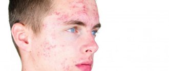
Harlequin ichthyosis is a rare skin disease that develops in children before birth. Individuals of both sexes develop this disorder. The approximate incidence of this type of ichthyosis is 1:500,000.
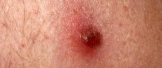
- Congenital ichthyosis is a hereditary skin disease. It is characterized by abnormally red, dry, hard skin with a large number of large and small white scales.
- Glued ichthyosis is a hereditary skin disease characterized by wide, dark, plate-like formations on the skin that are separated by deep cracks. Glue ichthyosis can also cause redness of the skin (erythroderma), thickening of the skin on the palms and soles, and decreased sweating with heat intolerance.
Harlequin ichthyosis (1)
Harlequin ichthyosis (2)
Harlequin ichthyosis (3)
Infants with harlequin ichthyosis are covered in thick, plate-like growths. These plates, due to their weight, pull the skin around the eyes and mouth downwards, as a result, most newborns will have eyelids and lips turned inside out. Your baby's chest and stomach may also feel severely tight, which can make breathing difficult.
Most babies are born prematurely, which increases the risk of complications. Narrowing and swelling of the mouth may make sucking difficult and these babies may need a special feeding tube.
The thick lamellar skin in children with harlequin ichthyosis will gradually begin to divide and peel off (over several weeks). Treatment with antibiotics may be necessary to prevent infections. Etretinate (1 mg/kg body weight) may accelerate the loss of thick scales. Most babies will need constant care during the first few weeks of life.
After the thick plates fall off, what is left in their place is dry and red skin, which may be covered with thin plates. The skin should be regularly lubricated with emollients. This can be especially effective after swimming while the skin is still damp. Topical medications containing ceramides or cholesterol, moisturizers with petroleum jelly or lanolin, and keratolytics can keep the skin hydrated, which can save your child from skin cracking and infections.
Severe cases of arliquin ichthyosis may require the use of retinoids. Retinoids are only used in the most severe cases of ichthyosis due to their toxicity and other known complications.
The reasons for the appearance are not clear. However, the presence of sharp differences in the color of the skin on both halves of the baby’s body, when he is positioned on his side, is associated with birth injuries, which are accompanied by damage to the diencephalic region (that is, the diencephalon), as well as infection of the newborn with staphylococci.
Ichthyosis in children. Symptoms
Adults do not always inherit the mutating gene; they can get sick when their immunity decreases or from old age. Ichthyosis in children is always hereditary and develops against the background of a slow metabolism. The main symptoms include the following:
- violation of thermoregulation;
- increased skin dryness;
- dehydration;
- cracks in the dermis;
- dystrophic changes in hair structure, dryness, loss;
- mucoid formations on the skin in the form of scales;
- eyelids turned out;
- stretched or narrowed mouth (severe form);
- deformation of the ears;
- webbing between the fingers (harlequin shape);
- splitting of nails;
- multiple caries.
With congenital ichthyosis of the skin in newborns, constant care is required. The plates can tighten the baby's organs tightly, causing their deformation. If the skin plates have severely tightened the baby's mouth, feeding is only possible through a tube.
A child suffering from mild ichthyosis does not require such intensive care. It is enough to take vitamin courses and choose the right diet. Creams and water procedures are carried out daily.
The result of the disease is a change in the structure of the skin. Due to malfunction of the cell, due to the loss of memory of a mutating gene (or the presence of another memory in it), the stratum corneum does not line up as it should.
The skin and its areas are covered with formations in the form of scales. The disease can be hereditary or acquired. Characteristic signs of ichthyosis include:
- dry dermis;
- peeling;
- presence of scales;
- the presence of spines, plates on the stratum corneum of gray, brown color;
- flour-like formations;
- thickening of the skin;
- film covering the body in newborns (disappears over time);
- nail damage;
- turned out mouth;
- brittle, dry hair;
- multiple caries;
- myopia;
- thermoregulation is impaired.
Ichthyosiform skin changes
Ichthyosiform skin changes (the so-called acquired I.) can be observed with vitamin A deficiency, aging (senile I.), Hodgkin's disease, Down's disease, toxicerma, mycosis fungoides, leprosy, hypothyroidism, cancer of internal organs, etc. Hyperkeratosis is usually mild, according to the type of xerosis (see).
In 1906, T. Toyama described a special form of acquired ichthyosiform condition called "Pityriasis circinata", the etiology and pathogenesis of which is unclear; This disease was later called “discoid ichthyosis.” The disease is observed in Japan, South Africa, etc.
Pathohistology:
hyperkeratosis, acanthosis, slightly enlarged granular elephant, parakeratosis. There is a small inflammatory infiltrate in the dermis.
Clinically manifests itself in childhood in the form of roughness, dryness of the skin of the extensor surfaces of the extremities and torso; the head, face, genitals, mucous membranes, palms and soles, and nail plates are not affected. Subsequently, round or oval rough brown plaques are formed on the lesions, the number of which may vary. The course is chronic, with spontaneous remissions, more often in the summer.
The diagnosis is usually not difficult; the diagnosis of a specific form of I. is made on the basis of a wedge, symptoms in combination with the characteristics of histol, pictures and identification of etiology and type of inheritance.
Differential diagnosis of I. and other forms of keratoses - see Keratoses, table.
Pathophysiology
Harlequin ichthyosis is accompanied by disturbances of microcirculation and peripheral blood flow due to neurological disorders, which consist of unilateral damage or asymmetry of the functions of the nervous sympathetic system. And, as mentioned above, it is a pathognomonic sign of asphyxia or TBI in newborns.
There is an assumption that the thickening (induration) of the stratum corneum, characteristic of this pathology, develops as a result of a defect in lipid synthesis or dephosphorylation of proteins, resulting in their secretion into the skin and, as a result, the formation of the latter is disrupted.
Treatment of ichthyosis
What is ichthyosis and how to treat it? Science has not yet found a way to treat skin ichthyosis that can lead to complete healing.
But modern medicine can prolong the life of those suffering from severe illness and improve their well-being. A group of doctors works with patients. The medical team includes:
- Dermatologist (basic courses of treatment).
- Family doctor, therapist, pediatrician (comprehensive treatment aimed at improving immunity).
- Psychologist (relieving stress, depressive states, getting rid of complexes, pressures inherent in patients).
- Otorhinolaryngologist (observes the condition of hearing and eardrums).
- Dentist (treats teeth).
The group may include those specialists whose help is required in the fight against the underlying disease and complications. Patients receive complexes of therapeutic measures aimed at alleviating the condition of patients with skin ichthyosis.
This includes ointments, creams, and gels that are applied to the skin. They help clear areas of scales and ease the course of the disease.
The skin needs to be moisturized and softened, as cracks form on it, which can become infected. Therefore, antimicrobial and antifungal drugs are chosen.
Also, moisturizing creams, shower gels with a therapeutic effect, and ointments with urea are selected.
How else can you treat ichthyosis of the skin? Baths have a good effect. Typically used:
- saline;
- starch;
- with a solution of potassium parchment;
- marine;
- carbonic.
Applications using peat and silt mud give good results. As a result of treatment, metabolism in the body and in the skin itself improves.
Vitamin therapy
Patients are offered vitamin and mineral complexes. They are available in capsule form. Therapy is aimed at increasing immunity. The drugs act as general strengthening agents.
Long-term oral use of vitamin A, its analogues (retinoids) B, C, E, PP leads to softening of scales, cleansing of the skin, and increasing its elasticity. The drug is prescribed by a doctor in a small dosage, since side effects are possible.
The patient’s diet should include foods rich in vitamins A, B, E, C. They will relieve hypovitaminosis, which is one of the causes of ichthyosis. The menu should include:
- Dairy products.
- Cheese.
- Meat.
- Yellow, red fruits, vegetables.
- Seafood.
- Porridge.
- Dried fruits.
Vegetables and fruits should not oversaturate the menu. This may cause allergies.
Heliotherapy
Treatment with the sun and its infrared radiation is indicated for those suffering from the disease. For this purpose, there are special devices that collect the sun's rays into a beam. He is sent to sore spots.
Thalassotherapy
Treatment with a marine climate involves not only sea bathing and walks in the air. The use of algae and seafood will enrich the patient’s menu and saturate the body with many useful substances.
Wrapping in therapeutic mud is also indicated for this diagnosis.
Climatotherapy refers to alternative methods of treatment. This method was proposed to the world by naturopaths. Treatment not only relieves symptoms, but also strengthens the psyche, elevates mood, and improves condition.
The cell is built by DNA chains. When a mutation occurs in the gene, the skin is left without a healthy stratum corneum, since the gene does not remember how it is built.
But he remembers how scales are built, that is, the skin of the inhabitants of the underwater world. This is how the skin is built for an underwater, amphibious inhabitant.
At home, herbs are used to treat ichthyosis. Therapy is aimed at relieving symptoms and improving well-being:
- Infusions help relieve itching and pain. They do lotions and rubdowns. Suitable raw materials are: motherwort, nettle, oats, rowan (berries), plantain, ivy.
- An ointment is prepared from pine resin, beeswax, celandine, chalk, St. John's wort oil, butter, and propolis.
- Collections are prepared from tansy, motherwort, horsetail, plantain, and wheatgrass. Apply orally, drink in courses of 30 days.
- Herbs from the collection can be combined with vegetable and olive oil and smeared on the affected areas.
How else can you treat skin ichthyosis at home? You can do herbal, salt baths, rinses with borax.
Herbal treatment can relieve pain and relieve symptoms only temporarily. But even with this effect, the dermis receives more moisture, nutrients, and is saturated with necessary elements. With this method, herbal infusions are prepared, an ointment is prepared from pig fat and herbs.
The following raw materials can be used: horsetail, tansy, rowan, oats. The herb is also added to baths. After water procedures, the body is lubricated with Vaseline. The water should not be too hot. Sea buckthorn and vegetable oil are added to the herbs and lotions are made.
Forecast
The prognosis depends on the form of I. With ordinary, lamellar, X-linked I., spiny I., and circumflex I., the prognosis is, as a rule, favorable for life. Dispensary observation, preventive courses of treatment in the autumn-winter periods with a complex of vitamins in combination with ultraviolet irradiation help maintain the skin of patients in a relatively good condition.
With congenital I., epidermolytic I., hereditary syndromes, lethal outcomes are possible due to impaired development of vital organs and systems.
Bibliography:
Kryazheva S.S., Vedrova I.N. and Eletsky A.Yu. Clinical and genetic features of various forms of ichthyosis, Vestn, derm, i ven., No. 9, p. 17, 1977; Kuklin V.T., Galchenko L.I. and Lombenko Yu.N. Determination of lipid absorption through the gastrointestinal tract in patients with ichthyosis, ibid., No. 10, p. 53, 1977; Pototsky I.I. Hyperkeratoses, Kyiv, 1977, bibliogr.; Suvorova K. N. and Antonev A. A. Hereditary dermatoses, M., 1977, bibliogr.; Brocq L. Erythrodermia congenitale ichthyosiforme avec hyperepidermotrophie, Ann. Derm. Sypb. (Paris), t. 3, p. 1, 1902; Gullen SI ao Congenital unilateral ich-thyosiform erythroderma, Arch. Derm. (Chicago), v. 99, p. 724, 1969; Nebe Hu Schwarz R. Erfahrungen mit der Vitamin-A-Saiire-Behandlung der ichthyosis congenitalarvata und der Erythrodermie ichthyosiforme congenitale, Derm. Mschr., Bd 160, S. 219, 1974; Netherton EW A unique case of trichorrhexis nodosa—“bamboo hairs,” Arch. Derm. (Chicago), v. 78, p. 483, 1958, bibliogr.; Radhakrishnamurthy K. a. Ratnakumari C. Pityriasis rotunda, discoid ichthyosis, Indian J. Derm. Venerol., v. 39, p. 167, 1973; Rud E. Case of infantilism with tetany, epilepsy, polyneuritis, ichthyosis and pernicious anemia, Hospitalstidende, v. 70, p. 525, 1927; Wells RS a. Kerr S.V. Genetic classification of ichthyosis, Arch. Derm. (Chicago), v. 92, p. 1, 1965.
S. S. Kryazheva.
History and clinic
The onset of the disease, that is, pathological thickening of the skin and its cracking with the formation of deep defects, is observed already at birth.
The characteristic symptoms of this syndrome are spots that can completely cover one half of the body, while the baby’s legs and arms are only partially covered with such spots. In case of hyperthermia or overheating of the baby, the spots acquire a bright burgundy hue, and in case of hypothermia, they turn blue. When the child is in normal condition, they are practically invisible.
In addition, Harlequin syndrome is accompanied by hypertrophy of the legs and arms on one side, redness (hyperemia) of one half of the face and increased sweating of the same half.
Local symptoms. Skin manifestations of Harlequin syndrome manifest as thick and rigid skin with the appearance of triangular/hexagonal/polygonal plaques that form when the affected skin cracks. In this case, the skin has a yellow or gray color with plaques and bright red deep cracks on the surface.
In addition, there is pronounced ectropion (that is, eversion) of the eyelids, eclabion (deformation in the form of a fish mouth), flat deformed ears and, in some cases, microcephaly.
General symptoms. The majority of babies with Harlequin syndrome are stillborn or die immediately after birth. Such infants are malnourished and suffer from electrolyte imbalance and heat exchange disorders. Due to skin rigidity, breathing and movement difficulties are noted.
Deep fissures often become infected, which leads to the development of sepsis.
Threat to child's life
Immediately after birth, a child with harlequin ichthyosis has an increased risk of a fatal outcome of the disease, which is explained by several factors:
- dehydration;
- electrolyte imbalance;
- temperature instability;
- problems with nutrition (fraught with decreased blood sugar, dehydration and kidney failure);
- the highest risk of bacterial infection;
- respiratory failure (due to limited chest expansion and skeletal deformities).
Hemorrhoids kill the patient in 79% of cases.
It is for the above reasons that children with Harlequin ichthyosis often die in the first days of life.
Diagnostics
Even at the stage of planning a child, future parents need to visit a geneticist. To reduce the risk of genetic diseases, the doctor carefully collects family history, analyzes existing diseases in the family, including cases of various skin diseases. Couples with a high risk of developing genetic pathologies in the baby may be recommended to undergo in vitro fertilization with preimplantation genetic diagnosis. This method allows for genetic research of embryos before transplantation into the uterine mucosa.
- electron microscopy of fetal skin particles (sampling of material for research is carried out under ultrasound or fetoscope control);
- diagnosis based on chorionic villi (this study is carried out at a period of 7-16 weeks under ultrasound control);
- amniocentesis (analysis of amniotic fluid);
- 3D or 4D ultrasound examination.
If Harlequin ichthyosis is confirmed in the fetus, the woman is clearly indicated for artificial termination of pregnancy. To date, doctors cannot offer any alternative for parents of children with this disease.
The diagnosis of “Harlequin syndrome” is made in accordance with the characteristic clinical manifestations and data from neurological studies, which includes MRI of the spinal cord and brain.
Differential diagnosis of this syndrome should be carried out with a milder form of congenital ichthyosis, the so-called “colloidal fetus”.
Histopathology. In the skin biopsy of patients with Harlequin syndrome, there are signs characteristic of this pathology: compact orthokeratosis and thickening of the stratum corneum and absence of the granular layer, obstruction (obstruction) of the ducts of the exocrine (sweat) glands and the presence of hyperkeratotic detritus in the sebaceous glands.
Ultramicroscopy. When examining the granular layer, pathologically transformed lamellar bodies are observed in it, and in the stratum corneum there is a complete absence (lack) of lipid platelets and/or lipid inclusions.
Ichthyosiform congenital erythroderma
Ichthyosiform congenital erythroderma (syn.: Broca's congenital ichthyosiform erythroderma, Widal's generalized hyperepidermotrophy, Unna's congenital hyperkeratosis, generalized or total Darrieus ichthyosiform hyperkeratosis, Riel's congenital keratosis, Cockayne's congenital exfoliative keratosis) was described by L. Brock (1902); clinically divided into two types - non-bullous and bullous. Many authors consider the nonbullous variety to be identical to lamellar I.; modern authors more often call the bullous variety epidermolytic hyperkeratosis, or ichthyosis.
Prevention of ichthyosis
If the parents of an unborn baby have mutating genes, they should consult a geneticist. He will calculate whether the future newborn will appear with a pathology or not. One parent can be a carrier.
If a woman is already pregnant, she can do an ultrasound. Some doctors send for a biopsy, where a scraping is taken from the skin of the fetus. This may be unsafe for the unborn baby. The disease is diagnosed in a child while he is still in the womb.
Consultation with a geneticist can help future parents. The test will show whether any of them is a carrier of the mutating gene and what kind of offspring they will have. If a woman is pregnant, doctors can diagnose the baby's disease while it is still in the womb, starting from the 16th week of pregnancy.
Specialists do a biopsy of fetal skin scraping, but the procedure is not safe. If the mother knows that the child is already seriously ill, the decision remains with her. There are no other preventive measures against the pathology.
Preventive measures in the presence of Harlequin syndrome are reduced to early detection and adequate treatment of pathological conditions of the fetus and pregnant woman. In addition, careful prevention of child hypoxia during labor is necessary (that is, adequate delivery).
Therapy
Treatment of this pathology is carried out by a neurologist.
Increased survival is achieved through the administration of systemic retinoids, such as Acitretin and Isotretinoin, but pathological changes in the skin persist, which adversely affects the quality of life of such patients.
In addition, systemic intensive supportive treatment is carried out to prevent infectious complications, maintain electrolyte-water balance and normal temperature, and ensure normal nutrition of the infant.
In the case of a mild course of the syndrome, parents are advised to maintain the air temperature at 20 degrees and train the child’s autonomic system and blood vessels, that is, the child’s gradual adaptation to cold and heat.